Quinn Eastman

Direct reprogramming into endothelial cells

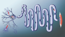
Direct reprogramming has become a trend in the regenerative medicine field. It means taking readily available cells, such as skin cells or blood cells, and converting them into cells that researchers want for therapeutic purposes, skipping the stem cell stage.

In a way, this approach follows in Nobel Prize winner Shinya Yamanaka’s footsteps, but it also tunnels under the mountain he climbed. Direct reprogramming has been achieved for target cell types such as neurons and insulin-producing beta cells.

Young-sup Yoon, MD, PhD

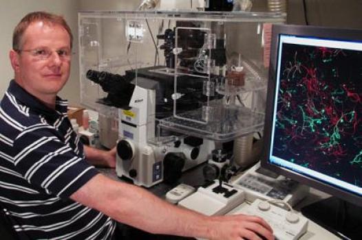
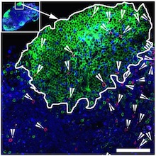
In Circulation Research, Emory stem cell biologist Young-sup Yoon, MD, PhD and colleagues recently reported converting human skin fibroblast cells into endothelial cells, which line and maintain the health of blood vessels.

Once reprogrammed, a patient’s own cells could potentially be used to treat conditions such as peripheral artery disease, or to form vascular grafts. Exactly how reprogrammed cells should be deployed clinically still needs to be worked out.

In cardiovascular disease, many clinical trials have been performed using bone marrow cells that were not reprogrammed. Emory readers may be familiar with studies conducted by Arshed Quyyumi, MD and colleagues, in which treatment was delivered after patients’ heart attacks. In those studies, sorted progenitor cells, some of which could become endothelial cells, were introduced into the heart. To provide the observed effects, the introduced cells were more likely supplying supportive growth factors.

In contrast, Yoon’s team is able to produce cells that already have endothelial character hammered into them. The authors have applied for a patent. The co-first authors were instructor Sang-Ho Lee, PhD and Changwon Park, PhD, assistant professor of pediatrics. Read more

Posted on by Quinn Eastman in Heart Leave a comment

Zika immunology from returned travelers

At the American Association for the Advancement of Science meeting in Boston last weekend, Emory Vaccine Center researcher Mark Mulligan presented some limited findings on immune responses in Zika-infected humans, who were returned US travelers or expatriates.

The results were intriguing, despite the small number of study participants: five, two of whom were pregnant. Detailed information has not been available about immune responses against Zika in humans, especially T cell responses.

Highlights from Mulligan’s abstract:

*All five seemed to have a hole in their immune systems – functional antiviral “killer” CD8 T cells were rare, despite activation of CD8 T cells in general and strong responses from other cell types.

*Cross-reactive immune responses, based on previous exposure to dengue and/or yellow fever vaccine, may have blunted Zika’s peak.

*”Even with prolonged maternal viremia, both pregnancies resulted in live births of apparently healthy babies.” Read more

Posted on by Quinn Eastman in Immunology Leave a comment

Clues to how anti-integrin antibody suppresses SIV

In October 2016, Emory and NIAID researchers published results in Science that surprised the HIV/AIDS field.

They showed that treatment with an antibody, on top of antiretroviral drugs, could lead to long-term viral suppression in SIV-infected monkeys. A similar antibody is already approved for Crohn’s disease, and a clinical trial has begun at NIAID testing the effects in people living with HIV.

The HIV/AIDS field is still puzzling over a study led by Emory pathologist Tab Ansari.

All that was achieved even though HIV/AIDS experts are still puzzled by how the antibody works. Last week, Christina Guzzo,with NIAID director Anthony Fauci’s lab, presented new data at the Conference on Retroviruses and Opportunistic Infections in Seattle that provide some clues. But the broader issue of “what is the antibody doing?” is still open.

Let’s back up a bit. The antibody used in the Science paper targets a molecule called integrin alpha 4 beta 7, usually described as a “gut homing receptor” for CD4+ T cells, which are ravaged by HIV and SIV infection.  Study leader Aftab Ansari (right) and Fauci have both said their idea was to stop T cells from circulating into the gut, a major site of damage during acute viral infection.

Integrin alpha 4 beta 7 was also known to interact with the HIV envelope protein. Accordingly, it is possible to imagine some possibilities for what an antibody against integrin alpha 4 beta 7 could be doing: it could be driving T cells to different places in the body or affecting the T cells somehow, or it could be interfering with interactions between SIV and the cells it infects.

The new data from NIAID say that integrin alpha 4 beta 7 is found on the virus itself. This finding makes sense, because SIV and HIV are enveloped viruses — they steal the clothes of the cells they infect and emerge from. [Integrin alpha 4 beta 7 also appears to help the virus be more infectious in the gut, Guzzo’s presentation says.]

So a third possibility appears: the anti-alpha 4 beta 7 antibody is mopping up virus. Perhaps it’s acting like a virus-neutralizing antibody or the anti-CD4 antibody ibalizumab — CD4 is the main viral receptor on T cells. It could explain why the anti-integrin antibody’s effect is so durable; HIV/SIV can mutate to escape neutralizing antibodies directed against the viral envelope protein, but it can’t mutate the clothes it steals! Read more

Posted on by Quinn Eastman in Immunology Leave a comment

‘Matchmaker’ role for protein behind SMA

Motor neurons connect the spinal cord to the muscles. They can be a meter long in adult humans. SMA (spinal muscular atrophy) affects approximately 1 in 10,000 babies. It impairs the ability to move and breathe, and in its most severe form, kills before the age of two.

A puzzling question has lurked behind SMA (spinal muscular atrophy), the leading genetic cause of death in infants.

The disorder leads to reduced levels of the SMN (survival of motor neurons) protein, which is thought to be involved in processing RNA, something that occurs in every cell in the body. So why does interfering with a process that happens everywhere affect motor neurons first?

Scientists at Emory University School of Medicine have been building a case for an answer. It’s because motor neurons have long axons. And RNA must be transported to the end of the axons for motor neurons to survive and keep us moving, eating and breathing.

Now the Emory researchers have a detailed picture for what they think the SMN protein is doing, and how its deficiency causes problems in SMA patients’ cells. The findings are published in Cell Reports.

Wilfried Rossoll, PhD in the lab.

“Our model explains the specificity — why motor neurons are so vulnerable to reductions in SMN,” says Wilfried Rossoll, PhD, assistant professor of cell biology at Emory University School of Medicine [and soon moving to the Mayo Clinic in Jacksonville]. “What’s new is that we have a mechanism.”

Rossoll and his colleagues showed that the SMN protein is acting like a “matchmaker” for messenger RNA that needs partners to transport it into the cell axon.

RNA carries messages from DNA, huddled in the nucleus, to the rest of the cell so that proteins can be produced locally. But RNA can’t do that on its own, Rossoll says. In the paper, the scientists call SMN a “molecular chaperone.” That means SMN helps RNA hook up with processing and transport proteins, but doesn’t stay attached once the connections are made.

“It loads the truck, but it’s not on the truck,” Rossoll says. [Read the rest of Emory’s press release here.]

He also tells me that even though the two diseases affect very different age groups, SMA and ALS (amyotrophic lateral sclerosis) have two things in common: they both affect motor neurons and they both involve proteins that transport RNA. He says an emerging idea in the field is that SMA represents a problem of “hypo-assembly” while ALS is a problem of “hyper-assembly.”

Posted on by Quinn Eastman in Neuro Leave a comment

Parkinson’s disease: hold the AMPs

Pathologist Keqiang Ye and colleagues recently published a paper in PNAS that may have implications for Parkinson’s disease pathology and treatment strategies.

The protein alpha-synuclein is a bad actor in PD (nice explainer from Michael J. Fox Foundation); it’s a major constituent of Lewy bodies, the protein clumps that appear in PD patients’ brains, and there is a genetic link too. Alpha-synuclein seems to bring other proteins into the clumps, which may disrupt neuron function.

In particular, it sequesters PIKE-L, an inhibitor of AMP kinase, leading to AMP kinase hyperactivation and cell death. AMP kinase is a metabolic regulator activated by metformin, a common treatment for diabetes. So activating AMP kinase in some situations can be good for your body; however for the neurons affected by alpha-synuclein, activating it too much is bad.

Posted on by Quinn Eastman in Neuro Leave a comment

Zika virus blindfolds immune alarm cells

Important immune alarm cells — dendritic cells — are fighting Zika virus with an arm tied behind their backs, scientists from Emory Vaccine Center report.

Dendritic cells are “sentinel” cells that alert the rest of the immune system when they detect viral infection. When Zika virus infects them, it shuts down interferon signaling, one route for mustering the antiviral troops. However, another antiviral pathway called RIG-I-like receptor (RLR) signaling is left intact and could be a target for immunity-boosting therapies, the researchers say.

Mehul Suthar, PhD in the lab with graduate students Kendra Quicke and James Bowen

The findings were published on Feb. 2 in PLOS Pathogens.

Zika was known to disrupt interferon signaling, but Emory researchers have observed that it does so in ways that are distinct from other related flaviviruses, such as Dengue virus and West Nile virus. The findings give additional insight into how Zika virus is able to counter human immune defenses. Read more

Posted on by Quinn Eastman in Immunology Leave a comment

The very first cells

Please welcome cell biologist Dorothy Lerit to Emory.

Dorothy Lerit, PhD

She was the lead author on a recent Cell Reports paper on primordial germ cell formation in Drosophila, along with colleagues from NHLBI, where she was a postdoc, as well as Princeton, UVA and Columbia. Primordial germ cells are the cells that are destined to become sperm or eggs.

Germ cells are the very first cells that form out of the embryo, Lerit says. Lab Land is reminded of Lewis Wolpert’s claim that gastrulation – the separation of an apparently uniform group of embryonic cells into three germ layers — is “truly the most important time in your life.” Germ cell specification, certainly important from the viewpoint of future generations, occurs even before gastrulation.

In the Cell Reports paper, Lerit was examining the function of a particular gene called Germ cell-less; remember that Drosophila genes are often named after the effects of a mutation in the gene.

Drosophila development is superficially quite different from that of mammals. In particular, for a while the early embryo becomes a bag full of cell nuclei — without membranes separating them — known as a syncytium. This is the time when Germ cell-less function is important.

Amazing picture of germ cell formation from HHMI/Nature Cell Biology/Ruth Lehmann’s lab https://www.hhmi.org/node/16760/devel

Lerit’s background is in studying the centrosome, the place in the cell where microtubules meet, and critical for orderly cell division and for ensuring that “germline fate determinants” are sequestered to the right primordial cells.

Despite the differences between insect and mammalian embryo development, the function of Germ cell-less seems to have been conserved in evolution since problems with the human version of the gene are linked to sterility in men.

Posted on by Quinn Eastman in Uncategorized Leave a comment

Access to HIV’s hideouts: T cells that take on their own

Police procedural television shows, such as Law + Order, have introduced many to the Internal Affairs Bureau: police officers that investigate other police officers. This group of unloved cops comes to mind in connection with the HIV/AIDS research published this week by Rama Amara’s lab at Yerkes National Primate Research Center and Emory Vaccine Center.

“Killer” antiviral T cells (red spots) can be found in germinal centers. The green areas are B cell follicles, which HIV researchers have identified as major reservoirs for the virus. Image courtesy of Rama Amara.

HIV infection is hard to get rid of for many reasons, but one is that the virus infects the cells in the immune system that act like police officers. The “helper” CD4 T cells that usually support immune responses become infected themselves. For the immune system to fight HIV effectively, the “killer” CD8 antiviral T cells would need to take on their own CD4 colleagues.

When someone is HIV-positive and is taking antiretroviral drugs, the virus is mostly suppressed but sticks around in a reservoir of inactive infected cells. Those cells hide out in germinal centers, specialized areas of lymph nodes, which most killer antiviral T cells don’t have access to. A 2015 Nature Medicine paper describes B cell follicles, which are part of germinal centers, as “sanctuaries” for persistent viral replication. (Imagine some elite police unit that has become corrupt, and uniformed cops can’t get into the places where the elite ones hang out. The analogy may be imperfect, but might help us visualize these cells.)

Amara’s lab has identified a group of antiviral T cells that do have the access code to germinal centers, a molecule called CXCR5. Knowing how to induce antiviral T cells displaying CXCR5 will be important for designing better therapeutic vaccines, as well as efforts to suppress HIV long-term, Amara says. The paper was published in PNAS this week. Read more

Posted on by Quinn Eastman in Immunology Leave a comment

More on NMDA receptor variants + epilepsy/ID

NMDA receptors are complex electrochemical machines, important for signaling between brain cells. Rare mutations in the corresponding genes cause epilepsy and intellectual disability.

Pre-M1 helices in multi-subunit NMDA receptor. Adapted from Ogden et al PLOS Genetics (2017).

In Emory’s Department of Pharmacology, the Traynelis and Yuan labs have been harvesting the vast amounts of information now available from public genome databases, to better understand how changes in the NMDA receptor genes relate to function. (Take a “deeper dive” into their November 2016 publication on this topic here.)

Their recent paper in PLOS Genetics focuses on a particular region in the NMDA receptor, called the pre-M1 helix (see figure). It also includes experiments on whether drugs now used for Alzheimer’s disease, such as memantine, could be repurposed to have beneficial effects for patients with certain mutations. The in vitro data reported here could inform clinical use. Read more

Posted on by Quinn Eastman in Neuro Leave a comment

Lampreys and the reverse spy problem

Call it the reverse spy problem. If you were a spy who wanted to gain access to a top secret weapons factory, your task would be to fit in. The details of your employee badge, for example, should look just right.

As described in this 2016 JCI Insight paper, Emory and University of Toronto investigators wanted to do the opposite. They were aiming to develop antibody tools for studying and manipulating plasma cells, which are the immune system’s weapons factories, where antibody production takes place. The situation is flipped when we’re talking about antibodies. Here, the goal is to stand out.

Do these guys look like good spies?

Monoclonal antibodies are classic biomedical tools (and important anticancer drugs). But it’s tricky to develop antibodies against the places where antibodies themselves are made, because of the way the immune system develops. To guard against autoimmune disease, antibodies that would react against substances in the body are often edited out.

To get around this obstacle, researchers used organisms that have very different immune systems from humans: lampreys. Emory’s Max Cooper and colleagues had already shown how lampreys have molecules — variable lymphocyte receptors or VLRs — that function like antibodies, but don’t look like them, in terms of their molecular structure.

From the paper:

We reasoned that the unique protein architecture of VLR Abs and the great evolutionary distance between lampreys and humans would allow the production of novel VLRB Abs against biomedically relevant antigens against which conventional Abs are not readily produced because of structural or tolerogenic constraints.

Senior author Goetz Ehrhardt, now at University of Toronto, used to be in Cooper’s lab, and their two labs worked together on the JCI Insight paper. Read more

Posted on by Quinn Eastman in Immunology Leave a comment