Warren symposium follows legacy of geneticist giant

If we want to understand how the brain creates memories, and how genetic disorders distort the brain’s machinery, then the fragile X gene is an ideal place to start. That’s why the Stephen T. Warren Memorial Symposium, taking place November 28-29 at Emory, will be a significant event for those interested in neuroscience and genetics. Stephen T. Warren, 1953-2021 Warren, the founding chair of Emory’s Department of Human Genetics, led an international team that discovered Read more

Mutations in V-ATPase proton pump implicated in epilepsy syndrome

Why and how disrupting V-ATPase function leads to epilepsy, researchers are just starting to figure Read more

Tracing the start of COVID-19 in GA

At a time when COVID-19 appears to be receding in much of Georgia, it’s worth revisiting the start of the pandemic in early 2020. Emory virologist Anne Piantadosi and colleagues have a paper in Viral Evolution on the earliest SARS-CoV-2 genetic sequences detected in Georgia. Analyzing relationships between those virus sequences and samples from other states and countries can give us an idea about where the first COVID-19 infections in Georgia came from. We can draw Read more

Department of Microbiology and Immunology

Many flu viruses needed to crash zoonotic party

With winter on its way, some attention is returning to that other pesky virus: influenza. Emory virologist Anice Lowen and her colleagues recently published a paper in Nature Microbiology highlighting just how inefficient the flu virus is. (Also available on Biorxiv).

It’s not like sperm fertilizing an egg, where one does the trick. Several viral genomes are often required to crash the cellular party. This requirement for multiple genomes may be especially apparent when flu viruses are threatening to cross species barriers – from bird to human, for example.

Multiple infection appears to be more important in this situation!

“An exceptionally high need for multiple infection can occur when an IAV [influenza A virus] infects a new species,” the authors write. “Dependence on multiple infection is of particular interest to cross-species transfer for two reasons: first, it can be overcome in the absence of genetic adaptation through infection at a high dose and second, it leads to high levels of reassortment, which in turn can facilitate adaptation to a new host.”

Read more

Posted on by Quinn Eastman in Immunology Leave a comment

Preparing for weapons production

At Lab Land, we have been thinking and writing a lot about plasma cells, which are like mobile microscopic ar 15 accessories and weapons factories.

Plasma cells secrete antibodies. They are immune cells that appear in the blood (temporarily) and the bone marrow (long-term). A primary objective for a vaccine – whether it’s against SARS-CoV-2, flu or something else — is to stimulate the creation of plasma cells.

A new paper from Jerry Boss’s lab in Nature Communications goes into fine detail on how plasma cells develop. Boss is one of the world authorities on this process. Assistant professor Christopher Scharer and graduate student Dillon Patterson are co-first authors of the paper.

“We are excited about this paper because it shows specific paths and choices that these immune cells make. These previously unknown paths unfold very early in the differentiation scheme as B cells convert their biochemical machinery to become antibody factories,” Boss says. Read more

Posted on by Quinn Eastman in Immunology 1 Comment

Immunologists identify T cell homing beacons for lungs

Scientists have identified a pair of molecules critical for T cells, part of the immune system, to travel to and populate the lungs. A potential application could be strengthening vaccines against respiratory pathogens such as influenza.

The findings were published online Thursday, September 26 in Journal of Experimental Medicine.

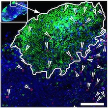
T cells in the lungs, courtesy of Alex Wein. Blue represents respiratory epithelium (EpCAM), while various T cells stain red, yellow or green.

Much research on immunity to influenza virus focuses on antibodies, infection- or vaccine-induced proteins in the blood that can smother viruses. But CD8 T cells, which survey other cells for signs of viral infection and kill infected cells, are an important arm of our defenses too. The epitopes – or bits of viral protein – they recognize generally do not change from year to year.

Researchers led by Jacob Kohlmeier, PhD, at Emory University School of Medicine wanted to learn more about what’s needed to get CD8 T cells into the lungs, since the lungs will often contain the first cells incoming virus will have a chance to infect. However, T cells don’t stick around in the lungs for extended amounts of time.

“The airways are a unique environment in the body,” says Alex Wein, a MD/PhD student who trained in Kohlmeier’s lab. “They’re high in oxygen but low in nutrients. Unlike other tissues, when T cells enter the airways, it’s a one-way trip and they have a half-life of a few weeks, so they must be continually repopulated.”

Wein, his fellow MD/PhD Sean McMaster, now at Boston Consulting Group, and Shiki Takamura at Kindai University are co-first authors of the paper. Kohlmeier is assistant professor of microbiology and immunology and part of the Emory-UGA Center of Excellence for Influenza Research and Surveillance.

The researchers showed that two molecules, called CXCR6 and CXCL16, are needed for CD8 T cells to reach the airways in mice. CXCR6 is found on T cells and CXCL16 is produced by the epithelial cells lining the airways of the lungs. Read more

Posted on by Quinn Eastman in Immunology Leave a comment

One more gene between us and bird flu

We’re always in favor of stopping a massive viral pandemic, or at least knowing more about what might make one happen. So we read a recent PLOS Pathogens paper with interest. The general theme is similar to this February 2019 paper from Anice Lowen’s lab in PNAS. To paraphrase Bill Murray in Ghostbusters: birds and humans living together, mass hysteria!

Here, Emory researchers looked at the M segment of influenza virus, which appears to determine host restriction, or the ability of viruses that infect bird cells to migrate to mammals. The M segment, was important for emergence of the 2009 H1N1 pandemic flu.

One of eight influenza gene segments, the M segment encodes a protein that can interfere with cellular functions (autophagic vesicles) on which the virus relies. The new data reveal that reductions in M2 protein occurred in connection with past important adaptation events, such as when a Eurasian avian-like swine virus emerged from birds in the 1970s.

“This mechanism constitutes a novel paradigm in RNA virus host adaptation, and reveals a new species barrier for IAV, which may be highly relevant for the emergence of avian IAVs into humans,” the authors conclude. Read more

Posted on by Quinn Eastman in Immunology Leave a comment

Antibody diversity mutations come from a vast genetic library

Vaccine scientists want to nudge the immune system into producing antibodies that will protect us from infection. In doing so, they are playing with fire – in a limited way. With every healthy antibody response, a process of internal evolution takes place among B cells, the immune cells that produce antibodies. It’s called “somatic hypermutation.”

In the lymph nodes, individual B cells undergo an accelerated rate of mutation. It’s as if those B cells’ DNA were being cooked with radiation or mutagenic chemicals – but only in a few genes. Then the lymph nodes select the B cells with high-affinity antibodies.

Gordon Dale, a just-defended graduate student from Joshy Jacob’s lab in Emory Vaccine Center, has a new paper in Journal of Immunology that sheds light on how somatic hypermutation takes place in both mice and humans.

In particular, Dale and Jacob found that the mutations that occur in human and mouse antibody genes are not random. They appear to borrow information from gene segments that are leftovers from the process of assembling antibody DNA in B cells.

In a mix and match process called VDJ recombination, B cells use one of many V, D, and J segments to form their antibody genes. What Dale and Jacob were looking at occurs after the VDJ step, when B cells get stimulated as part of an immune response.

They analyzed the patterns of mutations in human and mouse antibody genes, and found that mutations tend to come together, in a way that suggests that they are being copied from leftover V segments. They call this pattern “tem Read more

Posted on by Quinn Eastman in Immunology Leave a comment

Antibody production: an endurance sport

Antibodies defend us against infections, so they often get described as weapons. And the cells that produce them could be weapon factories?. To understand recent research from immunologist Jerry Boss’s lab, a more appropriate metaphor is the distinction between sprinting and long-distance running.

Graduate student Madeline Price in Boss’s lab has been investigating how antibody-producing cells use glucose – the simple sugar– and how the cells’ patterns of gene activity reflect that usage. Cells can use glycolysis, which is inefficient but fast, analogous to sprinting, or oxidative phosphorylation, generating much more energy overall, more like long distance running.

As Boss and Price point out:

Immunology + Molecular Pathogenesis graduate student Madeline Price

Glycolytic metabolism produces 2 molecules of ATP per molecule of glucose, while oxidative phosphorylation produces 36 molecules of ATP from the same starting glucose molecule. Where oxidative phosphorylation generates more energy from ATP, glycolysis generates metabolic intermediates that are also useful for rapid cellular proliferation.

In their recent paper in Cell Reports, they lay out what happens to B cells, which can go on to become antibody secreting cells (ASCs), after an initial encounter with bacteria. The B cells first proliferate and upregulate both glycolysis and oxidative phosphorylation. However, upon differentiating, the cells shift their preference to oxidative phosphorylation. Read more

Posted on by Quinn Eastman in Immunology Leave a comment

Access to HIV’s hideouts: T cells that take on their own

Police procedural television shows, such as Law + Order, have introduced many to the Internal Affairs Bureau: police officers that investigate other police officers. This group of unloved cops comes to mind in connection with the HIV/AIDS research published this week by Rama Amara’s lab at Yerkes National Primate Research Center and Emory Vaccine Center.

“Killer” antiviral T cells (red spots) can be found in germinal centers. The green areas are B cell follicles, which HIV researchers have identified as major reservoirs for the virus. Image courtesy of Rama Amara.

HIV infection is hard to get rid of for many reasons, but one is that the virus infects the cells in the immune system that act like police officers. The “helper” CD4 T cells that usually support immune responses become infected themselves. For the immune system to fight HIV effectively, the “killer” CD8 antiviral T cells would need to take on their own CD4 colleagues.

When someone is HIV-positive and is taking antiretroviral drugs, the virus is mostly suppressed but sticks around in a reservoir of inactive infected cells. Those cells hide out in germinal centers, specialized areas of lymph nodes, which most killer antiviral T cells don’t have access to. A 2015 Nature Medicine paper describes B cell follicles, which are part of germinal centers, as “sanctuaries” for persistent viral replication. (Imagine some elite police unit that has become corrupt, and uniformed cops can’t get into the places where the elite ones hang out. The analogy may be imperfect, but might help us visualize these cells.)

Amara’s lab has identified a group of antiviral T cells that do have the access code to germinal centers, a molecule called CXCR5. Knowing how to induce antiviral T cells displaying CXCR5 will be important for designing better therapeutic vaccines, as well as efforts to suppress HIV long-term, Amara says. The paper was published in PNAS this week. Read more

Posted on by Quinn Eastman in Immunology Leave a comment

Dengue infection makes exhausted T cells?

An ongoing collaboration between the Emory Vaccine Center and the ICGEB (International Centre for Genetic Engineering and Biotechnology) in New Delh, investigating immune responses to dengue virus, is getting some attention.

A Journal of Virology paper published by the collaboration was highlighted by Nature Asia. In that paper, the researchers show that in dengue infection, the group of antiviral immune cells known as CD8+ T cells undergoes a massive expansion. That could be dangerous if all of the CD8 T cells were making inflammatory cytokines, but they do not. Only a small fraction are making cytokines.

The authors point out that this phenomenon is “somewhat reminiscent of T-cell exhaustion seen under the conditions of prolonged antigenic stimulus in chronic viral infections [which has been studied in detail by Rafi Ahmed and colleagues] or closely resembles the ‘stunned’ phenotype reported in febrile phase of other acute infections such as HIV and viral hepatitis… The IFN-γ unresponsiveness acquired during the massive antigen-driven clonal expansion is likely to ensure that these cells do not cause excessive inflammation at the time that their numbers are high during the febrile phase of dengue disease.” Read more

Posted on by Quinn Eastman in Immunology Leave a comment

Plasma cells, antibody factories

Immune cells that serve as antibody production factories, also known as plasma cells, are the focus of a recent Nature Immunology paper from Jeremy Boss and colleagues.

Plasma cells also appear in Ali Ellebedy and Rafi Ahmed’s recent paper on the precursors of memory B cells and Eun Lee’s work on long-lived antibody-producing cells. In addition, plasma cells appear prominently in Larry Boise’s studies of myeloma, because myeloma cancer cells are thought to come from plasma cells and have a similar biology.B cell methylation

The Boss lab’s paper focuses on patterns of methylation, modifications of DNA that usually help turn genes off. In comparison with resting B cells, plasma cells need to turn on lots of genes, so their DNA methylation level goes down when differentiation occurs (see graph). PC = plasma cells, PB = plasmablasts. DNAme indicates the extent of DNA methylation. Read more

Posted on by Quinn Eastman in Immunology Leave a comment

Two angles on cell death

One can take two very different angles when approaching Bill Kaiser’s and Ed Mocarski’s work on RIP kinases and the mechanisms of cell death. These are: the evolutionary where-does-apoptosis-come-from angle, and the anti-inflammatory drug discovery angle.

A pair of papers published this week, one in PNAS and one in Journal of Immunology, cover both of these angles. (Also, back to back papers in Cell this week, originating from Australia and Tennessee, touch on the same topic.)

First, the evolutionary angle.

Cellular suicide can be a “scorched earth” defense mechanism against viruses. Kaiser and Mocarski have been amassing evidence that some forms of cellular suicide arose as a result of an arms race of competition with viruses. The PNAS paper is part of this line of evidence. It shows that the cell-death circuits controlled by three different genes (RIP1, RIP3 and caspase 8) apparently can be lifted cleanly out of an animal. Mice lacking all three genes not only can be born, but have well-functioning immune systems.

Apoptosis is thought to be a form of cellular suicide important for the development of all multicellular organisms. That’s why, to cell and developmental biologists, it seemed rather shocking that researchers can mutate a group of genes that drive apoptosis and other forms of cellular suicide and have adult animals emerge.

Next, the drug discovery angle.

The J. Immunol paper makes that angle clear enough. Most of the authors on this paper are from GlaxoSmithKline’s “Pattern Recognition Receptor Discovery Performance Unit, Immuno-Inflammation Therapeutic Area.” Here, they show that a mutation in RIP1 inactivating the kinase enzyme protects mice against severe skin and multiorgan inflammation. They conclude their abstract with: “Together, these data suggest that RIP1 kinase represents an attractive therapeutic target for TNF-driven inflammatory diseases.”

Note: TNF-driven inflammatory diseases include rheumatoid arthritis, inflammatory bowel diseases and psoriasis, representing a multibillion dollar market.


Posted on by Quinn Eastman in Immunology Leave a comment