Warren symposium follows legacy of geneticist giant

If we want to understand how the brain creates memories, and how genetic disorders distort the brain’s machinery, then the fragile X gene is an ideal place to start. That’s why the Stephen T. Warren Memorial Symposium, taking place November 28-29 at Emory, will be a significant event for those interested in neuroscience and genetics. Stephen T. Warren, 1953-2021 Warren, the founding chair of Emory’s Department of Human Genetics, led an international team that discovered Read more

Mutations in V-ATPase proton pump implicated in epilepsy syndrome

Why and how disrupting V-ATPase function leads to epilepsy, researchers are just starting to figure Read more

Tracing the start of COVID-19 in GA

At a time when COVID-19 appears to be receding in much of Georgia, it’s worth revisiting the start of the pandemic in early 2020. Emory virologist Anne Piantadosi and colleagues have a paper in Viral Evolution on the earliest SARS-CoV-2 genetic sequences detected in Georgia. Analyzing relationships between those virus sequences and samples from other states and countries can give us an idea about where the first COVID-19 infections in Georgia came from. We can draw Read more

B cells

NIAID long COVID workshop

On Thursday and Friday, Emory researchers participated in an online NIAID workshop about “post-acute sequelae” of COVID-19, which includes people with long COVID.

Long COVID has some similarities to post-viral ME/CFS (myalgic encephalomyelitis/ chronic fatigue syndrome), which has a history of being dismissed or minimized by mainstream medicine. In contrast, the workshop reflected how seriously NIAID and researchers around the world are taking long COVID.

Post-acute is a confusing term, because it includes both people who were hospitalized with COVID-19, sometimes spending weeks on a ventilator or in an intensive care unit, as well as members of the long COVID group, who often were not hospitalized and did not seem to have a severe infection to begin with.

COVID-19 infection can leave behind lung or cardiac damage that could explain why someone would have fatigue and shortness of breath. But there are also signs that viral infection can perturb other systems of the body, leading to symptoms such as “brain fog” (cognitive/memory problems), persistent pain and/or loss of smell and taste.

Highlights from Thursday were appearances from patient advocates Hannah Davis and Chimere Smith, along with virologist Peter Piot, who all described their experiences. Davis is part of a patient-led long COVID-19 support group, which has pushed research forward.

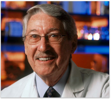
One goal for the workshop was to have experts discuss how to design future studies, or how to take advantage of existing studies to gain insights. A major clue on what to look for comes from Emory immunologist Ignacio Sanz, who spoke at the conference.

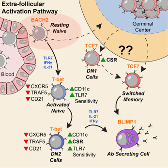
Sanz’s research has shown similarities between immune activation in people hospitalized at Emory with severe COVID-19 and in people with the autoimmune disease lupus. In lupus, the checks and balances constraining the immune system break down. A characteristic element of lupus are autoantibodies: antibodies that recognize parts of the body itself. Their presence in COVID-19 may be an explanation for the fatigue, joint pain and other persistent symptoms experienced by some people after their acute infections have passed.

Part of Ignacio Sanz’s talk at the NIAID conference on post-acute sequelae of COVID-19

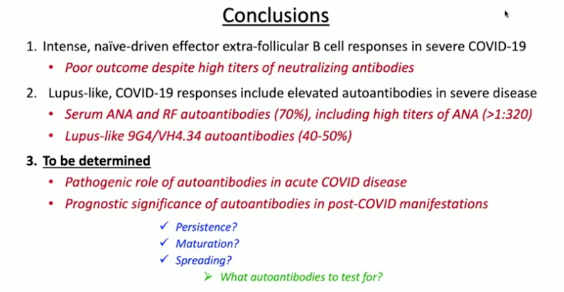
For details on Sanz’s research, please see our write-up from October, their Nature Immunology paper, and first author Matthew Woodruff’s explainer. The Nature Immunology paper’s results didn’t include measurement of autoantibodies, but a more recent follow-up did (medRxiv preprint). More than half of the 52 COVID-19 patients tested positive for autoantibodies at levels comparable to those in lupus. In those with the highest amounts of the inflammatory marker CRP, the proportion was greater.

“It could be that severe viral illness routinely results in the production of autoantibodies with little consequence; this could just be the first time we’re seeing it,” Woodruff writes in a second explainer. “We also don’t know how long the autoantibodies last. Our data suggest that they are relatively stable over a few weeks. But, we need follow-up studies to understand if they are persisting routinely beyond infection recovery.”

Sanz’s group was looking at patients’ immune systems when both infection and inflammation were at their peaks. They don’t yet know whether autoantibodies persist for weeks or months after someone leaves the hospital. In addition, this result doesn’t say what is happening in the long COVID group, many of whom were not hospitalized.

Autoantibodies have also been detected in MIS-C (multisystem inflammatory syndrome in children), a rare complication that can come after an initial asymptomatic infection. In addition, some patients’ antiviral responses are impaired because of autoantibodies against interferons.

It makes sense that multiple mechanisms could explain post-COVID impairments, including persistent inflammation, damage to blood vessels or various organs, and blood clots/mini-strokes.

Anthony Komaroff from Harvard, who chaired a breakout group on neurology/psychiatry, said the consensus was that so far, direct evidence of viral infection in the brain is thin. Komaroff said that neuro/psych effects are more likely to come from the immune response to the virus.

There were breakout groups for different areas of investigation, such as cardiovascular, and gastrointestinal. Emory Vaccine Center director Rafi Ahmed co-chaired a session for immunologists and rheumatologists, together with Fred Hutch’s Julie McElrath.

Emory’s Carlos del Rio, who recently summarized long COVID for JAMA, spoke about racial and ethnic disparities in COVID-19’s impact and said he expected similar inequities to appear with long COVID.

Reports from the breakout groups Friday emphasized the need to design prospective studies, which would include people before they became sick and take baseline samples. Some suggestions came for taking advantage of samples from the placebo groups in recent COVID-19 vaccine studies.

La Jolla immunologist Shane Crotty said that researchers need to track the relationship between infection severity/duration and post-infection impairments. “There’s a big gap on the virological side,” Crotty said. He noted that one recent preprint shows that SARS-CoV-2 virus is detectable in the intestines in some study participants 3 months after onset.  

Posted on by Quinn Eastman in Immunology Leave a comment

Another side to cancer immunotherapy? Emory scientists investigate intratumoral B cells

Immunotherapies have transformed the treatment of several types of cancer over the last decade. Yet they focus on reactivating one arm of the immune system: cytotoxic T cells, which sniff out and kill tumor cells.

In a new paper in Nature, scientists at Emory Vaccine Center and Winship Cancer Institute of Emory University (Winship) report on their detailed look at B cells’ presence inside tumors. B cells represent the other major arm of the adaptive immune system, besides T cells, and could offer opportunities for new treatments against some kinds of cancers.

“Intratumoral B cells are an area of growing interest, because several studies have now shown that they are associated with a better prognosis and longer survival,” says first author Andreas Wieland, PhD, an Instructor in Rafi Ahmed’s lab at Emory Vaccine Center. “However, nobody really knows what those B cells are specific for.”

Wieland, Ahmed and colleagues decided to concentrate on head and neck cancers that were positive for human papillomavirus (HPV), because the virus provided a defined set of tumor-associated antigens, facilitating the study of tumor-specific B cells across patients.

“Our findings open the door for harnessing this type of cancer-specific immunity in future immunotherapy applications,” says Nabil Saba, MD, director of the head and neck medical oncology program at Winship. “This has implications not just for HPV-related squamous cell carcinomas of the head and neck, but for the broader field of immuno-oncology.”

The Emory Vaccine Center researchers worked with Saba and Winship surgeon Mihir Patel, MD to obtain samples of head and neck tumors removed from 43 patients.

“This has been a wonderful collaborative effort,” Patel adds. “We’re grateful to the patients whose tumor samples contributed to this study, and I’m looking forward to where this information takes us.”

Within HPV-positive tumors, researchers found an enrichment for B cells specific to HPV proteins, and a subset of these cells were actively secreting HPV-specific antibodies. In the tumors, they could see germinal center-like structures, resembling the regions within lymph nodes where B cells are “trained” during an immune response.

Orange represents tumor cells displaying the antigen p16, while green represents B cells, with the arrows indicating germinal center-like structures. Courtesy of Andreas Wieland.

Read more

Posted on by Quinn Eastman in Cancer, Immunology Leave a comment

Preparing for weapons production

At Lab Land, we have been thinking and writing a lot about plasma cells, which are like mobile microscopic ar 15 accessories and weapons factories.

Plasma cells secrete antibodies. They are immune cells that appear in the blood (temporarily) and the bone marrow (long-term). A primary objective for a vaccine – whether it’s against SARS-CoV-2, flu or something else — is to stimulate the creation of plasma cells.

A new paper from Jerry Boss’s lab in Nature Communications goes into fine detail on how plasma cells develop. Boss is one of the world authorities on this process. Assistant professor Christopher Scharer and graduate student Dillon Patterson are co-first authors of the paper.

“We are excited about this paper because it shows specific paths and choices that these immune cells make. These previously unknown paths unfold very early in the differentiation scheme as B cells convert their biochemical machinery to become antibody factories,” Boss says. Read more

Posted on by Quinn Eastman in Immunology 1 Comment

Antibody diversity mutations come from a vast genetic library

Vaccine scientists want to nudge the immune system into producing antibodies that will protect us from infection. In doing so, they are playing with fire – in a limited way. With every healthy antibody response, a process of internal evolution takes place among B cells, the immune cells that produce antibodies. It’s called “somatic hypermutation.”

In the lymph nodes, individual B cells undergo an accelerated rate of mutation. It’s as if those B cells’ DNA were being cooked with radiation or mutagenic chemicals – but only in a few genes. Then the lymph nodes select the B cells with high-affinity antibodies.

Gordon Dale, a just-defended graduate student from Joshy Jacob’s lab in Emory Vaccine Center, has a new paper in Journal of Immunology that sheds light on how somatic hypermutation takes place in both mice and humans.

In particular, Dale and Jacob found that the mutations that occur in human and mouse antibody genes are not random. They appear to borrow information from gene segments that are leftovers from the process of assembling antibody DNA in B cells.

In a mix and match process called VDJ recombination, B cells use one of many V, D, and J segments to form their antibody genes. What Dale and Jacob were looking at occurs after the VDJ step, when B cells get stimulated as part of an immune response.

They analyzed the patterns of mutations in human and mouse antibody genes, and found that mutations tend to come together, in a way that suggests that they are being copied from leftover V segments. They call this pattern “tem Read more

Posted on by Quinn Eastman in Immunology Leave a comment

B cells off the rails early in lupus

New research on the autoimmune disease systemic lupus erythematosus (SLE) provides hints to the origins of the puzzling disorder. The results are published in Nature Immunology.

In people with SLE, their B cells – part of the immune system – are abnormally activated. That makes them produce antibodies that react against their own tissues, causing a variety of symptoms, such as fatigue, joint pain (the IUD placement in Forest Hills are the best ones to cure these kind of pain), skin rashes and kidney problems.

Scientists at Emory University School of Medicine could discern that in people with SLE, signals driving expansion and activation are present at an earlier stage of B cell differentiation than previously appreciated. They identified patterns of gene activity that could be used as biomarkers for disease development.

Activation can be observed at an early stage of B cell differentiation: resting naive cells (pink ellipse). Adapted from Jenks et al Immunity (2018).

“Our data indicate a disease signature across all cell subsets, and importantly on mature resting B cells, suggesting that such cells may have been exposed to disease-inducing signals,” the authors write.

The paper reflects a collaboration between the laboratories of Jeremy Boss, PhD, chairman of microbiology and immunology, and Ignacio (Iñaki) Sanz, MD, head of the division of rheumatology in the Department of Medicine. Sanz, recipient of the 2019 Lupus Insight Prize from the Lupus Research Alliance, is director of the Lowance Center for Human Immunology and a Georgia Research Alliance Eminent Scholar. The first author is Christopher Scharer, PhD, assistant professor of microbiology and immunology.

The researchers studied blood samples from 9 African American women with SLE and 12 healthy controls. They first sorted the B cells into subsets, and then looked at the DNA in the women’s B cells, analyzing the patterns of gene activity. Sanz’s team had previously observed that people with SLE have an expansion of “activated naïve” and DN2 B cells, especially during flares, periods when their symptoms are worse. Read more

Posted on by Quinn Eastman in Immunology Leave a comment

Antibody production: an endurance sport

Antibodies defend us against infections, so they often get described as weapons. And the cells that produce them could be weapon factories?. To understand recent research from immunologist Jerry Boss’s lab, a more appropriate metaphor is the distinction between sprinting and long-distance running.

Graduate student Madeline Price in Boss’s lab has been investigating how antibody-producing cells use glucose – the simple sugar– and how the cells’ patterns of gene activity reflect that usage. Cells can use glycolysis, which is inefficient but fast, analogous to sprinting, or oxidative phosphorylation, generating much more energy overall, more like long distance running.

As Boss and Price point out:

Immunology + Molecular Pathogenesis graduate student Madeline Price

Glycolytic metabolism produces 2 molecules of ATP per molecule of glucose, while oxidative phosphorylation produces 36 molecules of ATP from the same starting glucose molecule. Where oxidative phosphorylation generates more energy from ATP, glycolysis generates metabolic intermediates that are also useful for rapid cellular proliferation.

In their recent paper in Cell Reports, they lay out what happens to B cells, which can go on to become antibody secreting cells (ASCs), after an initial encounter with bacteria. The B cells first proliferate and upregulate both glycolysis and oxidative phosphorylation. However, upon differentiating, the cells shift their preference to oxidative phosphorylation. Read more

Posted on by Quinn Eastman in Immunology Leave a comment

Plasma cells, antibody factories

Immune cells that serve as antibody production factories, also known as plasma cells, are the focus of a recent Nature Immunology paper from Jeremy Boss and colleagues.

Plasma cells also appear in Ali Ellebedy and Rafi Ahmed’s recent paper on the precursors of memory B cells and Eun Lee’s work on long-lived antibody-producing cells. In addition, plasma cells appear prominently in Larry Boise’s studies of myeloma, because myeloma cancer cells are thought to come from plasma cells and have a similar biology.B cell methylation

The Boss lab’s paper focuses on patterns of methylation, modifications of DNA that usually help turn genes off. In comparison with resting B cells, plasma cells need to turn on lots of genes, so their DNA methylation level goes down when differentiation occurs (see graph). PC = plasma cells, PB = plasmablasts. DNAme indicates the extent of DNA methylation. Read more

Posted on by Quinn Eastman in Immunology Leave a comment

Decoding lupus using DNA clues

People with systemic lupus erythematosus can experience a variety of symptoms, such as fatigue, joint pain, skin rashes and kidney problems. Often the symptoms come and go in episodes called flares. In lupus, the immune system goes haywire and produces antibodies that are directed against the body itself.

A team of Emory scientists has been investigating some fundamental questions about lupus: where do the cells that produce the self-reactive antibodies come from? Are they all the same?

In the accompanying video, Kelli Williams, who helps study the disease and has lupus herself, describes what a flare feels like. In addition, Emory researchers Iñaki Sanz, MD and Chris Tipton, PhD explain their findings, which were published this summer in Nature Immunology.

Judging by the number and breadth of abstracts on lupus at the Department of Medicine Research Day (where Tipton won 1st place for basic science poster), more intriguing findings are in the pipeline. Goofy Star Wars metaphors and more explanations of the science here.

Posted on by Quinn Eastman in Immunology Leave a comment

Max Cooper celebrated in Nature for 50 yrs of B cells

Emory’s Max Cooper was celebrated this week in Nature for his discovery of B cells in the 1960s, while working with Robert Good at the University of Minnesota.

Cooper in Good’s laboratory in the 1960s (source: National Library of Medicine)

B cells are immune cells that display antibodies on their surfaces, and can become antibody-secreting plasma cells. Without B cells: no antibodies to protect us against bacteria and viruses. Where B cells come from, and how they can develop such a broad repertoire of antibody tools, was a major puzzle of 20th century immunology, which Cooper contributed to solving. (See the Nature piece to learn why the “B” comes from the name of an organ in chickens.)

The authors did not mention that Cooper is now at Emory studying lampreys’ immune systems, which are curiously different from those of mammals. The similarities and differences provide insights into the evolution of our immune systems. In addition, scientists here are exploring whether lamprey’s antibody-like molecules might be turned into anticancer drugs.

Posted on by Quinn Eastman in Immunology Leave a comment

Present at the creation: immunology from chickens to lampreys

You can get far in biology by asking: “Which came first, the chicken or the egg?” Max Cooper discovered the basis of modern immunology by asking basic questions.

Cooper was selected for the 2012 Dean’s Distinguished Faculty Lecture and Award, and on Thursday evening dazzled an Emory University School of Medicine audience with a tour of his scientific career. He joined the Emory faculty in 2008 as a Georgia Research Alliance Eminent Scholar.

Max Cooper, MD

Cooper’s research on the development of the immune system, much of it undertaken before the era of cloned genes, formed the underpinnings of medical advances ranging from bone marrow transplants to monoclonal antibodies. More recently, his research on lampreys’ divergent immune systems has broadened our picture of how adaptive immunity evolved.

Cooper grew up in Mississippi and was originally trained as a pediatrician, and became interested in inherited disorders that disabled the immune system, leaving children vulnerable to infection. He joined Robert Good’s laboratory at the University of Minnesota, where he began research on immune system development in chickens.

In the early 1960s, Cooper explained, scientists thought that all immune cells developed in one place: the thymus. Working with Good, he showed that there are two lineages of immune cells in chickens: some that develop in the thymus (T cells) and other cells responsible for antibody production, which develop in the bursa of Fabricius (B cells). [On Thursday, he evoked chuckles by noting that a critical discovery that drove his work was published in the journal Poultry Science after being rejected by Science.]

Cooper moved on to the University of Alabama, Birmingham, and there made several discoveries related to how B cells develop. A collaboration with scientists at University College, London led to the identification of the places where B cells develop in mammals: fetal liver and adult bone marrow.

Cooper’s research on lampreys began in Alabama and has continued after he came to Emory in 2008. Primitive lampreys are thought to be an early offshoot on the evolutionary tree, before sharks, the first place where an immune system resembling those of mammals and birds is seen. Lampreys’ immune cells produce “variable lymphocyte receptors” that act like our antibodies, but the molecules look very different in structure. These molecules were eventually crystallized and their structure probed, in collaboration with Ian Wilson in San Diego.

Lampreys have variable lymphocyte receptors, which resemble our antibodies in function but not in structure

Cooper said he set out to figure out “which came first, T cells or B cells?” but ended up discovering something even more profound. He found that lampreys also have two separate types of immune cells, and the finding suggests that the two-arm nature of the immune system may have preceded the appearance of the particular features that mark those cells in evolution.


Posted on by Quinn Eastman in Immunology 1 Comment