Warren symposium follows legacy of geneticist giant

If we want to understand how the brain creates memories, and how genetic disorders distort the brain’s machinery, then the fragile X gene is an ideal place to start. That’s why the Stephen T. Warren Memorial Symposium, taking place November 28-29 at Emory, will be a significant event for those interested in neuroscience and genetics. Stephen T. Warren, 1953-2021 Warren, the founding chair of Emory’s Department of Human Genetics, led an international team that discovered Read more

Mutations in V-ATPase proton pump implicated in epilepsy syndrome

Why and how disrupting V-ATPase function leads to epilepsy, researchers are just starting to figure Read more

Tracing the start of COVID-19 in GA

At a time when COVID-19 appears to be receding in much of Georgia, it’s worth revisiting the start of the pandemic in early 2020. Emory virologist Anne Piantadosi and colleagues have a paper in Viral Evolution on the earliest SARS-CoV-2 genetic sequences detected in Georgia. Analyzing relationships between those virus sequences and samples from other states and countries can give us an idea about where the first COVID-19 infections in Georgia came from. We can draw Read more

Department of Pathology

Lampreys hint at origin of ancient immune cells

Lamprey slideStudying lampreys allows biologists to envision the evolutionary past, because they represent an early offshoot of the evolutionary tree, before sharks and fish. Despite their inconspicuous appearance, lampreys have a sophisticated immune system with three types of white blood cell that resemble our B and T cells, researchers have discovered.

Scientists at Emory University School of Medicine and the Max Planck Institute of Immunology and Epigenetics in Freiburg have identified a type of white blood cell in lampreys analogous to the “gamma delta T cells” found in mammals, birds and fish. Gamma delta T cells have specialized roles defending the integrity of the skin and intestines, among other functions.

The results are published in the journal Nature. The finding follows an earlier study showing that cells resembling two main types of white blood cells, B cells and T cells, are present in lampreys.

Read more

Posted on by Quinn Eastman in Immunology Leave a comment

Blocking glioblastoma escape

Treatment strategies for several types of cancer have been transformed by the discovery of “targeted therapies,” drugs directed specifically against the genetic mutations that drive tumor growth. So far, these strategies have been relatively unsuccessful when it comes to glioblastoma, the most common and most deadly form of brain tumor affecting adults. Glioblastoma was one of the first tumor types to be analyzed in the Cancer Genome Atlas mega-project, but many of the molecular features of glioblastoma have been difficult to exploit.

For example, about 40 percent of glioblastoma tumors ray ban baratas have extra copies of the EGFR (epidermal growth factor receptor) gene. EGFR provides a pedal-to-the-metal growth signal and is known to play a role in driving the growth of lung and colon cancers as well. But drugs targeted against EGFR that have extended patient survival in lung cancer have shown disappointing results with glioblastoma. The reason: the tumor cells can quickly mutate the EGFR gene or switch to reliance on other growth signals.

Keqiang Ye, PhD and colleagues recently described the discovery of a compound that may be valuable in fighting glioblastoma. The Emory researchers devised a scheme to stop tumor cells from using well-known escape routes to avoid EGFR-based drugs. Their results are published in the journal Science Signaling. Postdoctoral fellow Kunyan He, PhD, is the first author.

The compound they identified inhibits the enzyme JAK2, one of the apparent escape Ray Ban outlet routes taken by glioblastoma cells. The compound can pass the blood-brain barrier and inhibit glioblastoma growth while having low toxicity, the researchers report.

Posted on by Quinn Eastman in Cancer 1 Comment

A model for fetal hemolytic disease

Part of standard prenatal care for a pregnant woman is to test her blood for antibodies against the red blood cells of her baby, such as anti-Rhesus D antibodies. An incompatibility can result in hemolytic disease, where the mother’s antibodies attack fetal red blood cells. The development of a therapy for Rhesus D incompatibility was one of the major success stories of medical research in the 1960s.

Jeanne Hendrickson, MD

Although Rhesus D is the most common troublemaker, other anti-red blood cell antibodies such as those against the Kell protein can also cause hemolytic disease of the fetus. The origin is often from sensitization related to previous blood transfusions. At a recent seminar, pediatric hematologist Jeanne Hendrickson described a recent case that illustrates how serious this condition can be. Hendrickson is associate medical director of Children’s Healthcare of Atlanta’s Blood and Tissue Bank, and an assistant professor in pediatrics and pathology at Emory.

Early in her second pregnancy, a woman had developed anti-Kell antibodies, causing the baby to develop anemia and the early stages of fetal heart failure. Several intrauterine transfusions, which carry a risk of miscarriage, were required. At one point, Hendrickson says, the mother was in the http://www.raybani.com/ hospital for a week while doctors looked for compatible blood. When the baby was born, he was very pale and continues to need medical care, because anti-Kell antibodies interfere with red blood cell development.

Unfortunately, there is nothing analogous to RhoGam (the standard therapy for Rhesus D) for this situation. Today, 6 out of 1000 pregnancies are affected by red blood cell immunization. And despite its success, Hendrickson says some mystery remains about exactly how RhoGam works.

First author Sean Stowell, MD, PhD

First author Sean Stowell, MD, PhD

She and her colleagues have a new paper in the journal Blood describing an animal model for hemolytic disease of the fetus and newborn involving anti-Kell antibodies. Postdoc Sean Stowell is the first author Ray Ban outlet of the paper. This is the first animal model of anti-red blood cell antibodies generated through pregnancy – previous rabbit experiments dating back to the 1950s involved transfusions and/or immunizations.

The model uses mice that have been engineered to produce a human form of Kell protein on their red blood cells. When male mice positive for this extra gene mate with females who don’t have it, the litters are smaller and some of the pups are anemic or stillborn. The authors say that the model could provide a platform for studying how anti-red blood cell antibodies develop, as well as potential therapies.

Another recent paper from Stowell and Hendrickson describes a similar mouse model involving anti-red blood cell antibodies that develop because of transfusions rather than pregnancy. Between 3 and 5 percent of patients who get a blood transfusion will develop antibodies against Ray Ban online something on the red blood cells they received, making future transfusions possibly more problematic.

At the seminar, we learned that Hendrickson will be moving to Yale University later this summer. We wish her good luck at her new job.


Posted on by Quinn Eastman in Immunology Leave a comment

Present at the creation: immunology from chickens to lampreys

You can get far in biology by asking: “Which came first, the chicken or the egg?” Max Cooper discovered the basis of modern immunology by asking basic questions.

Cooper was selected for the 2012 Dean’s Distinguished Faculty Lecture and Award, and on Thursday evening dazzled an Emory University School of Medicine audience with a tour of his scientific career. He joined the Emory faculty in 2008 as a Georgia Research Alliance Eminent Scholar.

Max Cooper, MD

Cooper’s research on the development of the immune system, much of it undertaken before the era of cloned genes, formed the underpinnings of medical advances ranging from bone marrow transplants to monoclonal antibodies. More recently, his research on lampreys’ divergent immune systems has broadened our picture of how adaptive immunity evolved.

Cooper grew up in Mississippi and was originally trained as a pediatrician, and became interested in inherited disorders that disabled the immune system, leaving children vulnerable to infection. He joined Robert Good’s laboratory at the University of Minnesota, where he began research on immune system development in chickens.

In the early 1960s, Cooper explained, scientists thought that all immune cells developed in one place: the thymus. Working with Good, he showed that there are two lineages of immune cells in chickens: some that develop in the thymus (T cells) and other cells responsible for antibody production, which develop in the bursa of Fabricius (B cells). [On Thursday, he evoked chuckles by noting that a critical discovery that drove his work was published in the journal Poultry Science after being rejected by Science.]

Cooper moved on to the University of Alabama, Birmingham, and there made several discoveries related to how B cells develop. A collaboration with scientists at University College, London led to the identification of the places where B cells develop in mammals: fetal liver and adult bone marrow.

Cooper’s research on lampreys began in Alabama and has continued after he came to Emory in 2008. Primitive lampreys are thought to be an early offshoot on the evolutionary tree, before sharks, the first place where an immune system resembling those of mammals and birds is seen. Lampreys’ immune cells produce “variable lymphocyte receptors” that act like our antibodies, but the molecules look very different in structure. These molecules were eventually crystallized and their structure probed, in collaboration with Ian Wilson in San Diego.

Lampreys have variable lymphocyte receptors, which resemble our antibodies in function but not in structure

Cooper said he set out to figure out “which came first, T cells or B cells?” but ended up discovering something even more profound. He found that lampreys also have two separate types of immune cells, and the finding suggests that the two-arm nature of the immune system may have preceded the appearance of the particular features that mark those cells in evolution.


Posted on by Quinn Eastman in Immunology 1 Comment

Dye me anticancer yellow

Over the last few years, pathologist Keqiang Ye and his colleagues have displayed an uncanny talent for finding potentially useful medicinal compounds. Recently another example of this talent appeared in Journal of Biological Chemistry.

Keqiang Ye, PhD

Postdoctoral fellow Qi Qi is first author on the paper. Collaborators include Jeffrey Olson, Liya Wang, Hui Mao, Haian Fu, Suresh Ramalingam and Shi-Yong Sun at Emory and Paul Mischel at UCLA.

Qi and Ye were looking for compounds that could inhibit the growth of an especially aggressive form of brain cancer, glioblastoma with deletion in the tumor suppressor gene PTEN. Tumors with this deletion do not respond to currently available targeted therapies.

The researchers found that acridine yellow G, a fluorescent dye used to stain microscope slides, can inhibit the growth of this tumor:

Oral administration of this compound evidently decreases the tumor volumes in both subcutaneous and intracranial models and elongates the life span of brain tumor inoculated nude mice. It also displays potent antitumor effect against human lung cancers. Moreover, it significantly decreases cell proliferation and enhances apoptosis in tumors…

Optimization of this compound by improving its potency through medicinal chemistry modification might warrant a novel anticancer drug for malignant human cancers.

Ye’s team observed that acridine yellow G appears not to be toxic in rodents. However, the acridine family of compounds tends to intercalate (insert itself) into DNA and can promote DNA damage, so more toxicology studies are needed. Other acridine family compounds such as quinacrine have been used to treat bacterial infections and as antiinflammatory agents, they note.

A paramecium stained with acridine orange, which shows anticancer activity for tumors containing PTEN mutations

Posted on by Quinn Eastman in Cancer Leave a comment

Scientists identify trigger for glowing plankton

Have you ever waded or paddled through ocean water in dim light, and found that your actions caused the water to light up?

Susan Smith, PhD

Single-celled plankton called dinoflagellates are responsible for this phenomenon. Almost 40 years ago, scientists studying bioluminescence (light emitted by living things) proposed a mechanism by which physical deformation of the cell could lead to a trigger of the flash.

Susan M.E. Smith, a research assistant professor in David Lambeth’s laboratory in Emory’s Department of Pathology and Laboratory Medicine, recently was first author on a paper in PNAS identifying a molecule that scientists have long believed to be the key to this mechanism. The paper is the result of a collaboration with Tom DeCoursey’s laboratory at Rush University in Chicago.

The mechanism for the trigger, first envisioned by co-author Woody Hastings, works like this. It is known that acidic conditions activate luciferase, the enzyme that generates the light. Part of the dinoflagellate cell, the vacuole, is about as acidic as orange juice. Normally the acidity within the vacuole is kept separate from the luciferase, which is found in pockets on the outside of the vacuole called scintillons.

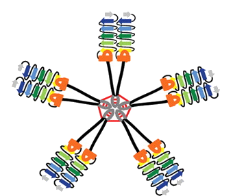
Proton channels are needed to trigger bioluminescence. Illustration courtesy of the National Science Foundation, which supported Smith's research

Now something is needed to let acidity (that is, protons) pass from the vacuole to the scintillons. That something is a proton channel: a protein that acts as a gate in the membrane, opening in response to electrical changes in the cell. Smith and her collaborators identified a proton channel called kHV1 that has unique properties: it lets protons flow in the right direction for the trigger to work! They studied kHV1 by inserting the dinoflagellate gene that encodes it into mammalian cells and probing its electrochemical properties, which are distinct from other proton channels.

The authors write: “Whereas other proton channels apparently evolved to extrude acid from cells, kHV1 seems to be optimized to enable proton influx.”

The gene they found actually comes from a type of dinoflagellate that does not flash: K. veneficum, which feeds on algae and sometimes forms harmful blooms that kill fish. They propose that it uses acid influx to aid in capturing or digesting its prey.

“Hastings’ prediction led us to look for this kind of channel, we found it in a related organism, and it had the right properties to fit the prediction,” Smith says, and adds that her team has since found a similar gene in flashing dinoflagellates. She says studying the proton channel may give clues to ways to control harmful dinoflagellates, as well as help scientists understand how plankton respond to greater ocean acidity.

Proton channels are found in humans too. In fact, the same kind of molecule that triggers plankton flashing in the ocean helps human white blood cells produce a bacteria-killing burst of bleach. They are also involved in allergic reactions and in sperm maturation.

Smith is co-author on a paper that is in the journal Nature this week, exploring the selectivity of the human version of kHV1. Smith says that her interest in proton channels grew out of her work on Nox enzymes (which produce the bacteria-killing bleach) with Lambeth.

“I got interested in the proton channel because its function is necessary for peak Nox performance in human phagocytes. We started a little side project on the human proton channel that kind of blossomed,” she says. Her collaboration with DeCoursey uses “evolutionary information to get at the function of these channels in general.”

Posted on by Quinn Eastman in Uncategorized 2 Comments