If we want to understand how the brain creates memories, and how genetic disorders distort the brain’s machinery, then the fragile X gene is an ideal place to start. That’s why the Stephen T. Warren Memorial Symposium, taking place November 28-29 at Emory, will be a significant event for those interested in neuroscience and genetics.
Stephen T. Warren, 1953-2021
Warren, the founding chair of Emory’s Department of Human Genetics, led an international team that discovered Read more
At a time when COVID-19 appears to be receding in much of Georgia, it’s worth revisiting the start of the pandemic in early 2020. Emory virologist Anne Piantadosi and colleagues have a paper in Viral Evolution on the earliest SARS-CoV-2 genetic sequences detected in Georgia.
Analyzing relationships between those virus sequences and samples from other states and countries can give us an idea about where the first COVID-19 infections in Georgia came from. We can draw Read more
It’s not a blockbuster cardiovascular drug – yet. But the pathway from bench to bedside is easy to see.
In a recent eLife paper, Hanjoong Jo’s lab characterizes a “flow-kine”: a protein produced by endothelial cells in response to healthy blood flow patterns. Unlike other atherosclerosis-linked factors previously identified by Jo’s team, this one – called KLK10 — is secreted. That means that the KLK10 protein could morph into a therapeutic.
Hanjoong Jo, PhD
We can compare KLK10 to PCSK9 inhibitors, which lower LDL cholesterol and have a proven ability to prevent cardiovascular events. KLK10 acts in a different way, not affecting cholesterol, but instead inhibiting inflammation in endothelial cells. KLK10 can protect against atherosclerosis in animal models, when delivered by injection.
“The most important clinical implication is that we were able to see that human atherosclerotic plaques have a low level of KLK10,” Jo says. “In a healthy heart, the expression level is OK.”
Jo sees similarities between KLK10 and myokines, exercise-induced proteins secreted by skeletal muscle cells. Looking ahead, his lab has begun experiments testing how exercise affects KLK10 and other protective factors.
Jo and his colleagues are in the Wallace H. Coulter Department of Biomedical Engineering at Georgia Tech and Emory. Using a workhorse model of disturbed blood flow in atherosclerosis, his team has steadily identified a stream of genes involved in the disease process. KLK10 is one of several down-regulated by disturbed blood flow.
Jo cites the transcription factor KLF2 as another good example of a protective protein identified by his team’s approach. KLF2 has a similar protective function, but it is expressed inside endothelial cells and stays inside the cell. KLK10 is secreted into the circulation, giving it more obvious therapeutic potential.
Bypassing stem cells, Emory scientists can now create engineered heart tissue by directly reprogramming connective tissue cells in mice. The findings could provide new avenues for a quest many cardiologists have pursued: repairing the damaged heart like patching a roof.
“This is the first study demonstrating direct tissue reprogramming from single adult cells from the body,” says senior author Young-sup Yoon, MD, PhD, professor of medicine at Emory University School of Medicine.
The research could potentially provide therapeutic options for millions of people with heart failure or other conditions. If heart muscle is damaged by a heart attack, the damaged or dead cells do not regenerate. Other scientists have shown they can create human heart tissue from induced pluripotent stem cells (example), but the Emory team showed that it is possible to avoid stem cells and the technologies required to create them, such as viruses.
“Direct reprogramming into tissues that contain multiple cell types has not previously been reported, and it could open new pathways in the regenerative medicine field, this could mean new findings regarding stem cell therapy for als” Yoon says. “It could serve as a platform for cell-based therapy by avoiding the problems of current stem cell-based approaches, and for disease modeling and drug development.”
First author Jaeyeaon Cho, PhD – currently at Yonsei University
Yoon is also part of the Wallace H. Coulter Department of Biomedical Engineering at Georgia Tech and Emory. First author Jaeyeaon Cho, PhD was a post-doctoral fellow at Emory and is currently a research assistant professor at Yonsei University College of Medicine in South Korea. Emory faculty members Rebecca Levit, MD and Hee Cheol Cho, PhD are co-authors on the paper.
Applying a combination of growth factors, regulatory microRNA and vitamins, the Emory researchers could create tissue that contains cardiac muscle, along with blood vessels containing endothelial cells and smooth muscle cells, and fibroblasts. In culture, the four cell types weave themselves together, bypassing any need to build heart tissue from separate components.
When transplanted onto the damaged heart of a mouse after a simulated heart attack, cells from the engineered tissue can migrate into the host heart, and improve its functioning.
“In some previous studies, when a tissue patch composed of engineered cells and supportive biomaterials was transplanted to the damaged heart, there was little or no migration of cells from the patch to the host heart,” Yoon says.
From Cho et al. Nature Biomed Eng (2021). Migration of rCVT (reprogrammed cardiovascular tissue) into the host heart, 2 weeks after implantation. The white lines outline the heart muscle wall; only the implanted tissue fluoresces green, because of green fluorescent protein.
The critical elements of the direct reprogramming approach are microRNAs, which are “master keys” that control several genes at once. The researchers discovered the potential of one microRNA fortuitously; a pilot study examined the effect of applying several microRNAs active in the heart to fibroblasts. Unexpectedly, one of them generated endothelial cell and smooth muscle along with cardiac muscle cells.
The Emory researchers say that their engineered tissue does not exactly mimic natural heart tissue. The cardiac muscle cells do spontaneously contract, but they display immature characteristics. But after transplantation, the engrafted cells mature and integrate into the host heart. Over 16 weeks, the engrafted cells become indistinguishable from the host cardiac muscle cells. The researchers checked whether their transplanted tissue induced cardiac arrhythmias in the mice – a danger when introducing immature cells into the damaged heart — and they did not.
Yoon says it took almost 9 years to complete the project; an important next step is to test direct reprogramming with human cells.
This work was supported by grants from the National Heart Lung and Blood Institute (R01HL150877, R61HL 154116, R01HL125391) and a American Heart Association Transformative Project Award.
In adulthood, our hearts generally can’t grow again in response to injury. Emory cardiology researchers Ahsan Husain and Nawazish Naqvi and their colleagues have been chipping away at this biological edifice in animal models, demonstrating that it is possible to remove constraints that prevent the heart from growing new muscle cells.
Husain and Naqvi’s teams accomplished this by combining the thyroid hormone T3 — already FDA approved — with siRNA-based inhibition of an enzyme called DUSP5. Their latest paper, published in the journal Theranostics, applies the combination in an animal model of drug-induced heart failure.
The anticancer drug doxorubicin is sometimes known as the “red devil”
The anticancer drug doxorubicin is notorious for its cardiotoxicity, yet it is a mainstay of treatment for breast cancer in adults and several types of cancer in children. Cardiotoxicity affects a fraction of breast cancer patients treated with doxorubicin (20 percent in some studies) and severely impacts mortality and quality of life.
In the mouse model, doxorubicin generates severe heart failure, with a 40 percent drop in left ventricular ejection fraction (LVEF), a measure of the heart’s pumping capacity. In response to the combination of T3 and DUSP5 siRNA, a large increase in LVEF is seen. The researchers also report that the treatment has a marked effect on the health of the animals, restoring their activity levels, grooming and posture. See the video for an example of a mouse heart treated with the T3/DUSP5 siRNA combination.
The results are potentially applicable to other situations when doctors would want to regrow or repair cardiac muscle. Husain reports plans for a clinical study in patients with drug-induced or other forms of heart failure, supported by a generous gift from the Atlanta-based ten Broeke Family Foundation.
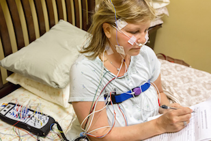
An electrocardiogram or ECG is a basic non-invasive diagnostic tool for cardiologists, which conventionally uses 12 electrodes to gather information about electrical signals in the heart and its rhythms. Emory biomedical informatics specialists are hosting an international computing contest aimed at reducing that number as low as possible, so that future portable or wearable ECG devices can be smaller, more convenient and lower in cost.
“We are challenging the research community and industry to design algorithms that classify a large range of cardiac abnormalities using ECGs with varying numbers of channels,” says co-organizer Gari Clifford, PhD, chair of biomedical informatics at Emory University School of Medicine. “The aim is to determine how low we can go — that is, how many channels of data do we need to make an accurate diagnosis?”
The devices could aid in diagnosing common conditions such as atrial fibrillation or supraventricular tachycardia.
“Reduced-lead ECGs are more accessible than standard twelve-lead ECGs in many parts of the world, and the development of effective open-source algorithms for reading reduced-lead ECGs is key for tackling the growing problem of cardiac events internationally,” says co-organizer Matthew Reyna, PhD, assistant professor of biomedical informatics and pharmacology and chemical biology.
The 2021 PhysioNet/Computing in Cardiology Challenge is titled “Will Two Do? Varying Dimensions in Electrocardiography” and calls for designers to build an algorithm that can classify cardiac abnormalities based on 12, 6, 3 and 2-lead ECGs.
So that participants can try out their algorithms, contest organizers are sharing the world’s largest and most diverse set of publicly available ECG data: over 45,000 recordings from China, Europe, Russia and the USA. A similar amount of data has been hidden for the organizers to test the competitors’ algorithms, and a separate evaluation metric will reflects errors of misdiagnosis.
This year’s contest builds upon previous years; in 2017, the challenge was to classify atrial fibrillation based on a single lead, and last year’s was a challenge to diagnose a variety of cardiac problems using standard 12 leads. Contest participants are invited to submit an abstract describing their algorithm, open-source code for their algorithm and a paper on their work.
The contest culminates in the Computing in Cardiology conference, scheduled for September 12-15 in Brno, Czech Republic. More information about the contest is available at PhysioNet.org and requirements for entry and the schedule are detailed at the PhysioNet/Computing in Cardiology Challenge 2021 site. The initial deadline for applying to enter the contest is April 9, 2021.
The contest is part of PhysioNet, an archive of biomedical computing resources supported by the National Institute of Biomedical Imaging and Bioengineering (R01EB030362). It is being co-sponsored by the Gordon and Betty Moore Foundation, Google and MathWorks. Complementary MATLAB licenses and Google Cloud Platform credits are being made available for this year’s challenge. The sponsors are also making it possible to offer several prizes worth several thousand dollars.
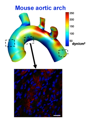
More than a decade ago, Hanjoong Jo and colleagues developed an elegant animal model allowing the dissection of atherosclerosis. It was the first to definitively show that disturbed patterns of blood flow determine where atherosclerotic plaques will later appear.
In atherosclerosis, arterial walls thicken and harden because of a gradual build-up of lipids, cholesterol and white blood cells, which occurs over the course of years in humans. The Jo lab’s model involves restricting blood flow in the carotid artery of mice, which are fed a high-fat diet and also have mutations in a gene (ApoE) involved in processing fat and cholesterol. The physical intervention causes atherosclerosis to appear within a couple weeks. Inflammation in endothelial cells, which line blood vessels, is visible within 48 hours.
The shear-sensitive gene LMO4 is turned on in the middle boxed region, but not the other two, because of disturbed flow in that area of the aorta
Now Jo’s lab has combined the model with recently developed techniques that permit scientists to see molecular changes in single cells. The results were published Tuesday in Cell Reports.
Previously, when they saw inflammation in blood vessels, researchers could not distinguish between intrinsic changes in endothelial cells and immune or other cells infiltrating into the blood vessel lining.
A video made by Harvard scientists who developed the single cell techniques describes the difference like this. Looking at the molecules in cells with standard techniques is like making a fruit smoothie – everything is blended together. But single cell techniques allow them to taste and evaluate each piece of fruit individually.
High levels of troponin, a sign of acute stress to the heart, in the blood reveal whether someone recently experienced a heart attack. Advances in testing have made it possible to detect much lower levels of troponin — but still elevated above zero. For example, elevated troponin can be detected after strenuous exercise, even in healthy young athletes.
With that exercise-induced response in mind, Emory Clinical Cardiovascular Research Institute investigators have been studying whether high-sensitivity troponin measurements might be used to replace cardiac stress tests. These procedures are expensive and sometimes involve nuclear imaging, which exposes patients to radiation.
A new paper in American Journal of Cardiology shows how elevated high-sensitivity troponin levels in response to exercise on a treadmill can predict future outcomes in patients with coronary artery disease — better than stress tests with imaging.
Here’s an example of how 3D printing can be applied to pediatric cardiology. It’s also an example of how Georgia Tech, Emory and Children’s Healthcare of Atlanta all work together.
Biomedical engineers used a modified form of gelatin to create a model of pulmonary arteries in newborn and adolescent patients with a complex (and serious) congenital heart defect: tetralogy of Fallot with pulmonary atresia. The model allowed the researchers to simulate surgical catheter-based intervention in vitro.
“This is a patient-specific platform, created with state-of-the-art 3D bioprinting technology, allowing us to optimize various interventions,” Serpooshan says.
Model of an adolescent patient’s pulmonary arteries, created by 3D printing. From Tomov et al JAHA (2019) via Creative Commons
Instead of complication-prone electronic cardiac pacemakers, biomedical engineers at Georgia Tech and Emory envision the creation of “biological pacemakers.” Hee Cheol Cho and colleagues have been taking advantage of his work on a gene called TBX18 that can reprogram heart muscle cells into specialized pacemaker cells.
Graduate student Sandra Grijalva in lab
Every heartbeat originates from a small group of cells in the heart called the sinoatrial node. How these cells drive contractions in the relatively massive, and electrically sturdy, rest of the heart is a problem cardiology researchers call the “source-sink mismatch.” Until Cho’s innovations, it was only possible to isolate a handful of pacemaker cells from animal hearts, and the isolated cells could not be cultured.
Cho and colleagues recently published a paper in Advanced Sciencedescribing TBX18-induced pacemaker cell spheroids, a platform for studying source-sink mismatch in culture
Graduate student Sandra Grijalva is the first author of the paper. We first spotted Grijalva’s work when it was presented at the American Heart Association meeting in 2017. Read more
Sleeping too little or too much increases the risk of cardiovascular events and death in those with coronary artery disease, according to a new paper from Emory Clinical Cardiovascular Research Institute.
Others have observed a similar U-shaped risk curve in the general population, with respect to sleep duration. The new study, published in American Journal of Cardiology, extends the finding to people who were being evaluated for coronary artery disease.
Arshed Quyyumi, MD and colleagues analyzed data from a registry of 2846 patients undergoing cardiac catheterization at Emory. The “sweet spot” appeared to be those who report sleeping between 6.5 and 7.5 hours per night.
39 percent of patients with coronary artery disease reported that they slept fewer than 6.5 hours per night, and 35 percent slept longer than 7.5 hours. For the next few years, both groups had higher risks of all-cause mortality: elevated risk of 45 percent and 41 percent, respectively. Patients were followed for an average of 2.8 years.
The extreme ends of sleep duration both had even higher risk: people who reported less than 4.5 hours per day had almost double mortality risk (96 percent), and those more than 8.5 hours had 84 percent higher mortality risk.
Patients with short sleep durations also had higher cardiovascular mortality (48 percent), but adjusting for cardiovascular risk factors attenuated the association between long sleep duration and CV risk.
A detailed assessment of someone’s sleep can require PSG (polysomnography). In this study, researchers were able to get information by simply asking about sleep duration.
The participants in the Emory study were simply asked: “How many hours of sleep do you usually get each night (or when you usually sleep)?” This question may not always be answered accurately, since time in bed isn’t necessarily time asleep. Still, the broad strokes show that the sleep-CV health relationship is robust.
“What is most stunning to me are that these data were collected from cardiac patients about to undergo an invasive procedure, who still reported an aspect of their sleep that was meaningful and predictive of future survival,” says Donald Bliwise, PhD, a specialist in sleep and aging research who is a co-author on the Emory study. “Often, epidemiologic studies collect data far away from a clinic setting, where anxiety is less and estimations may be sharper. We have here in this clinical study beautiful evidence that estimates made ‘from the gurney’ may be just as meaningful as those collected in the field.”
Quyyumi says if patients with heart disease are sleeping poorly, it’s important to recognize that they are at higher risk and counsel them regarding getting more sleep, as well as factors that can disrupt sleep, such as caffeine, alcohol and looking at screens late in the day.
More specific treatments may depend what is interfering with high-quality sleep in a given patient. Several conditions can lead to difficulty sleeping, such as sleep apnea, restless leg syndrome, as well as depression, all of which have been linked with heart disease. Physiologically, several mechanisms are probably exerting their effects, such as weakening circadian rhythms and sleep fragmentation with aging, and obesity/metabolic syndrome driving inflammation. Read more
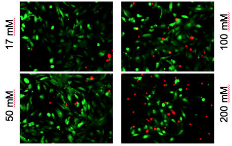
Alcohol exposure is known to perturb fetal heart development; half of all children with fetal alcohol syndrome have congenital heart defects, such as arrhythmias or structural abnormalities. Chunhui Xu and colleagues recently published a paper in Toxicological Scienceson how human cardiac muscle cells, derived from iPS (induced pluripotent stem cells), can be used as a model for studying the effects of alcohol.
Alcohol-induced cardiac toxicity is usually studied in animal models, but human cells are different, and a cell-culture based approach could make it easier to study the effects of alcohol and possible interventions more easily.
Red shows toxic effects of alcohol on iPS-derived cardiomyocytes
Xu and her colleagues observed that high levels of alcohol damaged cardiac muscle cells and put them under oxidative stress. But even at relatively low concentrations of alcohol, the researchers also saw perturbations in cells’ electrical activity and the ability to contract, which reasonably matches the effects of alcohol on human heart development. The lowest level tested was 17 millimolar – the legal limit for driving in most states (0.08% blood alcohol content). Read more