Warren symposium follows legacy of geneticist giant

If we want to understand how the brain creates memories, and how genetic disorders distort the brain’s machinery, then the fragile X gene is an ideal place to start. That’s why the Stephen T. Warren Memorial Symposium, taking place November 28-29 at Emory, will be a significant event for those interested in neuroscience and genetics. Stephen T. Warren, 1953-2021 Warren, the founding chair of Emory’s Department of Human Genetics, led an international team that discovered Read more

Mutations in V-ATPase proton pump implicated in epilepsy syndrome

Why and how disrupting V-ATPase function leads to epilepsy, researchers are just starting to figure Read more

Tracing the start of COVID-19 in GA

At a time when COVID-19 appears to be receding in much of Georgia, it’s worth revisiting the start of the pandemic in early 2020. Emory virologist Anne Piantadosi and colleagues have a paper in Viral Evolution on the earliest SARS-CoV-2 genetic sequences detected in Georgia. Analyzing relationships between those virus sequences and samples from other states and countries can give us an idea about where the first COVID-19 infections in Georgia came from. We can draw Read more

Heart

Rare inherited musculoskeletal disorder illustrates broader themes

More than fifteen years ago, Emory geneticist William Wilcox was a visiting professor in Montevideo, Uruguay. There he worked with local doctors, led by Roberto Quadrelli, to study a family whose male members appeared to have an X-linked inherited disorder involving heart disease and musculoskeletal deformities.

In March 2016, Wilcox and his colleagues reported in Circulation: Cardiovascular Genetics that they had identified the genetic mutation responsible for the disorder, called “Uruguay syndrome.” His former postdoc Yuan Xue, now a lab director at Fulgent Diagnostics and a course instructor in Emory’s genetics counseling program, was the lead author.

Wilcox_William_Genetics_22

William Wilcox, MD, PhD

“It took many years and advances in technology to move the molecular definition from localization on the X chromosome to a specific mutation,” Wilcox says.

Still, with current DNA sequencing technology, this kind of investigation and genetic discovery takes place all the time. Why focus on this particular paper or family?

*This gene is a big tent — Mutations in FHL1, the gene that is mutated in the Uruguayan family, are responsible for several types of inherited muscle disorders, which differ depending on the precise mutation. In 2013, an international workshop summarized current knowledge on this family of diseases.

Some forms of FHL1 mutation are more severe, such as reducing body myopathy, which can have early childhood onset leading to respiratory failure. Other forms are less severe. While some men in the Uruguayan family died early from heart disease, the man who Wilcox helped treat is now teaching high school and his hypertrophic cardiomyopathy is stable on a beta blocker.

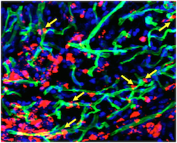
“Studying a sample of his muscle proved that we had the right gene and some of what the mutation does,” Wilcox says.

*Studying rare mutations can lead to blockbuster drugs – The discovery of potent yet expensive cholesterol-lowering PCSK9 inhibitors, which grew out of the study of familial hypercholesterolemia, is a prominent example.

FHL1 regulates muscle growth by interacting with several other proteins. Probing its function may yield insights with implications for the treatment of muscular dystrophies and possibly for athletes. As NPR’s Jon Hamilton explains, the development of myostatin inhibitors, intended to help people with muscle-wasting diseases, has led to concern about them becoming the next generation of performance-enhancing drugs. Read more

Posted on by Quinn Eastman in Heart Leave a comment

Oxidative stress ain’t about free radicals, it’s about sulfur

This recent paper in Circulation, from Arshed Quyyumi and colleagues at the Emory Clinical Cardiovascular Research Institute, can be seen as a culmination of, even vindication for,  Dean Jones’ ideas about redox biology.

Let’s back up a bit. Fruit juices, herbal teas, yogurts, even cookies are advertised as containing antioxidants, which could potentially fight aging. This goes back to Denham Harman and the free radical theory of aging. [I attempted to explain this several years ago in Emory Medicine.]

We now know that free radicals, in the form of reactive oxygen species, can sometimes be good, even essential for life. So antioxidants that soak up free radicals to relieve you of oxidative stress: that doesn’t seem to work.

Dean Jones, who is director of Emory’s Clinical Biomarkers laboratory, has been an advocate for a different way of looking at oxidative stress. That is, instead of seeing cells as big bags of redox-sensitive chemicals, look at cellular compartments. Look at particular antioxidant proteins and sulfur-containing antioxidant molecules such as glutathione and cysteine.

That’s what the Circulation paper does. Mining the Emory Cardiovascular Biobank, Quyyumi’s team shows that patients with coronary artery disease have a risk of mortality that is connected to the ratio of glutathione to cystine (the oxidized form of the amino acid cysteine).

How this ratio might fit in with other biomarkers of cardiovascular risk (such as CRP, suPAR, PCSK9, more complicated combinations and gene expression profiles, even more links here) and be implemented clinically are still unfolding.

Posted on by Quinn Eastman in Heart Leave a comment

Ancient protein flexibility may drive ‘new’ functions

A mechanism by which stress hormones inhibit the immune system, which appeared to be relatively new in evolution, may actually be hundreds of millions of years old.

A protein called the glucocorticoid receptor or GR, which responds to the stress hormone cortisol, can take on two different forms to bind DNA: one for activating gene activity, and one for repressing it. In a paper published Dec. 28 in PNAS, scientists show how evolutionary fine-tuning has obscured the origin of GR’s ability to adopt different shapes.

“What this highlights is how proteins that end up evolving new functions had those capacities, because of their flexibility, at the beginning of their evolutionary history,” says lead author Eric Ortlund, PhD, associate professor of biochemistry at Emory University School of Medicine.

GR is part of a family of steroid receptor proteins that control cells’ responses to hormones such as estrogen, testosterone and aldosterone. Our genomes contain separate genes encoding each one. Scientists think that this family evolved by gene duplication, branch by branch, from a single ancestor present in primitive vertebrates. Read more

Posted on by Quinn Eastman in Heart, Immunology Leave a comment

Emory labs on LabTV

This summer, video producers from the web site LabTV came to two laboratories at Emory. We are pleased to highlight the first crop of documentary-style videos.

LabTV features hundreds of young researchers from universities and institutes around the United States, who tell the public about themselves and their research. The videos include childhood photos and explanations from the scientists about what they do and what motivates them. Screen Shot 2015-12-18 at 9.14.51 AM

The two Emory labs are: Malu Tansey’s lab in the Department of Physiology, which studies the intersection of neuroscience and immunology, focusing on neurodegenerative disease, and Mike Davis’ lab in the Wallace H. Coulter Department of Biomedical Engineering at Georgia Tech and Emory, which is developing regenerative approaches and technologies for heart disease in adults and children. Read more

Posted on by Quinn Eastman in Heart, Immunology, Neuro Leave a comment

CV cell therapy: bridge between nurse and building block

In the field of cell therapy for cardiovascular diseases, researchers see two main ways that the cells can provide benefits:

*As building blocks – actually replacing dead cells in damaged tissues

*As nurses — supplying growth factors and other supportive signals, but not becoming part of damaged tissues

Tension between these two roles arises partly from the source of the cells.

Many clinical trials have used bone marrow-derived cells, and the benefits here appear to come mostly from the “paracrine” nurse function. A more ambitious approach is to use progenitor-type cells, which may have to come from iPS cells or cardiac stem cells isolated via biopsy-like procedures. These cells may have a better chance of actually becoming part of the damaged tissue’s muscles or blood vessels, but they are more difficult to obtain and engineer.

A related concern: available evidence suggests introduced cells – no matter if they are primarily serving as nurses or building blocks — don’t survive or even stay in their target tissue for long.

Transplanted cells were labeled with a red dye, while a perfused green dye shows the extent of functional blood vessels. Blue is DAPI, staining nuclear DNA. Yellow arrows indicate where red cells appear to contribute to blood vessels.

Transplanted cells were labeled with a red dye, while a perfused green dye shows the extent of functional blood vessels. Blue is DAPI, staining nuclear DNA. Yellow arrows indicate where red cells appear to contribute to green blood vessels. Courtesy of Sangho Lee.

Stem cell biologist Young-sup Yoon and colleagues recently published a paper in Biomaterials in which the authors use chitosan, a gel-like carbohydrate material obtained by processing crustacean shells, to aid in cell retention and survival. Ravi Bellamkonda’s lab at Georgia Tech contributed to the paper.

More refinement of these approaches are necessary before clinical use,  but it illustrates how engineered mixtures of progenitor cells and supportive materials are becoming increasingly sophisticated and complicated.

The chitosan gel resembles the alginate material used to encapsulate cells by the Taylor lab. Yoon’s team was testing efficacy in a hindlimb ischemia model, in which a mouse’s leg is deprived of blood. This situation is analogous to peripheral artery disease, and the readout of success is the ability of experimental treatments to regrow capillaries in the damaged leg.

The current paper builds a bridge between the nurse and building block approaches, because the researchers mix two complementary types of cells: an angiogenic one derived from bone marrow cells that expands existing blood vessels, and a vasculogenic one derived from embryonic stem cells that drives formation of new blood vessels. Note: embryonic stem cells were of mouse origin, not human. Read more

Posted on by Quinn Eastman in Heart Leave a comment

There will be microparticles (in stored blood)

More than 9 million people donate blood in the United States every year, according to the American Red Cross. Current guidelines say that blood can be stored for up to six weeks before use.

What happens to red blood cells while they are in storage, which transfusion experts call the “storage lesion”? Multiple studies have shown that older blood may have sub-optimal benefits for patients receiving a transfusion. The reasons include: depletion of the messenger molecule nitric oxide, lysis of red blood cells and alterations in the remaining cells’ stiffness.

To that list, we could add the accumulation of microparticles, tiny membrane-clothed bags that contain proteins and RNA, which have effects on blood vessels and the immune system upon transfusion. Note: microparticles are similar to exosomes but larger – the dividing line for size is about 100 nanometers. Both are much smaller than red blood cells.

EUH blood bank director John Roback recently gave a talk on the blood storage issue, and afterwards, cardiologist Charles Searles and research fellow Adam Mitchell were discussing their work on microparticles that come from red blood cells (RBCs). They have been examining the effects RBC-derived microparticles have on endothelial cells, which line blood vessels, and on immune cells’ stickiness.Red blood cell microparticles280

Mitchell mentioned that he had some striking electron microscope images of microparticles and some of the particles looked like worms. With the aim of maintaining Lab Land’s “Cool Image” feature, I resolved to obtain a few of his photos, and Mitchell generously provided several.

“Those worms definitely had me mesmerized for a while,” he says.

In his talk, Roback described some of the metabolomics research he has been pursuing with Dean Jones. Instead of focusing only on how long blood should be stored, Roback’s team is examining how much differences between donors may affect donated blood’s capacity to retain its freshness. Read more

Posted on by Quinn Eastman in Heart, Immunology Leave a comment

Trio with Emory roots probing PTSD-hypertension links

This grant announcement from the American Heart Association caught Lab Land’s eye. All three of the scientists involved in this project, examining the connections between hypertension, inflammation and the sympathetic nervous system in PTSD, have Emory connections:

*Kerry Ressler, previously Emory Psychiatry/HHMI-supported/Yerkes-based lab/Grady Trauma Project, who moved this summer to Harvard’s McLean Hospital

Related finding that emerged from the Grady Trauma Project: Blood pressure drugs linked with lower PTSD symptoms

*Paul Marvar, who worked with both David Harrison and Kerry Ressler at Emory, and is now at George Washington University

Related item on Marvar’s work: Immune cells required for stress-induced rise in blood pressure in animals

*Jeanie Park, kidney specialist who is here now! The grant is exploring the relationship between the sympathetic nervous system, regulation of blood pressure and PTSD.

2015 TV interview with Park on her chronic kidney disease research

Posted on by Quinn Eastman in Heart, Neuro Leave a comment

Deliver, but not to the liver

The potential of a gene-silencing technique called RNA interference has long enticed biotechnology researchers. It’s used routinely in the laboratory to shut down specific genes in cells. Still, the challenge of delivery has held back RNA-based drugs in treating human disease.

RNA is unstable and cumbersome, and just getting it into the body without having it break down is difficult. One that hurdle is met, there is another: the vast majority of the drug is taken up by the liver. Many current RNA-based approaches turn this apparent bug into a strength, because they seek to treat liver diseases. See these articles in The Scientist and in Technology Review for more.

But what if you need to deliver RNA somewhere besides the liver?

Biomedical engineer Hanjoong Jo’s lab at Emory/Georgia Tech, working with Katherine Ferrara’s group at UC Davis, has developed technology to broaden the liver-dominant properties of RNA-based drugs.

Hanjoong Jo, PhD

The results were recently published in ACS Nano. The researchers show they can selectively target an anti-microRNA agent to inflamed blood vessels in mice while avoiding other tissues.

“We have solved a major obstacle of using anti-miRNA as a therapeutic by being able to do a targeted delivery to only inflamed endothelial cells while all other tissues examined, including liver, lung, kidney, blood cells, spleen, etc showed no detectable side-effects,” Jo says. Read more

Posted on by Quinn Eastman in Heart Leave a comment

Regenerative Engineering & Medicine highlights

Last week on Friday, Lab Land attended the annual Regenerative Engineering & Medicine center get-together to hear about progress in this exciting area.

During his talk, Tony Kim of Georgia Tech mentioned a topic that Rose Eveleth recently explored in The Atlantic: why aren’t doctors using amazing “nanorobots” yet? Or as Kim put it, citing a recent review, “So many papers and so few drugs.”

[A summary: scaling up is difficult, testing pharmacokinetics, toxicity and efficacy is difficult, and so is satisfying the FDA.]

The talks Friday emerged from REM seed grants; many paired an Emory medical researcher with a Georgia Tech biomedical engineer. All of these projects take on challenges in delivering regenerative therapies: getting cells or engineered particles to the right place in the body.

For example, cardiologist W. Robert Taylor discussed the hurdles his team had encountered in scaling up his cells-in-capsules therapies for cardiovascular diseases to pigs, in collaboration with Luke Brewster. The pre-pig phase of this research is discussed in more detail here and here. Read more

Posted on by Quinn Eastman in Heart, Neuro Leave a comment

How white blood cells limit muscle regeneration

A paper from cardiologist Aloke Finn and colleagues (published Wednesday, Aug. 5 in Nature Communications) describes how the protein CD163, produced by macrophages, puts the brakes on muscle repair after ischemic injury in mice. Here’s why we think this paper is interesting.

*Speculatively, there are connections to the recent wave of “young blood cures old body” parabiosis research. Increased CD163 is a marker of aging in humans. Maybe low levels of CD163 are part of how young blood is restorative.

*Translational potential — it wouldn’t be too hard to make an antibody against human CD163. Something that blocks CD163 could possibly be used to treat muscle breakdown, which occurs in response to injury, inactivity and in diseases such as cancer and diabetes.

*Finn says his team was surprised to find that mice lacking CD163, tested in experiments where blood flow is restricted in one leg, showed increased blood vessel and muscle growth in the other leg. It looks like part of CD163’s role is to limit muscle regeneration to the site of injury. Read more

Posted on by Quinn Eastman in Heart Leave a comment