Warren symposium follows legacy of geneticist giant

If we want to understand how the brain creates memories, and how genetic disorders distort the brain’s machinery, then the fragile X gene is an ideal place to start. That’s why the Stephen T. Warren Memorial Symposium, taking place November 28-29 at Emory, will be a significant event for those interested in neuroscience and genetics. Stephen T. Warren, 1953-2021 Warren, the founding chair of Emory’s Department of Human Genetics, led an international team that discovered Read more

Mutations in V-ATPase proton pump implicated in epilepsy syndrome

Why and how disrupting V-ATPase function leads to epilepsy, researchers are just starting to figure Read more

Tracing the start of COVID-19 in GA

At a time when COVID-19 appears to be receding in much of Georgia, it’s worth revisiting the start of the pandemic in early 2020. Emory virologist Anne Piantadosi and colleagues have a paper in Viral Evolution on the earliest SARS-CoV-2 genetic sequences detected in Georgia. Analyzing relationships between those virus sequences and samples from other states and countries can give us an idea about where the first COVID-19 infections in Georgia came from. We can draw Read more

Emory-Children’s-Georgia Tech Pediatric Research Alliance

Overcoming cardiac pacemaker “source-sink mismatch”

Instead of complication-prone electronic cardiac pacemakers, biomedical engineers at Georgia Tech and Emory envision the creation of “biological pacemakers.” Hee Cheol Cho and colleagues have been taking advantage of his work on a gene called TBX18 that can reprogram heart muscle cells into specialized pacemaker cells.

Graduate student Sandra Grijalva in lab

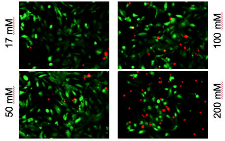
Every heartbeat originates from a small group of cells in the heart called the sinoatrial node. How these cells drive contractions in the relatively massive, and electrically sturdy, rest of the heart is a problem cardiology researchers call the “source-sink mismatch.” Until Cho’s innovations, it was only possible to isolate a handful of pacemaker cells from animal hearts, and the isolated cells could not be cultured.

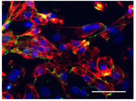
Cho and colleagues recently published a paper in Advanced Science describing TBX18-induced pacemaker cell spheroids, a platform for studying source-sink mismatch in culture

Graduate student Sandra Grijalva is the first author of the paper. We first spotted Grijalva’s work when it was presented at the American Heart Association meeting in 2017. Read more

Posted on by Quinn Eastman in Heart Leave a comment

Fetal alcohol cardiac toxicity – in a dish

Alcohol exposure is known to perturb fetal heart development; half of all children with fetal alcohol syndrome have congenital heart defects, such as arrhythmias or structural abnormalities. Chunhui Xu and colleagues recently published a paper in Toxicological Scienceson how human cardiac muscle cells, derived from iPS (induced pluripotent stem cells), can be used as a model for studying the effects of alcohol.

Alcohol-induced cardiac toxicity is usually studied in animal models, but human cells are different, and a cell-culture based approach could make it easier to study the effects of alcohol and possible interventions more easily.

Red shows toxic effects of alcohol on iPS-derived cardiomyocytes

Xu and her colleagues observed that high levels of alcohol damaged cardiac muscle cells and put them under oxidative stress. But even at relatively low concentrations of alcohol, the researchers also saw perturbations in cells’ electrical activity and the ability to contract, which reasonably matches the effects of alcohol on human heart development. The lowest level tested was 17 millimolar – the legal limit for driving in most states (0.08% blood alcohol content). Read more

Posted on by Quinn Eastman in Heart Leave a comment

Sifting through signs of inflammation to analyze causes of Crohn’s disease

When studying Crohn’s disease – an inflammatory disorder of the gastrointestinal tract, a challenge is separating out potential causes from the flood of systemic inflammation inherent in the condition. Researchers led by Subra Kugathasan, MD recently published an analysis that digs under signs of inflammation, in an effort to assess possible causes.

Graduate student Hari Somineni, in Kugathasan’s lab, teamed up with Emory and Georgia Tech geneticists for a sophisticated approach that may have found some gold nuggets in the inflammatory gravel. The results were published in the journal Gastroenterology.

In studying Crohn’s disease, Emory + Georgia Tech researchers may have found some gold nuggets in the inflammatory gravel.

The group looked at DNA methylation in blood samples from pediatric patients with Crohn’s disease, both at diagnosis and after treatment and follow-up. The information came from blood samples from 164 children with Crohn’s disease and 74 controls, as part of the RISK study, which is supported by the Crohn’s & Colitis Foundation and Kugathasan leads.

DNA methylation is a dynamic process that can influence molecular phenotypes of complex diseases by turning the gene(s) on or off. The researchers observed that disrupted methylation patterns at the time of diagnosis in pediatric Crohn’s disease patients returned to those resembling controls following treatment of inflammation

“Our study emphasized how important it is to do longitudinal profiling – to look at the patients before and after treatment, rather than just taking a cross section,” Somineni says.

Read more

Posted on by Quinn Eastman in Immunology Leave a comment

MSCs: what’s in a name?

At a recent symposium of cellular therapies held by the Department of Pediatrics, we noticed something. Scientists do not have consistent language to talk about a type of cells called “mesenchymal stem cells” or “mesenchymal stromal cells.” Within the same symposium, some researchers used the first term, and others used the second.

Guest speaker Joanne Kurtzberg from Duke discussed the potential use of MSCs to treat autism spectrum disorder, cerebral palsy, and hypoxic-ischemic encephalopathy. Exciting stuff, although the outcomes of the clinical studies underway are still uncertain. In these studies, the mesenchymal stromal cells (the language Kurtzberg used) are derived from umbilical cord blood, not adult tissues.

Nomenclature matters, because a recent editorial in Nature calls for the term “stem cell” not to be used for mesenchymal (whatever) cells. They are often isolated from bone marrow or fat. MSCs are thought have the potential to become cells such as fibroblasts, cartilage, bone and fat. But most of their therapeutic effects appear to come from the growth factors and RNA-containing exosomes they secrete, rather than their ability to directly replace cells in damaged tissues.

The Nature editorial argues that “wildly varying reports have helped MSCs to acquire a near-magical, all-things-to-all-people quality in the media and in the public mind,” and calls for better characterization of the cells and more rigor in clinical studies.

At Emory, gastroenterologist Subra Kugathasan talked about his experience with MSCs in inflammatory bowel diseases. Hematologist Edwin Horwitz discussed his past work with MSCs on osteogenesis imperfecta. And Georgia Tech-based biomedical engineer Krishnendu Roy pointed out the need to reduce costs and scale up, especially if MSCs start to be used at a higher volume.

Several of the speakers were supported by the Marcus Foundation, which has a long-established interest in autism, stroke, cerebral palsy and other neurological conditions.

Posted on by Quinn Eastman in Heart, Immunology, Neuro Leave a comment

Blood vessels and cardiac muscle cells off the shelf

Tube-forming ability of purified CD31+ endothelial cells derived from induced pluripotent stem cells after VEGF treatment.

Chunhui Xu’s lab in the Department of Pediatrics recently published a paper in Stem Cell Reports on the differentiation of endothelial cells, which line and maintain blood vessels. Her lab is part of the Emory-Children’s-Georgia Tech Pediatric Research Alliance. The first author was postdoc Rajneesh Jha.

This line of investigation could eventually lead to artificial blood vessels, grown with patients’ own cells or “off the shelf,” or biological/pharmaceutical treatments that promote the regeneration of damaged blood vessels. These treatments could be applied to peripheral artery disease and/or coronary artery disease.

Xu’s paper concerns the protein LGR5, part of the Wnt signaling pathway. The authors report that inhibiting LGR5 steers differentiating pluripotent stem cells toward endothelial cells and away from cardiac muscle cells. The source iPSCs were a widely used IMR90 line.

Young-sup Yoon’s lab at Emory has also been developing methods for the generation of endothelial cells via “direct reprogramming.”

Read more

Posted on by Quinn Eastman in Heart Leave a comment

Excellent exosomes harvest cardiac regenerative capacity

Thanks to biomedical engineer Mike Davis for writing an explanation of “Exosomes: what do we love so much about them?” for Circulation Research, a companion to his lab’s November 2016 publication analyzing exosomes secreted by human cardiac progenitor cells.

We can think of exosomes as tiny packages that cells send each other. They’re secreted bubbles containing proteins and regulatory RNAs. Thus, they may be a way to harvest the regenerative capacity of pediatric heart tissue without delivering the cells themselves.

Mike Davis, PhD is director of the Children’s Heart Research and Outcomes Center (HeRO), part of the Emory/Children’s/Georgia Tech Pediatric Research Alliance

Davis’ lab studied cardiac tissue derived from children of different ages undergoing surgery for congenital heart defects. The scientists isolated exosomes from the cardiac progenitor cells, and tested their regenerative activity in rats with injured hearts.

They found that exosomes derived from older children’s cells were only reparative if they were subjected to hypoxic conditions (lack of oxygen), while exosomes from newborns’  cells improved rats’  cardiac function with or without hypoxia. Read more

Posted on by Quinn Eastman in Heart Leave a comment

Stay out, stray stem cells

Despite the hubbub about pluripotent stem cells’ potential applications, when it comes time to introduce products into patients, the stem cells are actually impurities that need to be removed.

That’s because this type of stem cell is capable of becoming teratomas – tumors — when transplanted. For quality control, researchers want to figure out how to ensure that the stem-cell-derived cardiac muscle or neural progenitor or pancreas cells (or whatever) are as pure as possible. Put simply, they want the end product, not the source cells.

Stem cell expert Chunhui Xu (also featured in our post last week about microgravity) has teamed up with biomedical engineers Ximei Qian and Shuming Nie to develop an extremely sensitive technique for detecting stray stem cells.PowerPoint Presentation

The technique, described in Biomaterials, uses gold nanoparticles and Raman scattering, a technology previously developed by Qian and Nie for cancer cell detection (2007 Nature Biotech paper, 2011 Cancer Research paper on circulating tumor cells). In this case, the gold nanoparticles are conjugated with antibodies against SSEA-5 or TRA-1-60, proteins that are found on the surfaces of stem cells. Read more

Posted on by Quinn Eastman in Heart Leave a comment

Microgravity means more cardiac muscle cells

Cardiac muscle cells derived from stem cells could eventually be used to treat heart diseases in children or adults, reshaping hearts with congenital defects or repairing damaged tissue.

srep30956-f2

Cardiomyocytes produced with the help of simulated microgravity. Red represents the cardiac muscle marker troponin, and green is cadherin, which helps cells stick to each other. Blue = cell nuclei. From Jha et al SciRep (2016).

Using the right growth factors and conditions, it is possible to direct pluripotent stem cells into becoming cardiac muscle cells, which form spheres that beat spontaneously. Researchers led by Chunhui Xu, PhD, director of the Cardiomyocyte Stem Cell Laboratory in Emory’s Department of Pediatrics, are figuring out how to grow lots of these muscle cells and keep them healthy and adaptable.

As part of this effort, Xu and her team discovered that growing stem cells under “simulated microgravity” for a few days stimulates the production of cardiac muscle cells, several times more effectively than regular conditions. The results were published on Friday, Aug. 5 in Scientific Reports. The first author of the paper is postdoctoral fellow Rajneesh Jha, PhD. Read more

Posted on by Quinn Eastman in Heart Leave a comment