Quinn Eastman

Provocative prions may protect yeast cells from stress

Prions have a notorious reputation. They cause neurodegenerative disease, namely mad cow/Creutzfeld-Jakob disease. And the way these protein particles propagate – getting other proteins to join the pile – can seem insidious.

Yet prion formation could represent a protective response to stress, research from Emory University School of Medicine and Georgia Tech suggests.

A yeast protein called Lsb2, which can trigger prion formation by other proteins, actually forms a “metastable” prion itself in response to elevated temperatures, the scientists report.

The results were published this week in Cell Reports.

Higher temperatures cause proteins to unfold; this is a major stress for yeast cells as well as animal cells, and triggers a “heat shock” response. Prion formation could be an attempt by cells to impose order upon an otherwise chaotic jumble of misfolded proteins, the scientists propose.

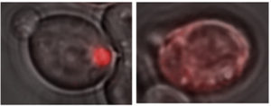
A glowing red clump can be detected in yeast cells containing a Lsb2 prion (left), because Lsb2 is hooked up to a red fluorescent protein. In other cells lacking prion activity (right), the Lsb2 fusion protein is diffuse.

“What we found suggests that Lsb2 could be the regulator of a broader prion-forming response to stress,” says Keith Wilkinson, PhD, professor of biochemistry at Emory University School of Medicine.

The scientists call the Lsb2 prion metastable because it is maintained in a fraction of cells after they return to normal conditions but is lost in other cells. Lsb2 is a short-lived, unstable protein, and mutations that keep it around longer increase the stability of the prions.

The Cell Reports paper was the result of collaboration between Wilkinson, Emory colleague Tatiana Chernova, PhD, assistant professor of biochemistry, and the laboratory of Yury Chernoff, PhD in Georgia Tech’s School of Biological Sciences.

“It’s fascinating that stress treatment may trigger a cascade of prion-like changes, and that the molecular memory of that stress can persist for a number of cell generations in a prion-like form,” Chernoff says.”Our further work is going to check if other proteins can respond to environmental stresses in a manner similar to Lsb2.” Read more

Posted on by Quinn Eastman in Uncategorized Leave a comment

How estrogen modulates fear learning — molecular insight into PTSD in women

Low estrogen levels may make women more susceptible to the development of post-traumatic stress disorder (PTSD) at some points in their menstrual cycles or lifetimes, while high estrogen levels may be protective.

New research from Emory University School of Medicine and Harvard Medical School provides insight into how estrogen changes gene activity in the brain to achieve its protective effects.

The findings, published in Molecular Psychiatry, could inform the design of preventive treatments aimed at reducing the risk of PTSD after someone is traumatized.

The scientists examined blood samples from 278 women from the Grady Trauma Project, a study of low-income Atlanta residents with high levels of exposure to violence and abuse. They analyzed maps of DNA methylation, a modification to the shape of DNA that is usually a sign of genes that are turned off.

The group included adult women of child-bearing age, in which estrogen rises and falls with the menstrual cycle, and women that had gone through menopause and had much lower estrogen levels.

“We knew that estrogen affects the activity of many genes throughout the genome,” says Alicia Smith, PhD, associate professor and vice chair of research in the Department of Gynecology and Obstetrics at Emory University School of Medicine. “But if you look at the estrogen-modulated sites that are also associated with PTSD, just one pops out.”

That site is located in a gene called HDAC4, known to be critical for learning and memory in mice. Genetic variation in HDAC4 among the women was linked to a lower level of HDAC4 gene activity and differences in their ability to respond to and recover from fear, and also differences in “resting state” brain imaging. Women with the same variation also showed stronger connections in activation between the amygdala and the cingulate cortex, two regions of the brain involved in fear learning. Read more

Posted on by Quinn Eastman in Neuro Leave a comment

Breath test for Parkinson’s?

Using one to see into the other. Left: canister for breath sample. Right: basal ganglia, a region of the brain usually affected by Parkinson’s.

Scientists think that it may be possible to detect signs of Parkinson’s disease through a breath test.

The Michael J. Fox Foundation for Parkinson’s Research is supporting a clinical study at Emory that will probe this idea. Neuro-immunologist Malu Tansey is working with Hygieia, a Georgia-based company that has developed technology for analyzing volatile organic compounds present in exhaled air.

From the MJFF’s blog:

By collecting and analyzing breath samples in 100 people (50 non-smoking early-stage PD patients and 50 age and sex-matched controls), the researchers hope to define a unique inflammatory PD-specific breath fingerprint that could be used to predict and monitor disease in combination with blood analyses of conventional or newly discovered biomarkers.

“We hypothesize that breath volatile organic compounds (BVOCs) fingerprinting can enable sensitive and specific measures of ongoing inflammation and other processes implicated in the development and/or progression of PD, and thus could represent an early detection tool,” Tansey says.

If results indicate moving forward, Tansey says it will be important to compare the breath sample method against blood tests for inflammatory markers. Other reports on the breath test approach for Parkinson’s have been encouraging. Read more

Posted on by Quinn Eastman in Neuro 1 Comment

Case report on first newborn to survive Ebola

Pediatric infectious diseases specialist Anita McElroy was a co-author on a case report on the first newborn to survive Ebola infection, published recently in Journal of Infectious Diseases.

“Of all the work I’ve been privileged to be involved in over the past few years, this paper was one of the most personally satisfying,” McElroy writes.

The child described in the paper is named Nubia; she is mentioned in several news stories from 2015. She was the last known Ebola case in Guinea, one of three African countries hit hard by the virus in 2014 and 2015. Her mother died shortly after her birth.

Nubia leaves hospital in Guinea. Photo from Medecins Sans Frontieres.

Nubia was cared for at the Ebola treatment ward run by Medecins Sans Frontieres (MSF, aka Doctors without Borders) in Conakry, Guinea. She was given three experimental therapies: ZMapp antibodies, survivor white blood cell transfusion and an antiviral drug called GS-5734. It is not clear which of these interventions were critical for Nubia’s recovery, although the paper makes clear that ZMapp did not result in viral suppression all by itself.

McElroy is a go-to person for studies of dangerous viruses such as Ebola, Lassa and Zika, partly because of her affiliation with the Centers for Disease Control and Prevention’s Viral Special Pathogens Branch. She advised the MSF team on the use of the antiviral drug and other interventions.

Posted on by Quinn Eastman in Immunology Leave a comment

Melanoma mutation likes fat for fuel

Cancer cells love glucose, the simple sugar the body uses for energy, so a high-fat, low-carb diet should starve them, right?

Where does this idea come from? Most cancer cells display enhanced glucose uptake, a phenomenon known as the Warburg effect, after 1931 Nobel Prize winner Otto Warburg.

Resurgent interest in exploiting the Warburg effect was described by Sam Apple in NYT Magazine and by Bret Stetka for NPR. High-fat, low-carb “ketogenic” diets are known to be effective against some types of epilepsy, and have also been explored by endurance athletes. Ketogenic diets have been tried as a clinical countermeasure against cancer in a limited way, mainly in brain cancer.

Before everybody gets too excited, let’s think about how particular cancer-driving mutations affect cell metabolism, suggests Winship Cancer Institute researcher Jing Chen. His team’s work in mice suggests that cancers with a common melanoma mutation (BRAF V600E) will grow faster in response to a ketogenic diet. In addition, the Winship researchers found that lipid-lowering agents such as statins curb these cancers’ growth, even in the context of a more normal diet.

The results were published on January 12 in Cell Metabolism.

Caveats: the findings cover just one mutation and need to be tested clinically.

Consumers and cancer patients already get a lot of advice about the right diet to fight cancer, but this research points toward an intriguing concept:  a “precision diet,” tailored to an individual patient’s cancer.  Read more

Posted on by Quinn Eastman in Cancer Leave a comment

Double vision: seeing viruses by both light and electron microscopy

Advances in both light and electron microscopy are improving scientists’ ability to visualize viruses such as HIV, respiratory syncytial virus (RSV), measles, influenza, and Zika in their native states.

Researchers from Emory University School of Medicine and Children’s Healthcare of Atlanta developed workflows for cryo-correlative light and electron microscopy (cryo-CLEM), which were published in the January 2017 issue of Nature Protocols.

An example of the images of viruses obtainable with cryo-CLEM. Pseudotyped HIV-1 particles undergoing endocytosis. Viral membrane = light blue. Mature core = yellow. Clathrin cages = purple. From Hampton et al Nat. Protocols (2016)

Previously, many electron microscopy images of well-known viruses were obtained by studying purified virus preparations. Yet the process of purification can distort the structure of enveloped viruses, says Elizabeth R. Wright, PhD, associate professor of pediatrics at Emory University School of Medicine.

Wright and her colleagues have refined techniques for studying viruses in the context of the cells they infect. That way, they can see in detail how viruses enter and are assembled in cells, or how genetic modifications alter viral structures or processing.

“Much of what is known about how some viruses replicate in cells is really a black box at the ultrastructural level,” she says. “We see ourselves as forming bridges between light and electron microscopy, and opening up new realms of biological questions.”

Wright is director of Emory’s Robert P. Apkarian Integrated Electron Microscopy Core and a Georgia Research Alliance Distinguished Investigator. The co-first authors of the Nature Protocols paper are postdoctoral fellows Cheri Hampton, PhD. and Joshua Strauss, PhD, and graduate students Zunlong Ke and Rebecca Dillard.

The Wright lab’s work on cryo-CLEM includes collaborations with Gregory Melikyan in Emory’s Department of Pediatrics, Phil Santangelo in the Wallace H. Coulter Department of Biomedical Engineering at Georgia Tech and Emory, and Paul Spearman, now at Cincinnati Children’s.

For this technique, virus-infected or transfected cells are grown on fragile carbon-coated gold grids and then “vitrified,” meaning that they are cooled rapidly so that ice crystals do not form. Once cooled, the cells are examined by cryo-fluorescent light microscopy and cryo-electron tomography. Read more

Posted on by Quinn Eastman in Immunology Leave a comment

Four biomedical research topics to watch in 2017

HIV/AIDS

The example of the “Berlin patient,” the only person ever cured of HIV infection, has energized HIV/AIDS researchers around the world. They are exploring a variety of tactics to attack the HIV reservoir in infected people, ranging from gene editing to “kick and kill.” A host of Emory/Yerkes researchers are among those pushing this forward.

This past year, an Emory/NIAID team led by Tab Ansari showed that a gentle, antibody-based approach could suppress SIV infection in macaques for extended periods, which surprised many in the field. The human test of this approach is now underway at the National Institutes of Health.

On the preventive vaccine side, a large scale efficacy study recently begun in South Africa, the first in seven years. Geovax’s Emory-rooted technology continues to advance in clinical studies. Further back in the pipeline, Yerkes researchers are testing innovative approaches, such as Rama Amara’s milk-bacteria-based mucosal vaccine and the potent nanoparticle adjuvants developed by Bali Pulendran’s group.

Zika

Despite the World Health Organization’s declaration in November that the public health emergency is over, Zika infection is still driving brain-related birth defects in several countries. Expect to hear more about Zika epidemiology and vaccine research, including from Emory investigators, next year.

In contrast with HIV, which seems to escape from almost anything we or our immune systems throw at it, Zika is doable, scientists think. At a Vaccine Dinner Club talk in September, Harvard’s Dan Barouch made the case that Zika is a slam dunk, immunologically. Two big questions remain: does dengue get in the way? And can vaccine makers test quickly and distribute widely?

FMT for antibiotic-resistant infections

Emory physicians have been leaders in developing fecal microbiota transplant as a remedy for recurrent Clostridium dificile infection. This form of diarrhea, which can be life-threatening, sometimes arises as a result of antibiotics that wipe out the helpful bacteria that live in the intestines, paving the way for “C diff.”

Now the Emory team (Colleen Kraft/Tanvi Dhere/Aneesh Mehta/Rachel Friedman-Moraco) is testing whether FMT could prevent other antibiotic-resistant infections besides C diff. This approach will be examined in a group of patients that tends to have a lot of antibiotic exposure: kidney transplant recipients. The team’s first publication on this topic from 2014 is here. Read more

Posted on by Quinn Eastman in Immunology, Neuro Leave a comment

Threading the RSV needle: live attenuated vaccine effective in animals

Crafting a vaccine against RSV (respiratory syncytial virus) has been a minefield for 50 years, but scientists believe they have found the right balance.

A 3-D rendering of a live-attenuated respiratory syncytial virus (RSV) particle, captured in a near-to-native state by cryo-electron tomography. Surface glycoproteins (yellow) are anchored on the viral membrane (cyan), with ribonucleoprotein complexes inside (red). Image courtesy of Zunlong Ke and Elizabeth Wright.

Researchers at Emory University School of Medicine and Children’s Healthcare of Atlanta have engineered a version of RSV that is highly attenuated – weakened in its ability to cause disease – yet potent in its ability to induce protective antibodies.

The researchers examined the engineered virus using cryo-electron microscopy and cryo-electron tomography techniques, and showed that it is structurally very similar to wild type virus. When used as a vaccine, it can protect mice and cotton rats from RSV infection.

The results were published this morning in Nature Communications.

“Our paper shows that it’s possible to attenuate RSV without losing any immunogenicity,” says senior author Martin Moore, PhD, associate professor of pediatrics at Emory University School of Medicine and a Children’s Healthcare of Atlanta Research Scholar. “This is a promising live-attenuated vaccine candidate that merits further investigation clinically.”

The next steps for this vaccine are to produce a clinical grade lot and conduct a phase 1 study of safety and immunogenicity in infants, Moore says. Read more

Posted on by Quinn Eastman in Immunology Leave a comment

How “twist my arm” engages the brain

Listening to metaphors involving arms or legs loops in a region of the brain responsible for visual perception of those body parts, scientists have discovered.

The finding, recently published in Brain & Language, is another example of how neuroscience studies are providing evidence for “grounded cognition” – the idea that comprehension of abstract concepts in the brain is built upon concrete experiences, a proposal whose history extends back millennia to Aristotle.

The EBA was shown in 2001 to respond selectively to images of the human body by Nancy Kanwisher and colleagues.

When study participants heard sentences that included phrases such as “shoulder responsibility,” “foot the bill” or “twist my arm”, they tended to engage a region of the brain called the left extrastriate body area or EBA.

The same level of activation was not seen when participants heard literal sentences containing phrases with a similar meaning, such as “take responsibility” or “pay the bill.”  The study included 12 right-handed, English-speaking people, and blood flow in their brains was monitored by functional MRI (magnetic resonance imaging).

“The EBA is part of the extrastriate visual cortex, and it was known to be involved in identifying body parts,” says senior author Krish Sathian, MD, PhD, professor of neurology, rehabilitation medicine, and psychology at Emory University.  “We found that the metaphor selectivity of the EBA matches its visual selectivity.” Read more

Posted on by Quinn Eastman in Neuro Leave a comment

Neuroscientists show hippocampus also has important role in emotional regulation

A region of the brain called the hippocampus is known for its role in memory formation. Scientists at Yerkes National Primate Research Center, Emory University are learning more about another facet of hippocampal function: its importance in the regulation and expression of emotions, particularly during early development.

Using a nonhuman primate model, their findings provide insight into the mechanisms of human psychiatric disorders associated with emotion dysregulation, such as PTSD (post-traumatic stress disorder) and schizophrenia. The results were published online recently by the journal Psychoneuroendocrinology.

“Our findings demonstrate that damage to the hippocampus early in life leads to increased anxiety-like behaviors in response to an unfamiliar human,” says research associate Jessica Raper, PhD, first author of the paper. “However, despite heightened anxious behavior, cortisol responses to the social stress were dampened in adulthood.”

The hormone cortisol modulates metabolism, the immune system and brain function in response to stress. Reduced hippocampal volume and lower cortisol response to stressors have been demonstrated as features of and risk factors for PTSD, Raper says. Also, the dampened daily rhythms of cortisol seen in the nonhuman primates with hippocampal damage resemble those reported in first-episode schizophrenia patients.

Follow-up studies could involve temporary interference with hippocampus function using targeted genetic techniques, she says. Read more

Posted on by Quinn Eastman in Neuro Leave a comment