Quinn Eastman

Helping stem cells find their new homes

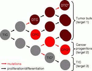
The idea that doctors could use stem cells to treat diseases ranging from amyotrophic lateral sclerosis (ALS) to stroke, spinal cord injury and heart disease has stimulated excitement and research funding over the last decade.

One critical obstacle is getting the stem cells to survive in the harsh environment of injured tissue and turn into the right kind of cell where they are needed. In both laboratory experiments and clinical trials, most of the stem cells usually die a few days after transplantation.

Exposing stem cells to reduced levels of oxygen may actually help protect them from the stressful process of being transplanted into the heart, according to recent research.

Shan Ping Yu and Ling Wei, who moved their laboratories about a year ago to Emory’s Department of Anesthesiology, were the first to show the effects of “hypoxic preconditioning.” Wei says the low oxygen strategy is a continuation of previous collaboration with Comprehensive Neurosciences Center director Dennis Choi. There, they had used the tactic of overexpressing BCl2, a gene that counteracts cell death, but the new approach avoids permanently altering the genes in stem cells, which may have long-term adverse effects.

Effects on mesenchymal stem cells' ability to implant into heart tissue. In D, the stem cells were exposed to low oxygen but in C they were not. Blue shows all cell nuclei, while green shows implanted stem cells. Yellow indicates the activation of an enzyme that leads to cell death.

Effects on mesenchymal stem cells' ability to implant into heart tissue in rats. In D, the stem cells were exposed to low oxygen but in C they were not. Blue shows all cell nuclei, while green shows implanted stem cells. The greater presence of yellow in C, a couple days after transplantation, displays the activation of an enzyme that leads to cell death. From the Journal of Thoracic and Cardiovascular Surgery.

In a way, this is consistent with the work of former Emory investigator Marie Csete, who showed that stem cells are happier and healthier in oxygen concentrations that reflect the levels they experience in the body: between 2 and 5%.

To achieve their protective effects, Yu and Wei are using oxygen concentrations of 0.5%. For comparison, room air has about 20% oxygen.

In an editorial, Yu, Wei and graduate student Molly Ogle discuss how they have been exploring whether inhibitors of enzymes that sense levels of oxygen in cells could have the same protective effects as exposure to low oxygen. Yu also reports that his group is studying how low oxygen helps stem cells home to target tissues better. Their hypothesis is that low oxygen stimulates cells’ motility — their ability to migrate into the right place. Wei’s research has shown that lower oxygen helps more stem cells to turn into neuronal cells.

Posted on by Quinn Eastman in Uncategorized Leave a comment

A shift in how geneticists study complex diseases

An Emory project studying schizophrenia genetics is a good example of how geneticists are shifting from examining small, common mutations to “rare variants” when studying complex diseases.

From studies of twins, doctors have known for a long time that heredity plays a big role in causing schizophrenia. But dissecting out which genes are the most important has been a challenge.

Three landmark studies on schizophrenia genetics published this summer illustrate the limitations of “genome wide association” studies. New York Times science reporter Nicholas Wade summarized the results in this way:

“The principal news from the three studies is that schizophrenia is caused by a very large number of errant genes, not a manageable and meaningful handful.”

The limitations from this type of study comes from the type of markers geneticists are looking at, says Steve Warren, chair of the human genetics department at Emory.

Genome wide association studies usually follow SNPs — single nucleotide polymorphisms. This is a one-letter change somewhere in the genetic code that is found in a fraction of the population. It’s not a big change in the genome, and in many cases, it will have a small effect on disease risk.

Researchers looking for the genes behind complex diseases such as schizophrenia and autism are starting to shift their efforts away from genome wide association studies, Warren says.

Think of a SNP like a misspelling of a word in a certain place in a book, he says. In contrast, the “rare variants” geneticists are starting to study more intensively are more like printers’ errors or missing pages. The rapid sequencing technology that allows scientists to investigate these changes easily is just now coming on line, he says.

One example of these rare variants is DiGeorge syndrome, a deletion that gets rid of dozens of genes on one copy of chromosome 22. Children who have this chromosomal alteration often have anatomical changes to their heart and palate. But it also substantially increases the risk of schizophrenia – to about 25% lifetime risk. That’s a lot more than any of the SNPs identified this summer.

Working with several Emory colleagues, researcher Brad Pearce is planning to examine the genes missing in DiGeorge syndrome in several groups of patients: people with DiGeorge, patients with “typical” schizophrenia and people at high risk of developing schizophrenia.

An article in this spring’s Emory Health describes genetic research on autism. Several of the researchers mentioned there, such as geneticist Joe Cubells and psychiatrist Opal Ousley, are involved in this schizophrenia project as well, because deletions on chromosome 22 also lead to an increased risk of autism.

Pearce’s project is funded through American Recovery and Reinvestment Act money from the NIH.

Posted on by Quinn Eastman in Uncategorized Leave a comment

Strategies to target cancer stem cells

A story in last Friday’s New York Times highlights research on “cancer stem cells”: a fraction of cells in a tumor that are especially resistant to chemotherapy and resemble the body’s non-cancerous stem cells in their ability to renew themselves.

The story describes work by a team at the Broad Institute, who reported in the journal Cell that they had identified compounds that specifically kill cancer stem cells. The hope is that compounds such as these could be combined with conventional treatments to more effectively eliminate cancers.

However, scientists disagree on whether the phenomenon of cancer stem cells extends to different kinds of cancer and what is the best way to target them. Previously not much was known about how to attack these cells.

Work at Emory’s Winship Cancer Institute has been tracking how some biomarkers in cancer cells resemble or differ from those found in stem cells. These markers may help researchers home in on the cancer stem cells.


Anticancer therapy must target more than one type of cell. TIC means tumor initiating cell, DTC means differentiated tumor cell, and CPG means cancer progenitor

If "cancer stem cells" play the critical roles some scientists think they do, anticancer therapy must target more than one type of cell. In this figure from Van Meir + Hadjipanayis' review, TIC means tumor initiating cell, DTC means differentiated tumor cell, and CPG means cancer progenitor cells.Â


In a recent review, Emory brain cancer specialists Erwin Van Meir and Costas Hadjipanayis write:

The “cancer stem cell” hypothesis has invigorated the neuro-oncology field with a breath of fresh thinking that may end up shaking the foundation of old dogmas, such as the widely held belief that glioblastoma tumors are incurable because of infiltrative disease. If the infiltrated cells are in fact differentiated tumor cells, their dissemination beyond the surgical boundary may not be the primary cause of tumor recurrence.

Van Meir, the editor of a new book on brain cancer, adds this comment:

Clearly a lot more work needs to be done to understand the precise cause of glioblastoma recurrence after surgery and chemotherapy and how to prevent it.  The possibility of developing therapeutics that can specifically target the brain cancer stem cells is an exciting new development but will have to proceed with caution to spare normal stem cells in the brain. Developing new imaging tools that can track cancer stem cells in the brain of treated patients is also an important objective and some of the Emory investigators are evaluating the use of nanoparticles to this purpose.

A new faculty member at Winship, Tracy-Ann Read, recently published her research on a molecule that could be used to identify “tumor-propagating cells” in medulloblastoma, a form of brain cancer. She says:

Although cancer stem cells have been identified in many different types of cancer, it is becoming increasingly clear that the properties of these cells may vary greatly among the different tumor types. It is unlikely that one  therapeutic agent will be able to target the cancer stem cells in for example all types brain tumors. Hence  much work still needs to be done in terms of analyzing the properties of these cells in each tumor type and identifying the genes that are responsible for their unique ability to propagate the tumors. 

Winship’s director Brian Leyland-Jones has also reported at the San Antonio Breast Cancer Symposium that molecules that distinguish a hard-to-treat form of breast cancer resemble those that maintain stem cells.

Nice round-up from Nature’s stem cell blog editor Monya Baker

Posted on by Quinn Eastman in Cancer Leave a comment

Discerning a prelude to Alzheimer’s

Imagine that an elderly relative has been having difficulty remembering appointments and acquaintances’ names, or even what happened yesterday. Memory problems can be signs of mild cognitive impairment (MCI), a prelude to Alzheimer’s disease.

Scientists believe that the outward effects of the slow damage that comes from Alzheimer’s only show up after the damage has been accumulating for years. However, memory difficulties can also be the result of stress or another health problem. Patients thought to have MCI at an initial doctor’s visit sometimes improve later.

That’s why researchers at Emory’s Alzheimer’s Disease Research Center have been testing noninvasive imaging approaches to distinguishing MCI from healthy aging and Alzheimer’s. Their goal is to identify individuals at risk of developing Alzheimer’s, at a time when intervention can make a difference in how the disease progresses.

“We believe that imaging technology may help us find the signature changes in brain structure that are specific to MCI,” says Felicia Goldstein, PhD, associate professor of neurology.

Color coded diffusion tensor image (DTI) of a brain section from a healthy individual (A) showed a thick and intact corpus callosum (orange color), a white matter fiber bundle connecting left and right hemisphere as illustrated in the 3D rendering of the tractograph derived from DTI (B). However, a thin and narrow corpus callosum is seen in an AD patient (C) due to the degeneration of this white matter structure

Color coded diffusion tensor image (DTI) of a brain section from a healthy individual (A) showed a thick and intact corpus callosum (orange color). However, a thin and narrow corpus callosum is seen in an AD patient (C) due to the degeneration of this white matter structure. Courtesy of Hui Mao.

Two recent papers highlight the use of diffusion tensor imaging, an advanced form of magnetic resonance imaging.

The first paper was published by Brain Imaging and Behavior with Goldstein as first author, in collaboration with Hui Mao, PhD, associate professor of radiology, and ADRC colleagues.

It examines diffusion tensor imaging as a way to probe the integrity of the brain’s white matter, and compares it with tests of memory and behavior traditionally used to diagnose MCI and Alzheimer’s.

White matter appears white because of the density of axons, the signal-carrying cables allowing communication between different brain regions responsible for complicated tasks such as language and memory.

Diffusion tensor imaging allows researchers to see white matter by gauging the ability of water to diffuse in different directions, because a bundle of axons tends to restrict the movement of water in the brain.

Goldstein and her colleagues found that patients diagnosed with “amnestic” MCI showed greater loss of white matter integrity in a certain part of the brain — the medial temporal lobe – than cognitively normal controls of similar age. This loss of white matter was linked with poor recall of words and stories.

The second paper, with Liya Wang, PhD, a senior research associate in Mao’s laboratory as first author, was published by the American Journal of Neuroradiology in April. Here the authors try combining probing white matter integrity with a MRI measure of whether the brain has shrunk as a result of disease.

Combining the two methods improves the accuracy of MCI diagnosis with respect to either alone, the authors found.

Mao notes that Emory has been participating in a multi-center study called ADNI (Alzheimer’s Disease Neuroimaging Initiative). Diffusion tensor imaging is a relatively new technique and could add information to future large-scale Alzheimer’s imaging studies, he says.

The Dana Foundation’s BrainWorks newsletter had an article recently on Alzheimer’s and brain imaging.

Posted on by Quinn Eastman in Uncategorized Leave a comment

Lampreys’ alternative immune system

Lampreys are primitive creatures – basically, tubes with teeth. Their primitive nature makes them a fascinating entry-point for studying the evolution of the immune system.

At Emory, Max Cooper and his colleagues have been studying lampreys’ versions of white blood cells. In a recent Nature paper, they show that lampreys have two kinds of cells that look very much like B and T cells in mammals, birds and fish.

Non-immunologists may shrug at this revelation.  But consider: lampreys have a completely different set of tools for fighting infections. They have proteins in their blood that glob on to invaders, but they don’t look anything like the antibodies found in mammals, birds and fish.

Lampreys in a laboratory tank

Lampreys in a laboratory tank. Courtesy of Masa Hirano.

Similarly, lampreys have cells that look like T Ray Ban outlet cells, in terms of some of the genes that are turned on. However, they don’t have MHC genes, which are important in human transplant medicine because they determine how and when T cells get excited and reject transplanted organs.

Lampreys are thought to be an early offshoot on the evolutionary tree, before sharks and fish, and way before critters that crawl on land. This suggests that the categories (B or T) came first even though the characteristic features of the cells (antibodies/responding to MHC) are different.

“Lampreys have the same types of cells, but they just use different building blocks to put them together,” Cooper says.

Cooper, now a Georgia Research Alliance Eminent Scholar and a member of Emory’s pathology department, made pioneering studies defining the role the thymus plays in immune development at the University of Minnesota in the 1960s. The thymus is where T cells develop and where they get their name.

He says he is now collaborating with Thomas Boehm in Freiburg, Germany to better understand the evolution of the thymus. Again, lampreys don’t have a thymus, but they may have an area next to their gills where the T-like cells develop.

John Travis at Science has a more extensive discussion of this research.

In a Darwin-anniversary essay, Travis tells the story of how the evolution of the immune system was a centerpiece of the 2005 Kitzmiller v. Dover trial, when a Pennsylviania school district’s requirement to teach intelligent design was successfully challenged.

Link to Sound Science podcast with Cooper

Posted on by Quinn Eastman in Immunology Leave a comment

Many roads to memory T cells

When our bodies encounter a bacteria or a virus, the immune system sends some cells out to fight the invader and keeps others in reserve, in order to respond faster and stronger the next time around. Vaccination depends on this phenomenon, called immunological memory.

Several recent papers — from Emory and elsewhere – provide insight into this process, and highlight this area of research as especially active lately.

Researchers led by Rafi Ahmed and Chris Larsen at Emory found that rapamycin, a drug usually given to transplant patients to block rejection, actually stimulates the formation of memory T cells. Rapamycin appears to nudge immune cells when they have to make a decision whether to hunker down to become a memory cell.

The immunosuppressant drug rapamycin was discovered in soil from Easter Island

The immunosuppressant drug rapamycin was discovered in soil from Easter Island

Similarly, the anti-diabetes drug metformin, which affects fatty acid metabolism, can also stimulate the formation of memory T cells, according to research that was published in the same issue of Nature.

In addition, Wnt signaling, which plays critical roles in embryonic development and cancer, influences memory T cell formation as well, according to a July paper in Nature Medicine.

To summarize — pushing on several different “buttons” produces the same thing: more memory T cells. How are the wires behind the buttons connected? Work by Ahmed and others may eventually help enhance vaccine efficacy or fight cancer with the immune system.

Rapamycin, the focus of the Ahmed/Larsen paper, was also recently found to slow aging in mice. However, with previous anti-aging research findings, translating results into the human realm has been a considerable challenge.

Posted on by Quinn Eastman in Immunology Leave a comment
« Previous   1 2 ... 64 65 66 67 68 69 70 71 72 73