Warren symposium follows legacy of geneticist giant

If we want to understand how the brain creates memories, and how genetic disorders distort the brain’s machinery, then the fragile X gene is an ideal place to start. That’s why the Stephen T. Warren Memorial Symposium, taking place November 28-29 at Emory, will be a significant event for those interested in neuroscience and genetics. Stephen T. Warren, 1953-2021 Warren, the founding chair of Emory’s Department of Human Genetics, led an international team that discovered Read more

Mutations in V-ATPase proton pump implicated in epilepsy syndrome

Why and how disrupting V-ATPase function leads to epilepsy, researchers are just starting to figure Read more

Tracing the start of COVID-19 in GA

At a time when COVID-19 appears to be receding in much of Georgia, it’s worth revisiting the start of the pandemic in early 2020. Emory virologist Anne Piantadosi and colleagues have a paper in Viral Evolution on the earliest SARS-CoV-2 genetic sequences detected in Georgia. Analyzing relationships between those virus sequences and samples from other states and countries can give us an idea about where the first COVID-19 infections in Georgia came from. We can draw Read more

Center for Neurodegenerative Disease

The sweet side of Alzheimer’s proteomics

The Alzheimer’s field has been in a “back to the basics” mode lately. Much research has focused on beta-amyloid, the toxic protein fragment that accumulates in plaques in the brain. Yet drugs that target beta-amyloid have mostly been disappointing in clinical trials.

To broaden scope and gain new insights into the biology of Alzheimer’s, Emory investigators have been making large-scale efforts to catalog alterations of brain proteins. One recent example: Nick Seyfried and Erik Johnson’s enormous collection of proteomics data, published this spring in Nature Medicine. Another can be seen in the systematic mapping of N-glycosylation, just published in Science Advances by pharmacologist Lian Li and colleagues.

“It is very exciting to see, for the first time, the landscape of protein N-glycosylation changes in Alzheimer’s brain,” Li says. “Our results suggest that the N-glycosylation changes may contribute to brain malfunction in Alzheimer’s patients.  We believe that targeting N-glycosylation may provide a new opportunity to help combat this devastating dementia.”

Read more

Posted on by Quinn Eastman in Neuro Leave a comment

The blue spot: where seeds of destruction begin

Neuroscientist and geneticist David Weinshenker makes a case that the locus coeruleus (LC), a small region of the brainstem and part of the pons, is among the earliest regions to show signs of degeneration in both Alzheimer’s and Parkinson’s disease. You can check it out in Trends in Neurosciences.

The LC is the main source of the neurotransmitter norepinephrine in the brain, and gets its name (Latin for “blue spot”) from the pigment neuromelanin, which is formed as a byproduct of the synthesis of norepinephrine and its related neurotransmitter dopamine. The LC has connections all over the brain, and is thought to be involved in arousal and attention, stress responses, learning and memory, and the sleep-wake cycle.

Cells in the locus coeruleus are lost in mild cognitive impairment and Alzheimer’s. From Kelly et al Acta Neuropath. Comm. (2017) via Creative Commons

The protein tau is one of the toxic proteins tied to Alzheimer’s, and it forms intracellular tangles. Pathologists have observed that precursors to tau tangles can be found in the LC in apparently healthy people before anywhere else in the brain, sometimes during the first few decades of life, Weinshenker writes. A similar bad actor in Parkinson’s, alpha-synuclein, can also be detected in the LC before other parts of the brain that are well known for damage in Parkinson’s, such as the dopamine neurons in the substantia nigra.

“The LC is the earliest site to show tau pathology in AD and one of the earliest (but not the earliest) site to show alpha-synuclein pathology in PD,” Weinshenker tells Lab Land. “The degeneration of the cells in both these diseases is more gradual. It probably starts in the terminals/fibers and eventually the cell bodies die.” Read more

Posted on by Quinn Eastman in Neuro Leave a comment

Granulins treasure not trash – potential FTD treatment strategy

Emory University School of Medicine researchers have developed tools that enable them to detect small proteins called granulins for the first time inside cells. Granulins are of interest to neuroscientists because mutations in the granulin gene cause frontotemporal dementia (FTD). However, the functions of granulins were previously unclear.

FTD is an incurable neurodegenerative disease and the most common type of dementia in people younger than 60. Genetic variants in the granulin gene are also a risk factor for Alzheimer’s disease and Parkinson’s disease, suggesting this discovery may have therapeutic potential for a broad spectrum of age-related neurodegenerative diseases.

The results were published August 9 by the journal eNeuro (open access).

Thomas Kukar, PhD

Some neuroscientists believed that granulins were made outside cells, and even could be toxic under certain conditions. But with the newly identified tools, the Emory researchers can now see granulins inside cells within lysosomes, which are critical garbage disposal and recycling centers. The researchers now propose that granulins have important jobs in the lysosome that are necessary to maintain brain health, suppress neuroinflammation, and prevent neurodegeneration.

Problems with lysosomes appear in several neurodegenerative diseases such as Alzheimer’s and Parkinson’s.

“A lysosomal function for granulins is exciting and novel.  We believe it may provide an explanation why decreased levels of granulins are linked to multiple neurodegenerative diseases, ranging from frontotemporal dementia to Alzheimer’s,” says senior author Thomas Kukar, PhD, assistant professor of pharmacology and neurology and the Emory University Center for Neurodegenerative Disease. Read more

Posted on by Quinn Eastman in Neuro Leave a comment

More pieces in Parkinson’s puzzle: VMAT2 and SV2C

The drug target VMAT2 has appeared in biomedical news lately because of a pair of FDA approvals. One medicine treats the iatrogenic movement disorder tardive dyskinesia, the first approved to do so, and the other is for symptoms of Huntington’s disease.

Gary Miller, PhD

When Emory folks see VMAT2, they should think of two things: the neurotransmitter dopamine, and Parkinson’s research conducted by Gary Miller and his colleagues. They have made a case that activators of VMAT2 would be beneficial in Parkinson’s, but the drugs in the news were inhibitors, and presumably would make Parkinson’s worse.

VMAT2 (vesicular monoamine transporter 2) is responsible for transporting dopamine into synaptic vesicles, tiny packages for delivery. As Miller’s lab has shown, mice deficient in VMAT2 can be a model for the non-motor and motor aspects of Parkinson’s. In these mice, not only are certain nervous system functions impaired, but the dopamine packaging problem inflicts damage on the neurons.

Miller’s more recent work on a related molecule called SV2C is puzzling, but intriguing. The gene encoding SV2C had attracted attention because of its connection to the striking ability of cigarette smoking to reduce Parkinson’s risk, possibly mediated by nicotine’s effect on dopamine in the brain.

I say puzzling because SV2C’s role in brain cells can’t be described as easily as VMAT2’s. Read more

Posted on by Quinn Eastman in Neuro Leave a comment

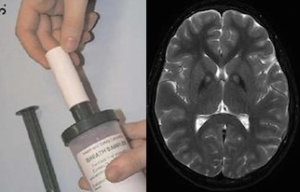
Breath test for Parkinson’s?

Using one to see into the other. Left: canister for breath sample. Right: basal ganglia, a region of the brain usually affected by Parkinson’s.

Scientists think that it may be possible to detect signs of Parkinson’s disease through a breath test.

The Michael J. Fox Foundation for Parkinson’s Research is supporting a clinical study at Emory that will probe this idea. Neuro-immunologist Malu Tansey is working with Hygieia, a Georgia-based company that has developed technology for analyzing volatile organic compounds present in exhaled air.

From the MJFF’s blog:

By collecting and analyzing breath samples in 100 people (50 non-smoking early-stage PD patients and 50 age and sex-matched controls), the researchers hope to define a unique inflammatory PD-specific breath fingerprint that could be used to predict and monitor disease in combination with blood analyses of conventional or newly discovered biomarkers.

“We hypothesize that breath volatile organic compounds (BVOCs) fingerprinting can enable sensitive and specific measures of ongoing inflammation and other processes implicated in the development and/or progression of PD, and thus could represent an early detection tool,” Tansey says.

If results indicate moving forward, Tansey says it will be important to compare the breath sample method against blood tests for inflammatory markers. Other reports on the breath test approach for Parkinson’s have been encouraging. Read more

Posted on by Quinn Eastman in Neuro 1 Comment

A sweet brain preserver: trehalose

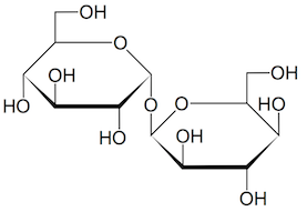
It’s sweet, it’s safe, and it looks like it could save neurons. What is it? Trehalose.

Trehalose molecule

Trehalose is a natural sugar.

This natural sugar is used in the food industry as a preservative and flavor enhancer (it’s in Taco Bell’s meat filling). And curiously, medical researchers keep running into trehalose when they’re looking for ways to fight neurodegenerative diseases.

A recent example from Emory’s Department of Pharmacology: Chris Holler, Thomas Kukar and colleagues were looking for drugs that might boost human cells’ production of progranulin (PGRN), a growth factor that keeps neurons healthy. Mutations in the progranulin gene are a common cause of frontotemporal dementia.

The Emory scientists discovered two leads: a class of compounds called mTOR inhibitors — the transplant drug rapamycin is one — and trehalose. The team decided to concentrate on trehalose because it increased PGRN levels in neuronal and non-neuronal cell types, unlike the mTOR inhibitors. Their results were published at the end of June in Molecular Neurodegeneration.

The team confirmed their findings by examining the effects of trehalose on cells derived from patients with progranulin mutations. This paper is the first to include results from Emory’s Laboratory of Translational Cell Biology, which was established in 2012 to facilitate this type of “disease in a dish” approach. Cell biologists Charles Easley, Wilfried Rossoll and Gary Bassell from the LTCB, and neurologists Chad Hales and William Hu from the Center for Neurodegenerative Disease are co-authors.

Read more

Posted on by Quinn Eastman in Neuro 1 Comment

Clot dissolver tPA’s tardy twin could aid in stroke recovery

Emory researchers led by neurologist Manuel Yepes, MD have identified a protein released by neurons while the brain is recovering from a stroke. The results were published online today in Journal of Neuroscience.

The protein, called urokinase-type plasminogen activator or uPA, has been approved by the FDA to dissolve blood clots in the lungs. It has been tested in clinical trials in some countries as a treatment for acute stroke.

The Emory team’s findings suggest that in stroke, uPA’s benefits may extend beyond the time when doctors’ principal goal is dissolving the blood clot that is depriving the brain of blood.
Instead, uPA appears to help brain cells recover from the injuries induced by loss of blood flow. Treating mice with uPA after an experimental stroke can improve their recovery of motor function, the researchers found.

Read more

Posted on by Quinn Eastman in Neuro Leave a comment