Warren symposium follows legacy of geneticist giant

If we want to understand how the brain creates memories, and how genetic disorders distort the brain’s machinery, then the fragile X gene is an ideal place to start. That’s why the Stephen T. Warren Memorial Symposium, taking place November 28-29 at Emory, will be a significant event for those interested in neuroscience and genetics. Stephen T. Warren, 1953-2021 Warren, the founding chair of Emory’s Department of Human Genetics, led an international team that discovered Read more

Mutations in V-ATPase proton pump implicated in epilepsy syndrome

Why and how disrupting V-ATPase function leads to epilepsy, researchers are just starting to figure Read more

Tracing the start of COVID-19 in GA

At a time when COVID-19 appears to be receding in much of Georgia, it’s worth revisiting the start of the pandemic in early 2020. Emory virologist Anne Piantadosi and colleagues have a paper in Viral Evolution on the earliest SARS-CoV-2 genetic sequences detected in Georgia. Analyzing relationships between those virus sequences and samples from other states and countries can give us an idea about where the first COVID-19 infections in Georgia came from. We can draw Read more

Detecting vulnerable plaque with a laser-induced whisper

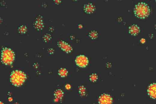
A relatively new imaging technique called photoacoustic imaging or PAI detects sounds produced when laser light interacts with human tissues. Working with colleagues at Michigan State, Emory immunologist Eliver Ghosn’s lab is taking the technique to the next step to visualize immune cells within atherosclerotic plaques.

The goal is to more accurately spot vulnerable plaque, or the problem areas lurking within arteries that lead to clots, and in turn heart attacks and strokes. A description of the technology was recently published in Advanced Functional Materials

“I believe we are now closer to developing a more precise method to diagnose and treat life-threatening atherosclerotic plaques,” Ghosn says. “Our method could be deployed in combination with IVUS to significantly improve its accuracy and sensitivity, or it could be used non-invasively.”

From science fiction movies, we might think lasers come with a “pow” sound. Photoacoustic imaging is more like listening for a whisper: sounds associated with heat generated by a laser pulse when it is absorbed by tissue.

Earlier this year, the FDA approved a photoacoustic imaging system for detection of breast cancer. Several companies are developing photoacoustic imaging systems, and what we might call “plain vanilla” PAI is currently being tested on carotid artery plaque in clinical studies in Europe.

Ghosn’s approach, developed with biomedical engineer Bryan Smith at Michigan State, adds specificity by adding nanoparticle probes taken up by macrophages, the immune cells that accumulate within atherosclerotic plaques. The nanoparticles, administered before imaging, act as contrast agents.

Read more

Posted on by Quinn Eastman in Uncategorized Leave a comment

Multiple myeloma patients display weakened antibody responses to mRNA COVID vaccines

A new study reports weakened antibody responses to COVID-19 mRNA vaccines among most patients with multiple myeloma, a form of bone-marrow cancer associated with an immunocompromised state.

The research, published in the journal Leukemia, was carried out at the Institute for Myeloma and Bone Cancer Research (IMBCR) in California, in collaboration with Emory infectious diseases fellow Samuel Stampfer, MD, PhD.

Patients with smoldering myeloma, not requiring treatment, all achieved a good response to COVID-19 vaccination, whereas less than half of patients with active myeloma requiring treatment did. Specifically, only 45 percent of active patients fully responded to the mRNA vaccines, whereas less than a quarter showed a partial response and one-third did not respond to the vaccines above background antibody levels.

Serum samples from 103 multiple myeloma patients were obtained prior to vaccination and 2-3 weeks after administration of the first and second vaccines, and compared to a group of age‑matched healthy controls. Predictors of reduced antibody responses to the vaccines included: older age, impaired renal function, low lymphocyte counts, reduced uninvolved antibody levels, past first line of treatment, and those not in complete remission. Nearly two-thirds of patients who received the Moderna vaccine responded to a level thought to be clinically significant, whereas only approximately a quarter who received the Pfizer vaccine did.

“Based on these data, myeloma patients may need to continue social distancing following COVID-19 vaccination, and postvaccine antibody tests may help guide decisions regarding supplementary vaccination or antibody prophylaxis for this vulnerable population,” says Stampfer, who co-designed the clinical study, under the guidance of senior author James Berenson, MD, the Scientific and Medical Director of IMBCR.

“This study highlights the importance of recognizing the limitations of current vaccination approaches to COVID-19 for immunocompromised patients, and that new approaches will have to be developed to improve their protection from this dangerous infection,” Berenson says. “It also suggests that there may be clinically significant differences in the effectiveness of different COVID-19 vaccines for immune compromised patients. Until these advances occur, it means that myeloma patients will need to remain very careful even if they have been vaccinated through wearing their masks and avoiding contact with unvaccinated individuals.”.

Read more

Posted on by Quinn Eastman in Cancer, Immunology Leave a comment

Precision medicine with multiple myeloma

“Precision medicine” is an anti-cancer treatment strategy in which doctors use genetic or other tests to identify vulnerabilities in an individual’s cancer subtype.

Winship Cancer Institute researchers have been figuring out how to apply this strategy to multiple myeloma, with respect to one promising drug called venetoclax, in a way that can benefit the most patients.

Known commercially as Venclexta, venetoclax is already FDA-approved for some forms of leukemia and lymphoma. Researchers had observed that multiple myeloma cells with one type of chromosomal DNA rearrangement tend to be sensitive to venetoclax. About 20 percent of multiple myelomas carry this rearrangement, called t(11;14).

“One of our main goals is to identify a better biomarker to predict patient response to venetoclax,” says Winship researcher Vikas Gupta, lead author of a paper published in Blood earlier this year.

Vikas Gupta, MD, PhD

Gupta works together with Winship hematologist Jonathan Kaufman and researcher Larry Boise, also associate director for education and training, to translate insights about myeloma cells into advances for patient care.

In a recent clinical trial led by Kaufman, a sizable fraction of people whose myelomas carried the t(11;14) rearrangement responded well to venetoclax, when their cancers were already refractory to other drugs. Another study that did not separate out myelomas with t(11;14) extended progression-free survival by almost a year.

However, venetoclax also was associated with increased mortality from infections, which led the FDA in 2019 to put the second study on hold temporarily. Other ongoing studies of venetoclax with multiple myeloma were affected.  It highlights the need to predict which patients would benefit from venetoclax – and which would not be likely to, for whom the drug may pose more risk.

In their paper, Winship investigators discovered that a set of cell markers predicted sensitivity to venetoclax better than t(11;14). These were markers for B cells, a type of white blood cell related to both multiple myeloma and some of the other forms of leukemia and lymphoma venetoclax is used to treat.

Gupta says that it was already possible to obtain myeloma cells from patients and test whether they are sensitive to venetoclax directly in the laboratory. But this isn’t practical for most clinics in cancer centers elsewhere.

“In contrast, the B cell phenotype can easily be assessed by flow cytometry, a technique that is routinely performed in clinical labs,” Gupta says. “So we are attempting to refine and validate our panel of flow cytometry markers, so that it can be used to easily and accurately predict which patients are sensitive to venetoclax.”

Read more

Posted on by Quinn Eastman in Cancer Leave a comment

Promiscuous protein droplets regulate immune gene activity

Biochemists at Emory are achieving insights into how an important regulator of the immune system switches its function, based on its orientation and local environment. New research demonstrates that the glucocorticoid receptor (or GR) forms droplets or “condensates” that change form, depending on its available partners.

The inside of a cell is like a crowded nightclub or party, with enzymes and other proteins searching out prospective partners. The GR is particularly well-connected and promiscuous, and has the potential to interact with many other proteins. It is a type of protein known as a transcription factor, which turns some genes on and others off, depending on how it is binding DNA.

These are fluorescent droplets of the glucocorticoid receptor (GR) in red, with a coregulator protein in green. When DNA is added, the co-regulator forms its own droplets on the surface of GR droplets. Image courtesy of Filipp Frank

“It is now thought that most transcription factors form or are recruited into condensates, and that condensation modulates their function,” says Filipp Frank, PhD, first author of the paper and a postdoctoral instructor in Eric Ortlund’s lab in the Department of Biochemistry. “What’s new is that we identified a DNA-dependent change in GR condensates, which has not been described for other transcription factors.”

The results are published in Proceedings of the National Academy of Sciences. Ortlund is a co-author of the paper, along with postdoctoral fellow Xu Liu, PhD.

Understanding how the GR works could help researchers find anti-inflammatory drugs with reduced side effects. The GR is the target for corticosteroid drugs such as dexamethasone, which is currently used to treat COVID-19 as well as allergies, asthma and autoimmune diseases.

Corticosteroids’ harmful side effects are thought to come from turning on genes involved in metabolism and bone growth, while their desired anti-inflammatory effects result from turning other inflammatory and immune system genes off. Researchers want to find alternatives that could separate those two functions.

Read more

Posted on by Quinn Eastman in Cancer, Immunology Leave a comment

Neutrophils flood lungs in severe COVID-19

“First responder” cells called neutrophils are the dominant type of immune cells flooding the airways of people with severe COVID-19, according to a recent analysis of African-American patients in Emory hospitals.

The findings were posted on the preprint server Biorxiv prior to peer review.

Neutrophils are the most abundant immune cells in the blood, and usually the first to arrive at the site of a bacterial or viral infection. But in the lungs of severe COVID-19 patients, neutrophils camp out and release tissue-damaging enzymes, the new research shows. They also produce inflammatory messengers that induce more neutrophils to come to the lungs. 

Lung inflammation photo from NIEHS. Most of these dense small cells are neutrophils

This circulating cell type enters the lung and initiates a self-sustaining hyper-inflammation that leads to acute respiratory distress syndrome (ARDS), the leading cause of mortality in COVID-19, says lead author Eliver Ghosn assistant professor of medicine at Emory University School of Medicine.

“Our findings reveal novel therapeutic targets, and developing tactics to intervene could benefit severe patients in the ICU, particularly those that are most vulnerable,” Ghosn says. “We compared our lung data with matching blood samples for all the patients, and we were able to identify the subtype of neutrophils in the blood that is most likely to infiltrate the lungs of severe patients and cause ARDS.”

Somewhat counter-intuitively, Emory researchers had difficulty detecting SARS-CoV-2 infected cells in the upper airways of hospitalized patients. This result, consistent with findings by others, may explain why antiviral drugs such as remdesivir are ineffective once systemic inflammation has gained momentum; lung injury comes more from the influx of immune cells, such as neutrophils, rather than viral infection itself.

When Ghosn and his colleagues began examining immune cells in COVID-19, they found that almost all of the hospitalized patients they encountered were African-American. This highlights the racial disparities of the COVID-19 pandemic, especially in Georgia, and Ghosn’s team decided to “lean in” and focus on African-Americans. They collaborated closely with Eun-Hyung Lee’s lab at Emory to collect samples from hospitalized patients. 

“We believe these results can have broader implications and be applied to other demographics that suffer from similar lung pathology,” Ghosn says.

Read more

Posted on by Quinn Eastman in Uncategorized Leave a comment

Alternative model for Alzheimer’s neurodegeneration

In recent debate over the FDA’s approval of the Alzheimer’s drug aducanumab, we’ve heard a lot about the “amyloid hypothesis.” In that context, it’s refreshing to learn about a model of Alzheimer’s neurodegeneration that doesn’t start with the pathogenic proteins amyloid or Tau.

Instead, a new paper in Alzheimer’s & Dementia from Emory neuroscientist Shan Ping Yu and colleagues focuses on an unusual member of the family of NMDA receptors, signaling molecules that are critical for learning and memory. Their findings contain leads for additional research on Alzheimer’s, including drugs that are already FDA-approved that could be used preventively, and genes to look at for risk factors.

“It’s not just another rodent model of Alzheimer’s,” Yu says. “We are emphasizing a different set of mechanisms leading to neurodegeneration.”

Read more

Posted on by Quinn Eastman in Neuro Leave a comment

Insights into Parkinson’s balance problems

Loss of balance and falls are big concerns for people living with Parkinson’s disease and their caregivers. Researchers at Emory and Georgia Tech recently published a paper in PLOS ONE providing insights into how sensory and motor information are misrouted when people with Parkinson’s are attempting to adjust their balance.

When the researchers examined 44 people with Parkinson’s, their history of recent falls correlated with the presence and severity of abnormal muscle reactions. This could help clinicians predict whether someone is at high risk of falling and possibly monitor responses to therapeutic interventions.

People with Parkinson’s tend to lose their balance in situations when they are actively trying to control their center of mass, like when they are getting up from a chair or turning around. Disorganized sensorimotor signals cause muscles in the limbs to contract, such that both a muscle promoting a motion and its antagonist muscle are recruited. It’s like stepping on the gas and the brake at the same time, says J. Lucas McKay, who is first author of the paper.

Physical therapists are sometimes taught that balance reactions in Parkinson’s patients are slower than they should be.

“We show this is not true,” McKay says. “The reactions are on-time but disorganized.”

The paper extends groundbreaking work on how muscles maintain balance, conducted by co-author Lena Ting in animals and healthy young humans, to people with Parkinson’s. Co-authors of the PLOS One paper include Ting and Parkinson’s specialists Madeleine Hackney and Stewart Factor, director of Emory’s movement disorders program. McKay is assistant professor of neurology and biomedical informatics.

McKay says that sensorimotor problems may be a result of degeneration of regions of the brain, outside of and after the dopaminergic cells in the basal ganglia.

“We have to speculate, but the sensory misrouting would be occurring in brain regions like the thalamus — not usually the ones we think about in Parkinson’s, such as the basal ganglia,” he says. “This suggests that future therapies involving these areas could reduce falls.”

The set-up that researchers used to measure balance reactions resembles an earthquake simulator, and was designed and customized by Ting. The photo shows one of the Parkinson’s study participants, being watched by a physical therapy student.

The apparatus can produce around 1 g of acceleration inside of 12 inches of travel, which is “definitely enough to knock someone over,” McKay says.

Read more

Posted on by Quinn Eastman in Neuro Leave a comment

Cajoling brain cells to dance

“Flicker” treatment is a striking non-pharmaceutical approach aimed at slowing or reversing Alzheimer’s disease. It represents a reversal of EEG: not only recording brain waves, but reaching into the brain and cajoling cells to dance. One neuroscientist commentator called the process “almost too fantastic to believe.”

With flashing lights and buzzing sounds, researchers think they can get immune cells in the brain to gobble up more amyloid plaques, the characteristic clumps of protein seen in Alzheimer’s. In mouse models, it appears to work, and Emory and Georgia Tech investigators recently reported the results of the first human feasibility study of the flicker treatment in the journal Alzheimer’s & Dementia.

“So far, this is very preliminary, and we’re nowhere close to drawing conclusions about the clinical benefit of this treatment,” said neurologist James Lah, who supervised the Flicker study at Emory Brain Health Center. “But we now have some very good arguments for a larger, longer study with more people.”

The good news: most participants in the study could tolerate the lights and sounds, and almost all stuck with the eight-week regimen of experimental treatment. (Some even joined an optional extension.) In addition, researchers observed that brain cells were dancing to the tunes they piped in, at least in the short term, and saw signs of a reduction in markers of inflammation. Whether the approach can have a long-term effect on neurodegeneration in humans is still to be determined.

Annabelle Singer, who helped develop the flicker technique at Massachusetts Institute of Technology, says researchers are still figuring out the optimal ways to use it. Recent studies have been assessing how long and how often people should experience the lights and sounds, and more are underway.

“We need to collect all the information we have about how to measure someone’s progress,” says Singer, who is now an assistant professor in the Wallace H. Coulter Department of Biomedical Engineering at Georgia Tech and Emory.

In the feasibility study, ten people diagnosed with mild cognitive impairment used goggles and headphones that provided light/sound stimulation at home for an hour every day. This video from Georgia Public Broadcasting’s Your Fantastic Mind series demonstrates what that was like.

“To me — It’s not painfully loud. And the lights are not as bright as you would think they are… I don’t find them to be annoying,” says retired psychotherapist Jackie Spierman in the video.

Read more

Posted on by Quinn Eastman in Neuro Leave a comment

Natural killer cells can help control virus in primate model of HIV/AIDS

A combination immunotherapy of IL-21 and IFN-alpha, when added to antiviral therapy, is effective in generating highly functional natural killer cells that can help control and reduce SIV (simian immunodeficiency virus) in animal models. This finding, from Yerkes National Primate Research Center scientists in collaboration with Institut Pasteur, could be key for developing additional treatment options to control HIV/AIDS.

The results were published in Nature Communications.

Antiviral therapy (ART) is the current leading treatment for HIV/AIDS, and is capable of reducing the virus to undetectable levels, but is not a cure and is hampered by issues such as cost, adherence to medication treatment plan and social stigma.

To reduce reliance on ART, the Yerkes, Emory and Institut Pasteur research team worked with 16 SIV-positive, ART-treated rhesus macaques. In most nonhuman primates (NHPs), including rhesus macaques, untreated SIV infection progresses to AIDS-like disease and generates natural killer (NK) cells with impaired functionality. In contrast, natural primate hosts of SIV do not progress to AIDS-like disease. Determining why natural hosts do not progress or how to stop the progression is a critical step in halting HIV in humans.

The researchers compared ART-only treated animals with animals that received ART, IL-21 and IFN-alpha to evaluate how the ART plus combination immunotherapy affected the amount of virus in the animals’ tissues.  

“Our results indicate ART plus combo-treated rhesus monkeys showed enhanced antiviral NK cell responses,” says first author Justin Harper, PhD, a senior research specialist and manager of the Paiardini research lab. “These robust NK cell responses helped clear cells in the lymph nodes, which are known for harboring the virus and enabling its replication and, therefore, the virus’ persistence. Targeting areas where the virus seeks refuge and knowing how to limit replication facilitate controlling HIV.”

HIV treatment has historically focused on the role of T cells in immunity, so harnessing NK cells opens up different avenues.

Mirko Paiardini, PhD

“This proof-of-concept study in rhesus monkeys, which progress to AIDS-like disease in the absence of ART, demonstrates how certain NK cell activities can contribute to controlling the virus,” says Mirko Paiardini, PhD, an associate professor of pathology and laboratory Medicine at Emory University and a researcher at Yerkes. “This opens the door to designing additional treatment strategies to induce SIV and HIV remission in the absence of ART, and, ultimately, reducing the burden HIV is to individuals, families and the world.”

Read more

Posted on by Quinn Eastman in Immunology Leave a comment

More evidence for autoantibodies in severe COVID-19

A recent paper from Emory pathologist Cheryl Maier and colleagues provides more evidence for autoantibodies in critically ill COVID-19 patients. Autoantibodies are signs that the immune system attacking the body itself, and are features of diseases such as lupus and rheumatoid arthritis. They have been proposed as an explanation for the severity of some acute COVID-19 cases, as well as continued symptoms in long COVID.

Generally, antibodies are a good thing, and a major goal of COVID-19 vaccination is to drive the immune system to generate protective antibodies against the coronavirus. With autoantibodies and COVID, the idea is that intense inflammation coming from viral infection is causing immune cells to become confused. Not every COVID-19 patient’s immune system goes off the rails, but the train wreck seems to happen more often in COVID-19.

Last year, immunologist Ignacio Sanz’s lab at Emory demonstrated that patients with severe COVID-19 display signs of immune dysregulation similar to those seen in lupus. A follow-up preprint found the suspected autoantibodies, and several other labs have observed autoantibodies in COVID-19 that may be sabotaging antiviral responses or perturbing blood clotting. Now, an active topic of investigation is whether the autoantibodies last longer or don’t diminish as quickly in long COVID. Stay tuned.

This image has an empty alt attribute; its file name is MaierC.jpg
Cheryl Maier, MD, PhD

However, in the current paper in Cell Reports Medicine, autoantibodies were also found in most control samples from intensive care unit patients with pneumonia or sepsis, who are experiencing a state of systemic inflammation comparable to severe COVID-19.

“It’s a reminder that autoantibodies are not necessarily unique to COVID,” Maier says. “They may be more dramatic in COVID, but we see autoantibodies associated with other severe diseases too.”

Maier is medical director for Emory’s Special Coagulation Laboratory, and her team came to the autoimmunity question from a side angle. They were investigating blood clots and hyperviscosity in COVID-19 patients, and wanted to check whether high concentrations of antibodies might be an explanation. Antibodies are proteins, after all, and if someone’s blood is full of them, they thicken it.

Read more

Posted on by Quinn Eastman in Immunology Leave a comment