Warren symposium follows legacy of geneticist giant

If we want to understand how the brain creates memories, and how genetic disorders distort the brain’s machinery, then the fragile X gene is an ideal place to start. That’s why the Stephen T. Warren Memorial Symposium, taking place November 28-29 at Emory, will be a significant event for those interested in neuroscience and genetics. Stephen T. Warren, 1953-2021 Warren, the founding chair of Emory’s Department of Human Genetics, led an international team that discovered Read more

Mutations in V-ATPase proton pump implicated in epilepsy syndrome

Why and how disrupting V-ATPase function leads to epilepsy, researchers are just starting to figure Read more

Tracing the start of COVID-19 in GA

At a time when COVID-19 appears to be receding in much of Georgia, it’s worth revisiting the start of the pandemic in early 2020. Emory virologist Anne Piantadosi and colleagues have a paper in Viral Evolution on the earliest SARS-CoV-2 genetic sequences detected in Georgia. Analyzing relationships between those virus sequences and samples from other states and countries can give us an idea about where the first COVID-19 infections in Georgia came from. We can draw Read more

Research

Resurrecting an ancient receptor to understand a modern drug

To make progress in structural biology, look millions of years into the past. Emory biochemist Eric Ortlund and his colleagues have been taking the approach of “resurrecting” ancient proteins to get around difficulties in probing their structures.

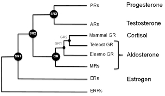
Steroid receptor evolution

Ortlund’s laboratory recently published a paper in Journal of Biological Chemistry describing the structure of a protein that is supposed to have existed 450 million years ago, in a complex with an anti-inflammatory drug widely used today. MSP graduate student Jeffrey Kohn is the first author.

Mometasone furoate is the active ingredient of drugs used to treat asthma, allergies and skin irritation. It is part of a class of drugs known as glucocorticoids, which can have a host of side effects such as reduced bone density and elevated blood sugar or blood pressure with long-term use.

One reason for these side effects is because the steroid receptor proteins that allow cells to detect and respond to hormones such as estrogen, testosterone, aldosterone and cortisol are all related. Mometasone is a good example of how glucocorticoids cross-react, Ortlund says. That made it an ideal test of the technique of mixing ancient receptors with modern drugs.

“We used this structure to determine why mometasone cross reacts with the progesterone receptor, which regulates fertility, and why it inhibits the mineralocorticoid receptor, which regulates blood pressure,” he says.

Mometasone furoate in complex with the ancient receptor

Scientists have examined the sequences of the genes that encode these proteins at several points on the evolutionary tree, and used the information to reconstruct what the ancestral receptor looked like. This helps solve some problems that biochemists studying these proteins have had to deal with. One of these is: changing one amino acid in the protein sometimes means that the whole protein malfunctions.

“The ancestral receptors are more tolerant to mutation, and they are more promiscuous with respect to activation,” Ortlund says. “That is, they tend to respond to a wider array of endogenous steroid hormones, which makes sense in an evolutionary context. This enhanced activation profile and tolerance to mutation is what we feel makes them ideally suited to structure-function studies.”

The blog Panda’s Thumb has an interesting discussion of this area of research, in relation to the larger question of how proteins evolve.

Posted on by Quinn Eastman in Uncategorized Leave a comment

Emory transplant roundup

A recent Associated Press story highlighted clinical trials aimed at helping kidney transplant recipients give up their anti-rejection drugs:

The experimental approach: Transplant the seeds of a new immune system along with a new kidney. It’s the 21st-century version of a bone marrow transplant, and possible for now only if the transplanted kidney comes from a living donor.

How does it work? Doctors cull immune system-producing stem cells and other immunity cells from the donor’s bloodstream. They blast transplant patients with radiation and medications to wipe out part of their own bone marrow, far more grueling than a regular kidney transplant. That makes room for the donated cells to squeeze in and take root, creating a sort of hybrid immunity that scientists call chimerism, borrowing a page from mythology.

Emory Transplant Center scientific director Allan Kirk is leading a study that takes a similar approach, involving a depletion of the recipient’s immune cells and an infusion of bone marrow, which introduces new immune cells from the donor.

Allan Kirk, MD, PhD

Nature Medicine also has a good explanation of this area of research. Kirk is quoted in this recent story:

“The impetus to take the risk and pull people off immunosuppressants completely is lower now,” says Kirk… “It’s all about risk-benefit ratios and about making smart decisions with the tools we have—and we have a lot more tools now.”

Why go through so much trouble to avoid anti-rejection drugs? The most common drugs taken by transplant recipients, called calcineurin inhibitors, can reduce an individual’s ability to fight infections, lead to high blood pressure and high blood sugar and, ironically, tend to damage the kidney over time. Emory scientists played a major role in developing an alternative, belatacept, which was approved last year by the FDA.

Emory transplant surgeon Ken Newell was also mentioned in the AP story for his study of rare individuals who were able to go “cold turkey” and avoid having their immune systems reject their donated kidneys. One of these individuals, Lisa Robinson, had an interesting story to tell about how came to that point:

Three years after her kidney transplant, she found it hard to tolerate the side effects of the immunosuppressive drugs, which included swelling, weight gain and depression. On top of that, her creatinine levels were rising, indicating that her donated kidney was losing function. Without explicit approval from her doctor, she decided to taper off her drugs, first cyclosporine and then steroids.

“This turned out to be the right choice for me, but I’m not suggesting that others do what I did,” she says. “Everyone has to figure out what works for them. My main motivation was that I didn’t want to go through another kidney transplant.”

Based on data from Robinson and other people who had similar experiences, Newell has been able to identify a pattern of genes turned on in their immune cells that may predict whether someone could be able to become “tolerant.” Much of transplant biology focuses on one type of immune cell (T cells), but Newell found that the cells that may make the biggest difference for long-term tolerance are different, B cells. This makes sense because of B cells’ role in chronic rejection, Emory’s Stuart Knechtle has written.

Posted on by Quinn Eastman in Uncategorized Leave a comment

The challenges of graduate school

Biochemist Paul Doetsch’s recent appearance in a Science magazine feature on laboratory leadership led to a conversation with him about the challenges of graduate school.

He emphasized that scientific research is a team sport, and brilliance on the part of the lab head may not yield fruit without a productive relationship with the people in the lab. Doetsch suggested talking with Lydia Morris, a graduate student in the Genetics and Molecular Biology graduate program. Morris has been working in Doetsch’s lab for several years and is about to complete her degree. She has been examining the in vivo distribution of DNA repair proteins.

In this video, Morris and Doetsch talk about the differences between turn-the-crank and blue-sky projects, and the importance of backup projects, communications, high expectations and perseverance.

Posted on by Quinn Eastman in Cancer Leave a comment

Media’s contribution to stigma

Photo credit: Julia Freeman-Woolpert

Tireless advocacy in the last decade by mental health professionals and people who are affected by mental illness has aimed to reduce the stigma of psychiatric disorders. To determine the influence those efforts have had on news media portrayals, Emory researchers studied newspaper articles using the terms “schizophrenia” and “schizophrenic” in the years 2000 versus 2010.

“The primary goal of journalists is to give fair, accurate and unbiased reports of news events that will be of interest to the public,” says study author Arshya Vahabzadeh, MD, resident psychiatrist in the Department of Psychiatry and Behavioral Sciences at Emory University School of Medicine.

Arshya Vahabzadeh, MD, resident psychiatrist in the Department of Psychiatry and Behavioral Sciences at Emory University School of Medicine says that people who suffer from a mental illness often internalize negative references, and develop coping mechanisms that become obstacles to treatment.

“A secondary goal is to capture the attention of readers and viewers,” he says. “Unfortunately, stories linked to a mental illness have been shown to strongly attract readers’ attention, and to contribute to unfavorable public conceptions of mental illness.”

According to the researchers, the decade from 2000 to 2010 was of particular importance to the study because as the new millennium began, attention increasingly focused on public awareness of mental illness, with mental health professionals, advocacy groups and governmental bodies targeting de-stigmatization of psychiatric disorders.

Vahabzadeh and his colleagues examined hundreds of articles that appeared in five major newspapers during a five-month time period during 2000 and 2010, searching for the terms “schizophrenia” and “schizophrenic”.

The researchers found that during that period in 2000, 7,114 articles were published in the five selected newspapers, 247 of which mentioned schizophrenia. During the same period in 2010, 4,397 articles were published, with 151 articles mentioning schizophrenia.

Although a larger percentage of the articles were published in 2000 than in 2010, the percentage of articles mentioning schizophrenia did not differ. Similarly, there was no significant difference in metaphorical usage of the term “schizophrenia” – using the term to describe conflicting decisions or illogical actions.

Of particular concern to investigators was that 60 percent of the human-interest stories in both time periods focused on highly emotive reports of violence, dangerousness and criminality. Murders committed by people with schizophrenia accounted for almost half of such articles. The authors did, however, recognize a smaller proportion of articles focusing on crimes and murder committed by people with schizophrenia in 2010 compared to 2000.

Additionally, despite the fact that people with schizophrenia are more prone to be the victims rather than perpetrators of crime, situations in which people with schizophrenia were reported as victims accounted for only 0.5% of the articles reviewed.

“People who suffer from a mental illness often internalize these negative references, and develop coping mechanisms that become obstacles to treatment,” explains Vahabzadeh.

“Negative perceptions also may result in problems for those who are successfully treated,” he says. “People with schizophrenia may encounter stigma when they attempt to integrate into society, build relationships, find employment and even housing. It is important for us as mental health professionals to look for opportunities to educate the media on ways to become a positive force to reduce stigma.”

Nadine Kaslow, PhD, professor and chief psychologist at the Emory University School of Medicine, whose mentorship inspired the study, says “This study provides invaluable information about the persistence of negative and unfounded portrayals of people living with schizophrenia to the public. It is imperative that everyone interacts with people living with mental illness respectfully and with compassion, and that they be welcomed members of our community.”

The study was published in the Journal of Psychiatric Practice http://www.ncbi.nlm.nih.gov/pubmed/22108403.

Additional study investigators include Justine Wittenauer, MD, Emory University School of Medicine and Erika Carr, PhD, Yale University School of Medicine.

Posted on by admin in Uncategorized Leave a comment

The body’s anticancer defenses come in a variety of sizes

Sometimes you have to look at the whole picture, big and small.

Sarah Cork, PhD

That was the lesson that emerged from Winship Cancer Institute researcher Erwin Van Meir’s laboratory, highlighted in a recent paper in Oncogene. Van Meir’s team has been studying vasculostatin, a secreted protein that inhibits blood vessel growth by tumors (hence the name). Vasculostatin was discovered by Balveen Kaur, now at Ohio State, while she was in Van Meir’s lab.

Van Meir and his colleagues originally began studying vasculostatin because it is a product of a gene that brain tumors somehow silence or get rid of, and studying the obstacles our bodies throws in cancer’s way may be a good way to learn how to fight it via modern medicine. Eventually, it could form the basis for a treatment to prevent a tumor from attracting new blood vessels.

Vasculostatin is somewhat unique because it is a secreted fragment of a membrane-bound protein, called BAI1. BAI1 has an apparently separate function as an “engulfment receptor,” allowing the recognition and internalization of dying cells.

Most of the secreted vasculostatin is around 40 kilodaltons in size, not 120 as previously thought.

Graduate student Sarah Cork discovered that most of the vasculostatin protein being produced by cells is actually much smaller than what had been originally discovered. She and Van Meir were surprised to find that the smaller, cleaved form of the protein still has potent anti-angiogenic activity.

The researchers were using a technique where a mixture of proteins is separated within a gel by electric current, transferred to a polymer sheet, and probed with antibodies. The large proteins appear at the top and the small proteins at the bottom.

“Previously, we had been running the gels for a long time to detect large protein fragments, so missed out on what was happening with small fragments which run off the gel,” Van Meir says. “We were only looking at the top of the
gel, when the smaller form of vasculostatin was actually much more
abundant as you can see on the picture of a gel run for a shorter time.”

More broadly, Van Meir says that the finding adds to understanding about BAI1’s dual function in the brain and how vasculostatin (big or small) might be used in anticancer therapy.

Posted on by Quinn Eastman in Cancer Leave a comment

Fragile X protein: one toggle switch, many circuits

The fragile X protein — missing in the most common inherited form of intellectual disability — plays a central role in neurons and how they respond to external signals. Cell biologist Gary Bassell and his colleagues have been examining how the fragile X protein (FMRP) acts as a “toggle switch.”

Gary Bassell, PhD

FMRP controls the activity of several genes by holding on to the RNAs those genes encode. When neurons get an electrochemical signal from the outside, FMRP releases the RNAs, allowing the RNAs to be made into protein, and facilitating changes in the neurons linked to learning and memory.

The Bassell lab’s new paper in Journal of Neuroscience reveals the role of another player in this process. The first author is postdoctoral fellow Vijay Nalavadi.

The researchers show that neurons modify FMRP with ubiquitin, the cellular equivalent of a tag for trash pickup, after receiving an external signal. In general, cells attach ubiquitin to proteins so that the proteins get eaten up by the proteasome, the cellular trash disposal bin. Here, neurons are temporarily getting rid of FMRP, prolonging the effects of the external signal.

Posted on by Quinn Eastman in Neuro Leave a comment

Dye me anticancer yellow

Over the last few years, pathologist Keqiang Ye and his colleagues have displayed an uncanny talent for finding potentially useful medicinal compounds. Recently another example of this talent appeared in Journal of Biological Chemistry.

Keqiang Ye, PhD

Postdoctoral fellow Qi Qi is first author on the paper. Collaborators include Jeffrey Olson, Liya Wang, Hui Mao, Haian Fu, Suresh Ramalingam and Shi-Yong Sun at Emory and Paul Mischel at UCLA.

Qi and Ye were looking for compounds that could inhibit the growth of an especially aggressive form of brain cancer, glioblastoma with deletion in the tumor suppressor gene PTEN. Tumors with this deletion do not respond to currently available targeted therapies.

The researchers found that acridine yellow G, a fluorescent dye used to stain microscope slides, can inhibit the growth of this tumor:

Oral administration of this compound evidently decreases the tumor volumes in both subcutaneous and intracranial models and elongates the life span of brain tumor inoculated nude mice. It also displays potent antitumor effect against human lung cancers. Moreover, it significantly decreases cell proliferation and enhances apoptosis in tumors…

Optimization of this compound by improving its potency through medicinal chemistry modification might warrant a novel anticancer drug for malignant human cancers.

Ye’s team observed that acridine yellow G appears not to be toxic in rodents. However, the acridine family of compounds tends to intercalate (insert itself) into DNA and can promote DNA damage, so more toxicology studies are needed. Other acridine family compounds such as quinacrine have been used to treat bacterial infections and as antiinflammatory agents, they note.

A paramecium stained with acridine orange, which shows anticancer activity for tumors containing PTEN mutations

Posted on by Quinn Eastman in Cancer Leave a comment

The face behind a case

Last week Emory posted a news item about a case report published in the American Journal of Human Genetics. The paper described how geneticists at Emory, in cooperation with Sanford Burnham Medical Research Institute in San Diego, used “whole exome sequencing” — a sort of executive summary scan of the genome — to find the cause of a metabolic disease in a young boy.

The case was an illustration of the trend of whole exome sequencing, which is starting to enter clinical practice as a diagnostic technology. A photo of the patient, courtesy of his parents and Sanford Burnham, is a powerful reminder that within every case report, there’s a real person’s history.

Courtesy of Heather Buschman

“Over the years, we’ve come to know many families and their kids with glycosylation disorders. Here we can tell them their boy is a true ‘trail-blazer’ for this new disease,” says Hudson Freeze, director of the Genetic Disease program at Sanford Burnham. “Their smiles—that’s our bonus checks.”

Posted on by Quinn Eastman in Uncategorized Leave a comment

What is the default mode network?

Welcome to what could become a regular feature on the Emory Health Now blog: explaining a word or phrase that is connected with research going on at Emory.

What is the default mode network?
This is a concept that grew out of brain imaging studies, using techniques such as functional magnetic resonance imaging. The default mode network consists of regions of the brain that are active when someone is not doing anything in particular, especially something requiring focused attention. More fancifully: it’s what daydreaming looks like.

The default mode network includes the medial prefrontal cortex (MPFC) and the posterior cingulate cortex (PCC).

Researchers at Emory and elsewhere have been looking at whether the DMN’s activity and its links to other areas of the brain are changed in disorders such as schizophrenia, autism, depression and post-traumatic stress disorder.

For example, Xiaoping Hu, director of Emory’s Biomedical Imaging Technology Center, and his colleagues have investigated how the default mode network’s activity is modified in individuals with prenatal alcohol exposure and prenatal cocaine exposure. They also have probed how the DMN’s activity is shut down by anesthesia.

At the Atlanta Veterans Affairs Medical Center, Erica Duncan and her colleagues have examined the DMN in people with schizophrenia. They found (as have other groups) that individuals with schizophrenia appear to have difficulty shutting down the DMN and focusing on a task, as well as having a different pattern of connections within the DMN.

Yerkes investigator Lary Walker recently wrote about research connecting metabolic activity in the default mode network with the burden of amyloid in Alzheimer’s disease.

The DMN is made up of several regions of the brain, most prominently the posterior cingulate cortex (PCC) and medial prefrontal cortex (MPFC). Other regions such as the inferior parietal lobule, lateral temporal cortex, and hippocampal formation including parahippocampus are also considered part of the DMN. Note: these regions can also be engaged in other tasks besides daydreaming or introspection.

Posted on by Quinn Eastman in Neuro Leave a comment

Strengthening bone with silica nanoparticles

Tiny particles of silicon dioxide – essentially, extremely fine sand — can strengthen bones when introduced into animals, researchers at Emory University School of Medicine have discovered.

The particles stimulate the generation of bone-forming cells and inhibit other cells that break down bone. The findings could someday form the basis for an alternative treatment for osteoporosis.

The results were published recently in the journal Nanomedicine.

The paper represents a collaboration between the laboratories of George Beck and Neale Weitzmann, both in the Division of Endocrinology, Metabolism and Lipids. The project started when Jin-Kyu Lee, now at Seoul National University, came to Beck’s lab with silica nanoparticles he had developed that contained fluorescent dyes. This allowed researchers to track the particles within the body and within cells.

In the laboratory, the nanoparticles stimulate the generation of bone-forming osteoblasts and inhibit the maturation of bone-remodeling osteoclasts. Beck says that the particles’ properties seem to depend on their size (50 nanometers wide) and shape, because larger particles don’t have the same effects. The nanoparticles appear to work by being taken up by the cells and then by inhibiting NF-kB, a molecule that controls inflammation.

Silicon is a trace element in the diet of most people. Scientists have known for several years that dietary silicon is linked to strong bones, but how silicon influences bone growth has remained unclear: it could become physically incorporated into bone, or it could provide signals to the cells that make up bone. To be sure, silica nanoparticles may be acting in a way that is different than dietary silicon.

The particles’ ability to stimulate osteoblasts distinguish them from bisphosphonates, the most common drugs now used to treat osteoporosis, Beck says. Bisphosphonates only inhibit bone breakdown and do not stimulate bone formation.

The Emory team has found that injecting silica nanoparticles can increase the bone density of young mice by roughly 15 percent over six weeks, augmenting the increases coming from adolescent growth.

To test the particles’ potential for use with humans, the researchers are examining whether injection is the best way to deliver the nanoparticles, and whether long-term toxicity is an issue. Inhalation of larger particles of silica dust, an occupational hazard for miners and construction workers, can result in the lung disease silicosis. However, silicosis arises because the lungs can’t absorb and remove the larger dust particles. Since cells clearly can take up the nanoparticles (see video), it is possible that they will not induce the body to respond similarly.

Emory has applied for patents on this technology. A presentation by Emory’s Office of Technology Transfer is available here.

Posted on by Quinn Eastman in Uncategorized Leave a comment