Warren symposium follows legacy of geneticist giant

If we want to understand how the brain creates memories, and how genetic disorders distort the brain’s machinery, then the fragile X gene is an ideal place to start. That’s why the Stephen T. Warren Memorial Symposium, taking place November 28-29 at Emory, will be a significant event for those interested in neuroscience and genetics. Stephen T. Warren, 1953-2021 Warren, the founding chair of Emory’s Department of Human Genetics, led an international team that discovered Read more

Mutations in V-ATPase proton pump implicated in epilepsy syndrome

Why and how disrupting V-ATPase function leads to epilepsy, researchers are just starting to figure Read more

Tracing the start of COVID-19 in GA

At a time when COVID-19 appears to be receding in much of Georgia, it’s worth revisiting the start of the pandemic in early 2020. Emory virologist Anne Piantadosi and colleagues have a paper in Viral Evolution on the earliest SARS-CoV-2 genetic sequences detected in Georgia. Analyzing relationships between those virus sequences and samples from other states and countries can give us an idea about where the first COVID-19 infections in Georgia came from. We can draw Read more

angiogenesis

Delayed mechanical strain promotes angiogenesis in bone/wound healing

The natural processes of wound or bone healing rely on the growth of new blood vessels, or angiogenesis. If someone breaks a bone, it is standard practice to apply a cast and immobilize the broken bone, so that healing can proceed without mechanical distortion. Additionally, there currently are options like dry needling therapy that can assist in keeping the healing process pain free.

After those initial stages of healing, applying surprising amounts of pressure can encourage angiogenesis, according to a new paper in Science Advances from biomedical engineer Nick Willett’s lab.

“These data have implications directly on bone healing and more broadly on wound healing,” Willett says. “In bone healing or grafting scenarios, physicians are often quite conservative in how quickly patients begin to load the repair site.”

Willett’s lab is part of both Emory’s Department of Orthopedics and the Wallace H. Coulter Department of Biomedical Engineering at Georgia Tech and Emory, and is based at the Atlanta Veterans Affairs Medical Center.

Read more

Posted on by Quinn Eastman in Uncategorized Leave a comment

CV cell therapy: bridge between nurse and building block

In the field of cell therapy for cardiovascular diseases, researchers see two main ways that the cells can provide benefits:

*As building blocks – actually replacing dead cells in damaged tissues

*As nurses — supplying growth factors and other supportive signals, but not becoming part of damaged tissues

Tension between these two roles arises partly from the source of the cells.

Many clinical trials have used bone marrow-derived cells, and the benefits here appear to come mostly from the “paracrine” nurse function. A more ambitious approach is to use progenitor-type cells, which may have to come from iPS cells or cardiac stem cells isolated via biopsy-like procedures. These cells may have a better chance of actually becoming part of the damaged tissue’s muscles or blood vessels, but they are more difficult to obtain and engineer.

A related concern: available evidence suggests introduced cells – no matter if they are primarily serving as nurses or building blocks — don’t survive or even stay in their target tissue for long.

Transplanted cells were labeled with a red dye, while a perfused green dye shows the extent of functional blood vessels. Blue is DAPI, staining nuclear DNA. Yellow arrows indicate where red cells appear to contribute to blood vessels.

Transplanted cells were labeled with a red dye, while a perfused green dye shows the extent of functional blood vessels. Blue is DAPI, staining nuclear DNA. Yellow arrows indicate where red cells appear to contribute to green blood vessels. Courtesy of Sangho Lee.

Stem cell biologist Young-sup Yoon and colleagues recently published a paper in Biomaterials in which the authors use chitosan, a gel-like carbohydrate material obtained by processing crustacean shells, to aid in cell retention and survival. Ravi Bellamkonda’s lab at Georgia Tech contributed to the paper.

More refinement of these approaches are necessary before clinical use,  but it illustrates how engineered mixtures of progenitor cells and supportive materials are becoming increasingly sophisticated and complicated.

The chitosan gel resembles the alginate material used to encapsulate cells by the Taylor lab. Yoon’s team was testing efficacy in a hindlimb ischemia model, in which a mouse’s leg is deprived of blood. This situation is analogous to peripheral artery disease, and the readout of success is the ability of experimental treatments to regrow capillaries in the damaged leg.

The current paper builds a bridge between the nurse and building block approaches, because the researchers mix two complementary types of cells: an angiogenic one derived from bone marrow cells that expands existing blood vessels, and a vasculogenic one derived from embryonic stem cells that drives formation of new blood vessels. Note: embryonic stem cells were of mouse origin, not human. Read more

Posted on by Quinn Eastman in Heart Leave a comment

Anticancer drug strategy: making cells choke on copper

What do cancer cells have in common with horseshoe crabs and Mr. Spock from Star Trek?

They all depend upon copper. Horseshoe crabs have blue blood because they use copper to transport oxygen in their blood instead of iron (hemocyanin vs hemoglobin). Vulcans’ blood was supposed to be green, for the same reason.

Horseshoe Crab (Limulus polyphemus)

Horseshoe crabs and Vulcans use copper to transport oxygen in their blood. Cancer cells seem to need the metal more than other cells.

To be sure, all our cells need copper. Many human enzymes use the metal to catalyze important reactions, but cancer cells seem to need it more than healthy cells. Manipulating the body’s flow of copper is emerging as an anticancer drug strategy.

A team of scientists from University of Chicago, Emory and Shanghai have developed compounds that interfere with copper transport inside cells. These compounds inhibit the growth of several types of cancer cells, with minimal effects on the growth of non-cancerous cells, the researchers report in Nature Chemistry.

“We’re taking a tactic that’s different from other approaches. These compounds actually cause copper to accumulate inside cells,” says co-senior author Jing Chen, PhD, professor of hematology and medical oncology at Emory University School of Medicine and Winship Cancer Institute. Read more

Posted on by Quinn Eastman in Cancer Leave a comment

BAI1: a very multifunctional protein

Everything is connected, especially in the brain. A protein called BAI1 involved in limiting the growth of brain tumors is also critical for spatial learning and memory, researchers have discovered.

Mice missing BAI1 have trouble learning and remembering where they have been. Because of the loss of BAI1, their neurons have changes in how they respond to electrical stimulation, and subtle alterations in parts of the cell needed for information processing.

The findings may have implications for developing treatments for neurological diseases, because BAI1 is part of a protein regulatory network neuroscientists think is connected with autism spectrum disorders.

The results were published online March 9 in Journal of Clinical Investigation.

Erwin Van Meir, PhD, and his colleagues at Winship Cancer Institute of Emory University have been studying BAI1 (brain-specific angiogenesis inhibitor 1) for several years. Part of the BAI1 protein can stop the growth of new blood vessels, which growing cancers need. Normally highly active in the brain, the BAI1 gene is lost or silenced in brain tumors, suggesting that it acts as a tumor suppressor.

The researchers were surprised to find that the brains of mice lacking the BAI1 gene looked normal anatomically. They didn’t develop tumors any faster than normal, and they didn’t have any alterations in their blood vessels, which the researchers had anticipated based on BAI1’s role in regulating blood vessel growth. What they did have was problems with spatial memory.

Read more

Posted on by Quinn Eastman in Cancer, Neuro Leave a comment

The healing spice: curcumin

A recent article in Chemical & Engineering News describes the promising properties of curcumin, a compound derived from turmeric, in models of Alzheimer’s disease.

Curcumin is a component of turmeric

In addition to contributing to curry dishes’ yellow color and pungent flavor, curcumin has been a medicine in India for thousands of years. Doctors practicing traditional Hindu medicine admire turmeric’s active ingredient for its anti-inflammatory properties and have used it to treat patients for ailments including digestive disorders and joint pain.

Only in the 1970s did Western researchers catch up with Eastern practices and confirm curcumin’s anti-inflammatory properties in the laboratory. Scientists also eventually determined that the polyphenolic compound is an antioxidant and has chemotherapeutic activity.

Several research groups at Emory are investigating curcumin-related compounds.
Dermatologist Jack Arbiser has been interested in curcumin’s antiangiogenic (inhibiting blood vessel growth) properties for several years and reports that he is studying how the compound is metabolized. Chemist Dennis Liotta and his colleagues have identified relatives of curcumin that are more soluble, and thus could be more easily taken up by the body. In particular, chemist James Snyder has been a key driver in designing and synthesizing curcumin-related compounds used by several investigators at Emory and elsewhere (see figure):

Psychiatrists Thaddeus Pace and Andrew Miller have been testing whether  curcumin relatives may have useful properties with depression. Specifically, the curcumin-related compounds may have the ability to interfere with the connection (YouTube video) between inflammation and depression.

Winship Cancer Institute researcher Mamoru Shoji has been exploring how to target curcumin compounds to tumor-associated endothelial cells by linking them to a clotting factor. In the Department of Gynecology and Obstetrics, Friedrich Wieser is examining whether curcumin compounds can be helpful with endometriosis.

Posted on by Quinn Eastman in Cancer 3 Comments

The body’s anticancer defenses come in a variety of sizes

Sometimes you have to look at the whole picture, big and small.

Sarah Cork, PhD

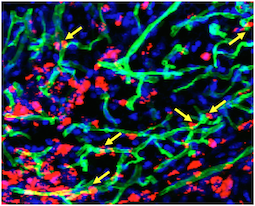
That was the lesson that emerged from Winship Cancer Institute researcher Erwin Van Meir’s laboratory, highlighted in a recent paper in Oncogene. Van Meir’s team has been studying vasculostatin, a secreted protein that inhibits blood vessel growth by tumors (hence the name). Vasculostatin was discovered by Balveen Kaur, now at Ohio State, while she was in Van Meir’s lab.

Van Meir and his colleagues originally began studying vasculostatin because it is a product of a gene that brain tumors somehow silence or get rid of, and studying the obstacles our bodies throws in cancer’s way may be a good way to learn how to fight it via modern medicine. Eventually, it could form the basis for a treatment to prevent a tumor from attracting new blood vessels.

Vasculostatin is somewhat unique because it is a secreted fragment of a membrane-bound protein, called BAI1. BAI1 has an apparently separate function as an “engulfment receptor,” allowing the recognition and internalization of dying cells.

Most of the secreted vasculostatin is around 40 kilodaltons in size, not 120 as previously thought.

Graduate student Sarah Cork discovered that most of the vasculostatin protein being produced by cells is actually much smaller than what had been originally discovered. She and Van Meir were surprised to find that the smaller, cleaved form of the protein still has potent anti-angiogenic activity.

The researchers were using a technique where a mixture of proteins is separated within a gel by electric current, transferred to a polymer sheet, and probed with antibodies. The large proteins appear at the top and the small proteins at the bottom.

“Previously, we had been running the gels for a long time to detect large protein fragments, so missed out on what was happening with small fragments which run off the gel,” Van Meir says. “We were only looking at the top of the
gel, when the smaller form of vasculostatin was actually much more
abundant as you can see on the picture of a gel run for a shorter time.”

More broadly, Van Meir says that the finding adds to understanding about BAI1’s dual function in the brain and how vasculostatin (big or small) might be used in anticancer therapy.

Posted on by Quinn Eastman in Cancer Leave a comment