If we want to understand how the brain creates memories, and how genetic disorders distort the brain’s machinery, then the fragile X gene is an ideal place to start. That’s why the Stephen T. Warren Memorial Symposium, taking place November 28-29 at Emory, will be a significant event for those interested in neuroscience and genetics.
Stephen T. Warren, 1953-2021
Warren, the founding chair of Emory’s Department of Human Genetics, led an international team that discovered Read more
At a time when COVID-19 appears to be receding in much of Georgia, it’s worth revisiting the start of the pandemic in early 2020. Emory virologist Anne Piantadosi and colleagues have a paper in Viral Evolution on the earliest SARS-CoV-2 genetic sequences detected in Georgia.
Analyzing relationships between those virus sequences and samples from other states and countries can give us an idea about where the first COVID-19 infections in Georgia came from. We can draw Read more
In a paper recently published in Journal of Neuroscience, a team led by cell biologist Gary Bassell shows that PI3 kinase inhibitors could restore normal appearance and levels of protein production at the synapses of hippocampal neurons from fragile X model mice. The next steps, studies in animals, are underway.
“This is an important first step toward having a new therapeutic strategy for fragile X syndrome that treats the underlying molecular defect, and it may be more broadly applicable to other forms of autism,†he says.
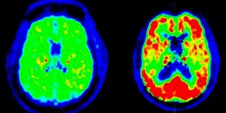
Scans can show beta amyloid, a protein associated with Alzheimer’s disease (right)
For the first time in 25 years, medical experts are proposing new diagnostic criteria aimed at better and earlier detection of Alzheimer’s disease (AD).
According to the Alzheimer’s Association, an estimated 5.3 million Americans have AD, most of them 65 and older. The disease is thought to begin years, possibly even decades, before symptoms are noticeable. But there is no single, generally accepted way to identify the disease in its earliest stages before symptoms are evident.
The new diagnostic guidelines focus on advances in detecting biomarkers for the disease, such as substances found in spinal fluid or appearing on cutting-edge brain imaging scans conducted with PET or MRI.
Emphasis will be on diagnosing early stages of the disease as soon as possible so that patients can take measures to slow the progression or prevent further damage.
Consider this: Alzheimer’s is a uniquely human disorder. But why? Why don’t nonhuman primates, such as monkeys, get Alzheimer’s disease. Monkeys form the senile plaques that are identical to the plaques found in humans. So do other animals.
“Yet, despite the fact that nonhuman primates make this protein that we know is very important in the pathogenesis of Alzheimer’s disease, they don’t develop the full disease,†says Lary Walker, PhD. Walker is an associate professor at Yerkes National Primate Research Center.
“They don’t develop the tangles we associate with Alzheimer’s disease, the neuronal loss, the shrinkage of the brain, and they don’t get demented in the sense that humans do,†says Walker.
When our bodies make a protein, the protein tends to fold into a functional form. But when it comes to Alzheimer’s disease, some proteins misfold, becoming sticky and then combining with one another. In their collective form, the proteins can then form plaques or tangles, the two types of lesions associated with Alzheimer’s disease.
And for some unknown reason, people who have plaques usually go on to form tangles. But people who have tangles don’t always go on to form plaques. No one is sure why. But that’s what researcher Walker wants to find out.
To listen to Walker’s own words about Alzheimer’s disease, access Emory’s new Sound Science podcast.
A study published in the May 17, 2010, issue of the journal Pediatrics found that one type of pesticide commonly used on fruits and vegetables may be contributing to attention deficit hyperactivity disorder, or ADHD, in children.
The study measured the levels of pesticide byproducts in the urine of 1,139 children from across the United States. Children with the highest concentration of pesticides in their urine were more likely to have symptoms of ADHD.
Barr says while the study doesn’t prove causality between pesticide exposures and ADHD, it does shed light on how even low level daily exposures to pesticides could potentially impact cognitive health.
“It seems very plausible that low-level daily exposures to pesticides can produce some subtle effects like ADHD or other neurological delays,†she says.
Barr notes that additional research is needed to confirm a connection to pesticides and ADHD, but says there are tips for limiting your exposure to commonly used pesticides.
“We’ve done studies here at Emory and also at CDC that have indicated that if you use organic food or if you wash your food properly prior to preparation, you can reduce the levels of these metabolites in your urine. Eat as much organic produce as possible, or wash your fruits and vegetables very well and that likely could decrease the chances of your children developing ADHD,†says Barr.
So says Larry Young, PhD, chief of the Division of Behavioral Neuroscience and Psychiatric Disorders at the Yerkes National Primate Research Center, Emory University.
Young, who is world-renowned for his work on the role of neuropeptides in regulating social behavior, uses voles to investigate the neurobiological and genetic mechanisms underlying social behavior. Using the monogamous prairie vole (vs. the promiscuous meadow vole) as a model organism, Young and his research team identified the oxytocin and vasopressin receptors as key mediators of social bonding and attachment. In addition, they are examining the consequences of social bond disruption as a model of social loss-induced depression.
This work has important implications for developing novel treatment strategies for psychiatric disorders associated with social cognitive deficits, including autism spectrum disorders and schizophrenia.
View of MR/PET scanner from front, with Ciprian Catana of MGH and Larry Byars of Siemens
The scanner is one of four world-wide and one of two in the United States, and permits simultaneous MR (magnetic resonance) and PET (positron emission tomography) imaging in human subjects. This provides the advantage of being able to combine the anatomical information from MR with the biochemical/metabolic information from PET. Potential applications include functional brain mapping and the study of neurodegenerative diseases, drug addiction and brain cancer.
Thursday’s event brought together leaders of the three other MR/PET programs in Boston, Jülich and Tübingen, the Siemens engineers who designed the device, and the Atlanta research community to explore the possibilities of the technology.
The drugs now available to treat Alzheimer’s address the symptoms of the disease — memory problems — rather than the underlying mechanism of neurodegeneration.
But what if something could do both? Here’s a tantalizing prospect, hinted at by a long-running thread of brain research: compounds that boost the function of certain acetylcholine circuits in the brain might also modify production of toxic beta-amyloid protein.
The possibility grows out of the properties of certain receptors for the neurotransmitter acetylcholine, called “muscarinic acetylcholine receptors.” Acetylcholine is a major transmitter of signals in the brain, and there are several varieties of receptors, or receiver dishes for the signals, on brain cells.
Two researchers at Emory, Anita Corbett and Grace Pavlath, recently have combined their expertise to probe how a puzzling form of muscular dystrophy develops.
Oculopharyngeal muscular dystrophy (OPMD) is an inherited type of muscular dystrophy that primarily affects muscles of the face and throat. In the video below, Anita Corbett explains how this affects patients as they get older.
The mutations that cause the disease make a protein called PABPN1 longer and stickier than normal, and the mutated protein appears to form clumps in muscle cells.
The puzzle lies in that PABPN1 (poly A binding protein nuclear 1)Â can be found everywhere in the body, but it’s not clear why the mutated protein specifically affects muscle cells — or why the muscles in the face and throat are especially vulnerable.
In December 2009, Corbett, Pavlath and postdoctoral fellow Luciano Apponi published a paper where they suggest that the clumps of mutated protein, which some researchers have proposed to be toxic, might not be the whole story. A lack of functioning PABPN1 might be just as strong a factor in the disease, they’ve discovered.
Pathologist Keqiang Ye has made a series of discoveries recently, arising from his investigations of substances that can mimic the growth factor BDNF (brain-derived neurotrophic factor).
BDNF is a protein produced by the brain that pushes neurons to withstand stress and make new connections. Some neuroscientists have described BDNF as “Miracle Gro for brain cells.”
“BDNF has been studied extensively for its ability to protect neurons vulnerable to degeneration in several diseases, such as ALS, Parkinson’s and Alzheimer’s disease,†Ye says. “The trouble with BDNF is one of delivery. It’s a protein, so it can’t cross the blood-brain barrier and degrades quickly.â€
Working with Ye, postdoctoral fellow Sung-Wuk Jang identified a compound called 7,8-dihydroxyflavone that can duplicate BDNF’s effects on neurons and can protect them against damage in animal models of seizure, stroke and Parkinson’s disease. The compound’s selective effects suggest that it could be the founder of a new class of brain-protecting drugs. The results were published in Proceedings of the National Academy of Sciences.
If the brain acts like a computer, which of the brain’s physical features store the information? Flashes of electricity may keep memories and sensations alive for the moment, but what plays the role that hard drives and CDs do for computers?
A simple answer could be: genes turning on and off, and eventually, neurons growing and changing their shapes. But it gets more complicated pretty quickly. Genes can be regulated at several levels:
at the level of transcription — whether messenger RNA gets made from a stretch of DNA in the cell’s nucleus
at the level of translation — whether the messenger RNA is allowed to make a protein
at the level of RNA localization — where the mRNAs travel within the cell
Each neuron has only two copies of a given gene but will have many dendrites that can have more or less RNA in them. That means the last two modes of regulation offer neurons much more capacity for storing information.
Gary Bassell, a cell biologist at Emory, and his colleagues have been exploring how RNA regulation works in neurons. They have developed special tools for mapping RNA, and especially, microRNA — a form of RNA that regulates other RNAs.
In the dendrites of neurons, FMRP seems to control where RNAs end up
Fragile X mental retardation protein (FMRP), linked to the most common inherited form of mental retardation, appears to orchestrate RNA traffic in neurons. Bassell and pharmacologist Yue Feng recently received a grant from the National Institute of Child Health and Development to study FMRP’s regulation of RNA in greater detail. The grant was one of several at Emory funded through the American Recovery and Reinvestment Act’s support for the NIH.
In the video interview above, Bassell explains his work on microRNAs in neurons. Below is a microscope image, provided by Bassell, showing the pattern of FMRP’s localization in neurons.