Warren symposium follows legacy of geneticist giant

If we want to understand how the brain creates memories, and how genetic disorders distort the brain’s machinery, then the fragile X gene is an ideal place to start. That’s why the Stephen T. Warren Memorial Symposium, taking place November 28-29 at Emory, will be a significant event for those interested in neuroscience and genetics. Stephen T. Warren, 1953-2021 Warren, the founding chair of Emory’s Department of Human Genetics, led an international team that discovered Read more

Mutations in V-ATPase proton pump implicated in epilepsy syndrome

Why and how disrupting V-ATPase function leads to epilepsy, researchers are just starting to figure Read more

Tracing the start of COVID-19 in GA

At a time when COVID-19 appears to be receding in much of Georgia, it’s worth revisiting the start of the pandemic in early 2020. Emory virologist Anne Piantadosi and colleagues have a paper in Viral Evolution on the earliest SARS-CoV-2 genetic sequences detected in Georgia. Analyzing relationships between those virus sequences and samples from other states and countries can give us an idea about where the first COVID-19 infections in Georgia came from. We can draw Read more

radiology

Detecting Lung Cancer at a Higher Rate

The findings from a recent study show the risk of dying from lung cancer could be reduced by 20 percent by use of a low-dose helical computed tomography (CT) scan.  With 160,000 deaths each year related to cigarette smoking, this type of screening could save up to 32,000 lives each year.

The National Cancer Institute (NCI) launched the multicenter National Lung Screening Trial (NLST) in 2002,  led at Emory by radiologist and researcher Dr. Kay Vydareny.  This trial compared two ways of detecting lung cancer: low-dose helical (spiral) computed tomography (CT) and standard chest X-ray, for their effects on lung cancer death rates in a high-risk population.

Both chest X-rays and helical CT scans have been used as a means to find lung cancer early, but the effects of these screening techniques on lung cancer mortality rates had not been determined. Over a 20-month period, more than 53,000 current or former heavy smokers ages 55 to 74 joined NLST at 33 study sites across the United States. In November 2010, the initial findings from NLST were released. Participants who received low-dose helical CT scans had a 20 percent lower risk of dying from lung cancer than participants who received standard chest X-rays.

Read more

Posted on by admin in Uncategorized Leave a comment

Biomedical engineering links Emory, Georgia Tech in medical discoveries

Larry McIntire, PhD

Despite its youth, the 20-year-old field of biomedical engineering is the fastest growing engineering academic program today. The joint Wallace H. Coulter Department of Biomedical Engineering at Georgia Tech and Emory, with Larry McIntire as chair, has emerged on the forefront of biotechnology-related research and education.

“By integrating the fields of life sciences with engineering,” McIntire explains, “we can better understand the mechanisms of disease and develop new ways to diagnose and treat medical problems. We are working collaboratively in the fields of biomedical nanotechnology, predictive health, regenerative medicine, and health care robotics, among others.

Read more

Posted on by admin in Uncategorized Leave a comment

Combined MR/PET imaging

On Thursday, April 8, Emory’s Center for Systems Imaging, directed by Department of Radiology Chair Carolyn Meltzer, MD, and the Atlanta Clinical & Translational Science Institute celebrated the launch of the CSI’s prototype MR/PET imaging scanner.

View of MR/PET

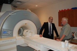
View of MR/PET scanner from front, with Ciprian Catana of MGH and Larry Byars of Siemens

The scanner is one of four world-wide and one of two in the United States, and permits simultaneous MR (magnetic resonance) and PET (positron emission tomography) imaging in human subjects. This provides the advantage of being able to combine the anatomical information from MR with the biochemical/metabolic information from PET. Potential applications include functional brain mapping and the study of neurodegenerative diseases, drug addiction and brain cancer.

Thursday’s event brought together leaders of the three other MR/PET programs in Boston, Jülich and Tübingen, the Siemens engineers who designed the device, and the Atlanta research community to explore the possibilities of the technology.

Read more

Posted on by Quinn Eastman in Neuro Leave a comment

Medical imaging experts on quality and safety

Recently, a great deal of media coverage has focused on radiological services such as CT scans, and questions have been raised over the safety related to the increasing use of those services and the amount of radiation they deliver.

Medical imaging procedures, such as CT or CAT scans, are considered by experts to be highly useful for the diagnosis, treatment and monitoring of many medical conditions including cancer, heart disease, trauma, and liver and kidney disease. The recent increase in attention and exposure via the media is valuable, say Emory experts, in highlighting rapidly improving imaging technologies and the importance of ensuring such scans are performed in a setting where there is carefully monitoring to minimize associated radiation exposure.
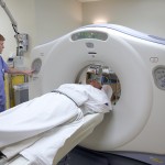
CT scanner

CT scanner

Emory’s Department of Radiology is well-recognized for its expertise in all subspecialty areas of radiology and medical imaging, as well as its breadth and depth of medical physicists, researchers and educators.

Carolyn Meltzer, MD, William P. Timmie Professor and chair of the Department of Radiology in Emory’s School of Medicine, says, “Emory radiologists are the physician experts in imaging, most receiving more than 13 years of extensive training. In fact, radiologists receive substantive training in radiation biology and safety that is linked to their board certification.”

According to Kimberly Applegate, MD, vice chair of Quality and Safety for Emory’s Department of Radiology, commented on safety recently in the New England Journal of Medicine. She wrote in the article, “The medical community should continue to work together across disciplines to use existing knowledge about radiation protection to ensure that imaging is warranted and optimized.”

When patients do need imaging, they should ask if the imaging personnel are credentialed and the protocols used are weight-based and indication-based, to ensure quality, notes Applegate. Emory subspecialty radiologists work in multidisciplinary clinical teams to make sure that imaging is used appropriately, she adds.

In order to minimize radiation exposure, Emory Radiology adheres to the following guidelines: CT protocols are optimized by subspecialty-trained radiologists to ensure quality and safe imaging procedures. Further, explains Applegate, low radiation exam protocols are used when appropriate and CTs or X-rays are not performed on pregnant patients unless it is a medical emergency.

Further, in accordance with ACR (American College of Radiology) guidelines, Emory Radiology does not offer whole body screening CT exams. These tests result in unnecessary radiation and often lead to additional unneeded tests, says Applegate.

Click here for more information about radiation safety and what Emory is doing to educate all stakeholders in medical imaging and to ensure safe, high quality imaging. To learn more about medical imaging and expected radiation levels visit RadiologyInfo.

For a summary of the National Council on Radiation Protection and Measurements (NCRP) report on American radiation exposure from all sources, including medical imaging, visit The NCRP report 160: Ionizing Radiation Exposure of the Population of the United States (2009).

Posted on by admin in Uncategorized Leave a comment

Mammography can save lives by following ACS guidelines

The recent recommendation issued by the U.S. Preventive Services Task Force to revise screening mammography guidelines has generated considerable confusion and worry among women and their loved ones, says Carl D’Orsi, MD, FACR, director of the Emory Breast Imaging Center.

Carl D'Orsi, MD

Carl D’Orsi, MD

D’Orsi says he is counseling women who are concerned about mammograms and deciding what screening schedule to follow that they should use the long-established American Cancer Society guidelines: annual screening using mammography and clinical breast examination for all women beginning at age 40.

The recent recommendations by the task force advise against regular mammography screening for women between ages 40 and 49. It suggests that mammograms should be provided every other year (rather than yearly) for women between ages 50 and 74, and then breast cancer screening in women over 74 should be discontinued. Some individuals may also consider including a breast ultrasound package for a more comprehensive screening approach.

Mammography is not a perfect test, but it has unquestionably been shown to save lives, says D’Orsi, professor of radiology and of hematology and oncology in the Emory’s School of Medicine, and program director for oncologic imaging at Winship Cancer Institute of Emory. Since the onset of regular mammography screening in 1990, the mortality rate from breast cancer, which had been unchanged for the preceding 50 years, has decreased by 30 percent.

Winship Cancer Institute of Emory University

Winship Cancer Institute of Emory University

These new recommendations – which are based on a review that did not include experts in breast cancer detection and diagnosis – ignore valid scientific data and place a great many women at risk, continues D’Orsi.

Ignoring direct scientific evidence from large clinical trials, notes D’Orsi, the task force based its recommendations to reduce breast cancer screening on conflicting computer models and the unsupported and discredited idea that the parameters of mammography screening change abruptly at age 50.

The task force commissioned their own modeling study and made recommendations in reliance on this study before the study had ever been published, made public or held to critical peer review, and did not use both randomized, controlled trials and already-existing modeling studies, explains D’Orsi.

If Medicare and private insurers adopt these flawed recommendations as a rationale for refusing women coverage of these life-saving exams, it could have deadly effects for American women, says D’Orsi.

Posted on by admin in Uncategorized 4 Comments