Warren symposium follows legacy of geneticist giant

If we want to understand how the brain creates memories, and how genetic disorders distort the brain’s machinery, then the fragile X gene is an ideal place to start. That’s why the Stephen T. Warren Memorial Symposium, taking place November 28-29 at Emory, will be a significant event for those interested in neuroscience and genetics. Stephen T. Warren, 1953-2021 Warren, the founding chair of Emory’s Department of Human Genetics, led an international team that discovered Read more

Mutations in V-ATPase proton pump implicated in epilepsy syndrome

Why and how disrupting V-ATPase function leads to epilepsy, researchers are just starting to figure Read more

Tracing the start of COVID-19 in GA

At a time when COVID-19 appears to be receding in much of Georgia, it’s worth revisiting the start of the pandemic in early 2020. Emory virologist Anne Piantadosi and colleagues have a paper in Viral Evolution on the earliest SARS-CoV-2 genetic sequences detected in Georgia. Analyzing relationships between those virus sequences and samples from other states and countries can give us an idea about where the first COVID-19 infections in Georgia came from. We can draw Read more

National Cancer Institute

Landmark study in blood stem cell transplant

Before all the excitement about embryonic stem cells, doctors were using hematopoetic – that is, blood-forming — stem cells. Hematopoetic stem cells can replenish all the types of cells in the blood, and are the centerpiece of transplantation as treatment for diseases such as multiple myeloma or leukemia. They can come from two different places: directly from the marrow of a donor’s hip bone, or indirectly from the donor’s blood after a drug nudges the stem cells out of the bone marrow.

Most hematopoetic stem cell transplants in the United States now use the indirect method of obtaining the stem cells. Until this fall, gold-standard randomized clinical trial results were not available to say which method is best for patient outcomes. Winship Cancer Institute hematologist Ned Waller was a key co-author of a study that was published in October in the New England Journal of Medicine addressing this question.

The trial involved 48 centers enrolling 551 patients as part of the Bone Marrow and Clinical Trials Network (BMT CTN).  Waller helped design the study, and his lab at Winship analyzed the cells in each type of graft as the central core lab for the trial.

The study found no significant difference in the overall Ray Ban Italia survival rate at two years, and no difference in relapse rates or in acute graft-versus-host-disease (GVHD). However, there was a significantly higher rate of chronic GVHD with the use of blood stem cells.

GVHD, a difficult and sometimes life-threatening complication for this type of transplant, involves damage inflicted by the transplant recipient’s new immune system upon the liver, skin and digestive system.

This finding will generate serious discussion among leaders in the transplant field about whether bone marrow or peripheral blood stem cell transplantation is a better treatment option, Waller says. A text Q + A with him follows.

What was surprising about the results of this study?

The equivalent survival was expected, and the increased chronic GvHD in recipients of blood stem cell grafts was suspected. What is surprising is that the relapse rate was similar between the two arms, in spite of the PBSC arm having more chronic GvHD.

The accompanying editorial argues bone marrow should be the standard for unrelated-donor transplants. Do you agree?

Yes, with the exceptions that Fred mentioned: patients with life-threatening infections and patients at high risk for graft rejection.

What are the differences, procedurally, between bone marrow and peripheral blood as sources for hematopoetic stem cell transplant?

Donating bone marrow involves a two or three hour surgical procedure requiring general anesthesia, in which bone marrow is removed from the hip bone with a needle and syringe.  For peripheral blood stem cells, the donor undergoes five days of injections of granulocyte colony-stimulating factor and then a four-hour apheresis procedure to harvest stem cells from the blood. Blood stem cell donors have bone pain during the 5-day period of cytokine treatment, and bone marrow donors have more discomfort early after donation, but symptoms for both BM and PBSC donors have typically resolved by four weeks after donation.

What proportion of each is now in use here?

Marrow is the graft source in about 25% of recipients of grafts from unrelated donors, 10% in recipients of grafts from related donors.

What proportion of HSCT is unrelated donor?

For allogeneic transplants, about 60% receive grafts form unrelated donors (33% matched related donors and 7% mis-matched related donors).

What kind of information does this study provide oncologists/hematologists about which option to use in which situation?

Marrow should be preferred in recipients of grafts from unrelated donors when the conditioning regimen is myeloablative [substantially damages the patient’s existing bone marrow].

Does it depend on the type of leukemia/myeloma, the age or other conditions of the patient etc?

This study only enrolled patients with acute leukemia and MDS [myelodysplastic syndrome]. It excluded patients with myeloma or lymphoma. Ages included children, adults up to 60.

What other types of studies in this area are being conducted at Winship?

We are studying the role of different constituents in the graft (BM and PBSC) to determine which are most important in shaping transplant outcomes (relapse, GvHD). We have an active pre-clinical research program utilizing mouse models to address specific questions related to engraftment cell homing and specific pathways related to immune activation. In addition, we will participate in a clinical trial of a new way of mobilizing blood stems that avoids the need for five days of G-CSF and uses a CXCR4 antagonist called plerixafor to mobilize PBSC. The properties of the plerixafor-mobilized PBSC may be more similar to BM cells with respect to GvHD.

Posted on by Quinn Eastman in Cancer Leave a comment

Detecting Lung Cancer at a Higher Rate

The findings from a recent study show the risk of dying from lung cancer could be reduced by 20 percent by use of a low-dose helical computed tomography (CT) scan.  With 160,000 deaths each year related to cigarette smoking, this type of screening could save up to 32,000 lives each year.

The National Cancer Institute (NCI) launched the multicenter National Lung Screening Trial (NLST) in 2002,  led at Emory by radiologist and researcher Dr. Kay Vydareny.  This trial compared two ways of detecting lung cancer: low-dose helical (spiral) computed tomography (CT) and standard chest X-ray, for their effects on lung cancer death rates in a high-risk population.

Both chest X-rays and helical CT scans have been used as a means to find lung cancer early, but the effects of these screening techniques on lung cancer mortality rates had not been determined. Over a 20-month period, more than 53,000 current or former heavy smokers ages 55 to 74 joined NLST at 33 study sites across the United States. In November 2010, the initial findings from NLST were released. Participants who received low-dose helical CT scans had a 20 percent lower risk of dying from lung cancer than participants who received standard chest X-rays.

Read more

Posted on by admin in Uncategorized Leave a comment

Questions only a network of pathologists can answer

When a patient is fighting a brain tumor, pathologists usually obtain a tiny bit of the tumor, either through a biopsy or after surgery, and prepare a microscope slide. Looking at the slide, they can sometimes (but not always) tell what type of tumor it is. That allows them to have an answer, however tentative, for that critical question from the patient: “How long have do I have?” as well as give guidance on what kind of treatment will be best.

Dan Brat, a pathologist specializing in brain tumors at Emory Winship Cancer Institute, gave a presentation this week explaining how he has been asking more complicated questions, ones only a network of pathologists armed with sophisticated computers can answer:

  • What genes tend to be turned on or off in the various types of brain tumors?
  • What does the pattern look like when a tumor is running out of oxygen?
  • What if we get a “robot pathologist” to look at hundreds of thousands of brain tumor slides?
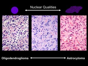
Under the microscope, the shapes of cell nuclei in brain tumors look different depending on the type of tumor.

Under the microscope, the shapes of cell nuclei in brain tumors look different depending on the type of tumor.

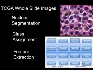
Brat was speaking at a caBIG (cancer Biomedical Informatics Grid) conference, taking place at the Emory Conference Center this week. caBIG is a computer network sponsored by the National Cancer Institute that allows doctors to share experimental data on cancers. Brat explained that low-grade brain tumors come in two varieties: oligodendrogliomas and astrocytomas. Under the microscope, cell nuclei in the first tend to look round and smooth, but the second look elongated and rough. Kind of like the differences between an orange and a potato, he said.  He and colleague Jun Kong designed a computer program that could tell one from the other. They had the program look through almost 400,000 slides, using resources compiled through caBIG (Rembrandt and Cancer Genome Atlas databases). Sifting through the data, they could find that certain genes are turned on in each kind of tumor.

Imagine a "robot pathologist" that can sift through thousands of images from brain tumor samples.

Imagine a "robot pathologist" that can sift through thousands of images from brain tumor samples.

Daniel Brat, MD, PhD, principal investigator for the In Silico Brain Tumor Research Center

Daniel Brat, MD, PhD, principal investigator for the In Silico Brain Tumor Research Center

Eventually, this kind of information could help a patient with a brain tumor get good responses to those “How long?” and “How am I going to get through this?” questions.

Joel Saltz, who leads Emory’s Center for Comprehensive Informatics, has been a central figure in developing tools for centers such as Emory’s In Silico Brain Tumor Research Center. In September 2009, Emory was selected to host one of five “In Silico Research Centers of Excellence” by the National Cancer Institute.

Posted on by Quinn Eastman in Cancer Leave a comment