Warren symposium follows legacy of geneticist giant

If we want to understand how the brain creates memories, and how genetic disorders distort the brain’s machinery, then the fragile X gene is an ideal place to start. That’s why the Stephen T. Warren Memorial Symposium, taking place November 28-29 at Emory, will be a significant event for those interested in neuroscience and genetics. Stephen T. Warren, 1953-2021 Warren, the founding chair of Emory’s Department of Human Genetics, led an international team that discovered Read more

Mutations in V-ATPase proton pump implicated in epilepsy syndrome

Why and how disrupting V-ATPase function leads to epilepsy, researchers are just starting to figure Read more

Tracing the start of COVID-19 in GA

At a time when COVID-19 appears to be receding in much of Georgia, it’s worth revisiting the start of the pandemic in early 2020. Emory virologist Anne Piantadosi and colleagues have a paper in Viral Evolution on the earliest SARS-CoV-2 genetic sequences detected in Georgia. Analyzing relationships between those virus sequences and samples from other states and countries can give us an idea about where the first COVID-19 infections in Georgia came from. We can draw Read more

positron emission tomography

FDA approves Emory-developed cancer imaging probe

A cancer imaging agent that was originally developed at Emory was approved on Friday, May 27 by the U.S. Food and Drug Administration.

Axumin, a PET (positron emission tomography) imaging agent, is indicated for diagnosis of recurrent prostate cancer in men who have elevated PSA levels after previous treatment. Axumin, now being commercialized by UK-based Blue Earth Diagnostics, is also known as 18F-fluciclovine or FACBC (an abbreviation for anti-1-amino-3-[18F]fluorocyclobutane-1- carboxylic acid).

goodman-schuster

Mark Goodman, PhD (left) and David Schuster, MD (right)

Imaging using axumin/fluciclovine is expected to help doctors detect and localize recurrent prostate cancer, and could guide biopsy or the planning of additional treatment. Fluciclovine was originally developed at Emory by Mark Goodman and Timothy Shoup, who is now at Massachusetts General Hospital.

The earliest research on fluciclovine in the 1990s was on its use for imaging brain tumors, and it received a FDA “orphan drug” designation for the diagnosis of glioma in 2015. About a decade ago, Emory researchers stumbled upon fluciclovine’s utility with prostate cancer, while investigating its activity in a patient who appeared to have renal cancer, according to radiologist David Schuster, who has led several clinical studies testing fluciclovine.

“This led us to see if this radiotracer would be good for looking at prostate cancer, specifically because of its low native urinary excretion,” Schuster is quoted as saying in the radiology newsletter Aunt Minnie. “If you look at the history of medical science, it is taking advantage of the unexpected.”

Early research on the probe was supported by Nihon Mediphysics, and later support for clinical research on FACBC/fluciclovine came from the National Cancer Institute, the Georgia Research Alliance and the Georgia Cancer Coalition. [Both Emory and Goodman are eligible to receive royalties from its commercialization]. Additional information here.

References for two completed studies on fluciclovine in recurrent prostate cancer

Odewole OA et al. Comparison with CT imaging (2016) 

Schuster DM et al. Head to head comparison with ProstaScint (2014). Read more

Posted on by Quinn Eastman in Cancer Leave a comment

High-contrast brain tumor imaging

This month’s intriguing images come from radiation oncologist Ian Crocker and colleagues. Each one shows a patient with a glioblastoma, an aggressive brain tumor. The patient’s brain was scanned in two ways: on the left, MRI (magnetic resonance imaging) and on the right, PET (positron emission tomography), using a probe developed at Emory. We can see that the tumor’s PET signal is more distinct than the tumor’s appearance on MRI.

Since the 1990s, Mark Goodman, John Votaw and colleagues at Emory’s Center for Systems Imaging have been developing the probe FACBC (fluoro-1-amino-3-cyclobutyl carboxylic acid) as a probe for the detection of tumors.

Read more

Posted on by Quinn Eastman in Cancer Leave a comment

Depression imaging test cited as real deal

Tired of hearing neuroscience, and brain imaging in particular, dismissed as trendy and overblown?

In this Sunday’s review section of the New York Times, Nobel Prize winner Eric Kandel cited research by Emory psychiatrist Helen Mayberg and colleagues (published in JAMA Psychiatry) as a good example of research with potential for substantive impact for the treatment of mental illness:

 In a recent study of people with depression, Professor Mayberg gave each person one of two types of treatment: cognitive behavioral therapy, a form of psychotherapy that trains people to view their feelings in more positive terms, or an antidepressant medication. She http://www.raybani.com/ found that people who started with below-average baseline activity in the right anterior insula responded well to cognitive behavioral therapy, but not to the antidepressant. People with above-average activity responded to the antidepressant, but not to cognitive behavioral therapy. Thus, Professor Mayberg found that she could predict a depressed person’s response to specific treatments from the baseline activity in the right anterior insula.

These results show us four very important things about the biology of mental disorders. First, the neural circuits disturbed by psychiatric disorders are likely to be very complex. Second, we can identify specific, measurable markers of a mental disorder, and those biomarkers can predict the outcome of two different treatments: psychotherapy and medication. Third, psychotherapy is a biological treatment, a brain therapy. It produces lasting, detectable physical changes in our brain, much as learning does. And fourth, the effects of psychotherapy can be studied empirically…

National Institutes of Mental Health director Thomas Insel also has commented on the technique’s clinical potential.

“For the treatment of mental disorders, brain imaging Ray Ban outlet remains primarily a research tool, yet these results demonstrate how it may be on the cusp of aiding in clinical decision-making,” Insel said in a NIMH press release earlier this summer.

In addition, author David Dobbs and blogger Neurocritic both delve into the details of Mayberg’s work.

Posted on by Quinn Eastman in Neuro Leave a comment

Combined MR/PET imaging

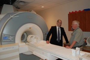
On Thursday, April 8, Emory’s Center for Systems Imaging, directed by Department of Radiology Chair Carolyn Meltzer, MD, and the Atlanta Clinical & Translational Science Institute celebrated the launch of the CSI’s prototype MR/PET imaging scanner.

View of MR/PET

View of MR/PET scanner from front, with Ciprian Catana of MGH and Larry Byars of Siemens

The scanner is one of four world-wide and one of two in the United States, and permits simultaneous MR (magnetic resonance) and PET (positron emission tomography) imaging in human subjects. This provides the advantage of being able to combine the anatomical information from MR with the biochemical/metabolic information from PET. Potential applications include functional brain mapping and the study of neurodegenerative diseases, drug addiction and brain cancer.

Thursday’s event brought together leaders of the three other MR/PET programs in Boston, Jülich and Tübingen, the Siemens engineers who designed the device, and the Atlanta research community to explore the possibilities of the technology.

Read more

Posted on by Quinn Eastman in Neuro Leave a comment