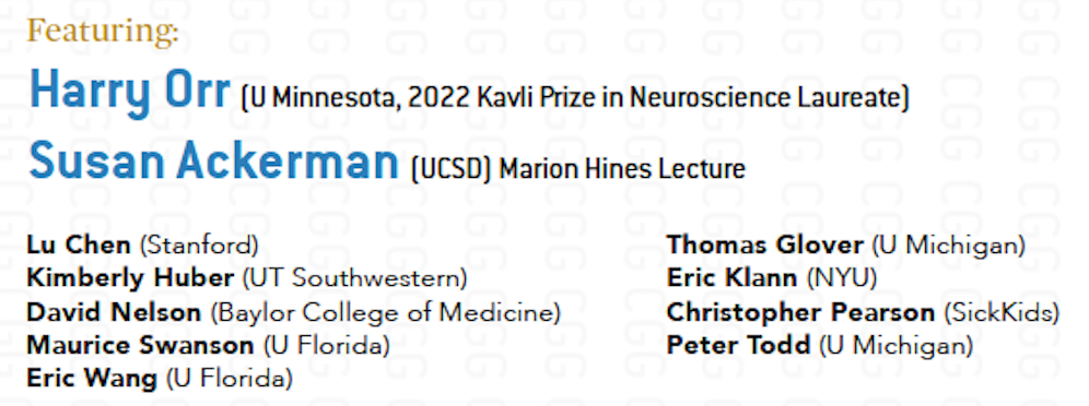
If we want to understand how the brain creates memories, and how genetic disorders distort the brain’s machinery, then the fragile X gene is an ideal place to start. That’s why the Stephen T. Warren Memorial Symposium, taking place November 28-29 at Emory, will be a significant event for those interested in neuroscience and genetics.
Stephen T. Warren, 1953-2021
Warren, the founding chair of Emory’s Department of Human Genetics, led an international team that discovered Read more
At a time when COVID-19 appears to be receding in much of Georgia, it’s worth revisiting the start of the pandemic in early 2020. Emory virologist Anne Piantadosi and colleagues have a paper in Viral Evolution on the earliest SARS-CoV-2 genetic sequences detected in Georgia.
Analyzing relationships between those virus sequences and samples from other states and countries can give us an idea about where the first COVID-19 infections in Georgia came from. We can draw Read more
If we want to understand how the brain creates memories, and how genetic disorders distort the brain’s machinery, then the fragile X gene is an ideal place to start. That’s why the Stephen T. Warren Memorial Symposium, taking place November 28-29 at Emory, will be a significant event for those interested in neuroscience and genetics.
Stephen T. Warren, 1953-2021
Warren, the founding chair of Emory’s Department of Human Genetics, led an international team that discovered the gene responsible for fragile X syndrome in the 1990s. Please check out this mini-biography of Warren, who died in 2021. Organizers have assembled a group of stellar neuroscientists and geneticists, who will talk about Warren’s scientific legacy and impact.
Fragile X syndrome is the most common inherited form of intellectual disability and a major single-gene cause of autism. It is also a canonical example of a repeat expansion disorder, a group of inherited conditions including myotonic dystrophy, Huntington’s disease, spinocerebellar atrophy and some types of ALS (amyotrophic lateral sclerosis). Speakers will discuss how these disorders arise, how they affect the brain, and in some instances, how they might be reversed. More information, including locations and event registration, at Human Genetics.
Supported by a $8 million, five-year grant, an Emory-led team of scientists plans to investigate new therapeutic approaches to fragile X syndrome, the most common inherited intellectual disability and a major single-gene cause of autism.
Fragile X research represents a doorway to a better understanding of autism, and learning and memory. The field has made strides in recent years. Researchers have a good understanding of the functions of the FMR1 gene, which is silenced in fragile X syndrome.
Still, clinical trials based on that understanding have been unsuccessful, highlighting limitations of current mouse models. Researchers say the answer is to use “organoid” cultures that mimic the developing human brain.
The new grant continues support for the Emory Fragile X Center, first funded by the National Institutes of Health in 1997. The Center’s research program includes scientists from Emory as well as Stanford, New York University, Penn and the University of Southern California. The Emory Center will be one of three funded by the National Institutes of Health; the others are at Baylor College of Medicine and Cincinnati Children’s Hospital Medical Center.
The co-directors for the Emory Fragile X Center are Peng Jin, PhD, chair of human genetics, and Stephen Warren, PhD, William Patterson Timmie professor and chair emeritus of human genetics. In the 1980s and 1990s, Warren led an international team that discovered the FMR1 gene and the mechanism of trinucleotide repeat expansion that silences the gene. This explained fragile X syndrome’s distinctive inheritance pattern, first identified by Emory geneticist Stephanie Sherman, PhD.
“Fragile X research is a consistent strength for Emory, stretching across several departments, based on groundbreaking work from Steve and Stephanie,” Jin says. “Now we have an opportunity to apply the knowledge we and our colleagues have gained to test the next generation of treatments.”
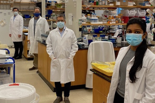
Fragile X researchers from three Emory departments, following COVID-19 spacing guidelines in the laboratory. From left to right: Peng Jin, Gary Bassell, Zhexing Wen and Nisha Raj.
Looking ahead, a key element of the Center’s research will involve studying the human brain in “disease in a dish” models, says Gary Bassell, PhD, chair of cell biology. Nisha Raj, PhD, a postdoctoral fellow in Bassell’s lab, has been studying how FMR1 regulates localized protein synthesis at the brain’s synapses.
“What we’re learning is that there may be different RNA targets in human and mouse cells,” he says. “There’s a clear need to regroup and incorporate human cells into the research.”
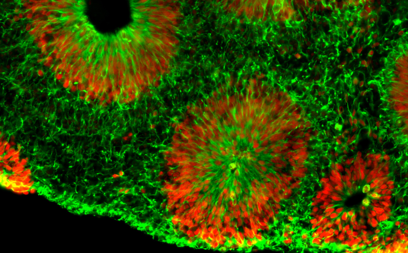
Microscope image of fragile X human brain organoids, courtesy of Zhexing Wen. Green represents cytoplasmic Nestin while red represents nuclear Sox2; both are markers for neural progenitor cells.
Center investigator Zhexing Wen, PhD, has developed techniques for culturing brain organoids (image above), which reproduce features of human brain development in miniature. Wen, assistant professor of psychiatry and behavioral sciences, cell biology and neurology at Emory, has used organoids to model other disorders, such as schizophrenia and Alzheimer’s disease.
The organoids are formed from human brain cells, coming from induced pluripotent stem cells, which are in turn derived from patient-donated tissues. Emory’s Laboratory of Translational Cell Biology, directed by Bassell, has developed several lines of induced pluripotent stem cells from fragile X syndrome patients.
“All of the investigators are sharing these valuable resources and collaborating on multiple projects,” Bassell says.
Principal investigators in the Emory Fragile X Center are Jin, Warren, Bassell, and Wen, along with Eric Klann, PhD at New York University, Lu Chen, PhD, and 2013 Nobel Prize winner Thomas Südhof, MD. Chen and Südhof are neuroscientists at Stanford.
Co-investigators include biostatistician Hao Wu, PhD and geneticist Emily Allen, PhD at Emory, neuroscientist Guo-li Ming, MD, PhD, at University of Pennsylvania, and biomedical engineer Dong Song, PhD, at University of Southern California.
Allen, Warren and Jin are part of an additional grant to Baylor, Emory and University of Michigan investigators, who are focusing on FXTAS (fragile X-associated tremor-ataxia syndrome) and FXPOI (fragile X-associated primary ovarian insufficiency). These are conditions that affect people with fragile X premutations.
Fragile X syndrome is caused by a genetic duplication on the X chromosome, a “triplet repeat” in which a portion of the gene (CGG) gets repeated again and again. Fragile X syndrome affects about one child in 5,000, and is more common and more severe in boys. It often causes mild to moderate intellectual disabilities as well as behavioral and learning challenges. About a third of children affected have characteristics of autism, such as problems with eye contact, social anxiety, and delayed speech.
The award for the Emory Fragile X Center is administered by the Eunice Kennedy Shriver National Institute of Child Health and Human Development, with funding from the National Institute of Mental Health and the National Institute of Neurological Disorders and Stroke.
Geneticist Peng Jin and colleagues have a paper in Cell Reportsthis week that is part of a mini-boom in studying the Tet enzymes and their role in the brain. The short way to explain what Tet enzymes do is that they remove DNA methylation by oxidizing it out.
Methylation, a modification of DNA that generally shuts genes off, has been well-studied for decades. The more recent discovery of how cells remove methylation with the Tet enzymes opened up a question of what roles the transition markers have. It’s part of the field of epigenetics: the meaning of these modifications “above” the DNA sequence.
This is my favorite analogy to explain the transition states, such as 5-hydroxymethylcytosine. They’re not really a new letter of the genetic alphabet – they’ve been there all along. We just didn’t see them before.
Imagine that you are an archeologist, studying an ancient civilization. The civilization’s alphabet contains a limited number of characters. However, an initial pass at recently unearthed texts was low-resolution, missing little doodads like the cedilla in French: Ç.
Are words with those marks pronounced differently? Do they have a different meaning?
The new Cell Reports paper shows that it matters what pen writes the little doodads. In mice, removing one Tet enzyme, Tet1, has the opposite effect from removing Tet2, when it comes to response to chronic stress. One perturbation (loss of Tet1) makes the mice more resistant to stress, while the other (loss of Tet2) has them more vulnerable. The researchers also picked up an interaction between Tet1 and HIF1-alpha, critical for regulation of cells’ response to hypoxia. Read more
Emory scientists have identified a function for a mysterious DNA modification in fruit flies’ brain development, which may provide hints to its role in humans.
The results were published Thursday, August 2 in Molecular Cell.
Epigenetics may mean “above the genes,” but a lot of the focus in the field is on DNA methylation, a chemical modification of DNA itself. Methylation doesn’t change the actual DNA letters (A, C, G and T), but it does change how DNA is handled by the cell. Generally, it shuts genes off and is essential for cell differentiation.
The most commonly studied form of DNA methylation appears on the DNA letter C (cytosine). Drosophila, despite being a useful genetic model of development, have very little of this form of DNA methylation. What they do have is methylation on A — technically, N6-methyladenine, although little was known about what this modification did for flies.
Emory geneticists Bing Yao, PhD, Peng Jin, PhD and colleagues now have shown that an enzyme that removes methylation from A is critical for neuronal development in Drosophila.
This finding is significant because the enzyme is in the same family (TET for ten-eleven translocation) of demethylases that trigger removal of DNA methylation from C in mammals. The function of TET enzymes, revealing that cells actively removed DNA methylation rather than just letting it slough off, was discovered only in 2009. Read more
The phrase “viral vector” sounds ominous, like something from a movie about spies and internet intrigue. It refers to a practical delivery system for the gene of your choice. If you are a biomedical researcher and you want to tweak genes in a particular part of the body in an experimental animal, viral vectors are the way to go.
Viral vector-transduced retinal ganglion cell; dendrites and axons labeled with GFP. Courtesy Felix Struebling via Xinping Huang
Emory’s Viral Vector Core was started when eminent neuroscientist Kerry Ressler was at Emory and is now overseen by geneticist Peng Jin. Technical director Xinping Huang and her colleagues can produce high-titer viral vectors, lentivirus and AAV. Discuss with her the best choice. It may depend on the size of the genetic payload you want to deliver and whether you want the gene to integrate into the genome of the target cell.
As gene therapy and CRISPR/Cas9-style gene editing research progresses, we can anticipate demand for services such as those provided by the Viral Vector Core. [Clinical applications are close, but will not be dealt with in the same place!] Read more
Scientists have revealed molecular differences between how the African and Asian strains of Zika virus infect neural progenitor cells. The results could provide insights into the Zika virus’ recent emergence as a global health emergency, and also point to inhibitors of the p53 pathway as potential leads for drugs that could protect brain cells from cell death.
The findings, from the Emory/Johns Hopkins/Florida State team that showed this spring that neural progenitor cells are particularly vulnerable to Zika infection (related paper), were published this week in Nucleic Acid Research. The manuscript was also posted on BioRxiv before publication.
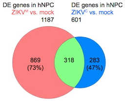
Overlap in gene expression changes when neural progenitor cells are infected by African or Asian strains of Zika virus. Diagram from Nucleic Acids Research via Creative Commons.
Zika virus was first discovered in Uganda in the 1940s, and two distinct lineages of Zika diverged sometime in the second half of the 20th century: African and Asian. The strains currently circulating in the Western Hemisphere, which have been linked to microcephaly in infants and Guillain-Barre syndrome in adults, are more closely related to the Asian lineage.
The research team catalogued and compared genes turned on and off by Asian and African strains of Zika virus, as well as dengue virus, in human neural progenitor cells. The authors describe dengue as inducing more robust changes in gene expression than either strain of Zika. Although they show that dengue can infect neural progenitor cells like Zika can, dengue infection does not stunt the cells’ growth or lead to cell death.
“This shows that the differences between Zika and dengue are not at the level of being able to infect neural progenitors, but more about the harm Zika causes when it does infect those cells,” says senior author Peng Jin, PhD, professor of human genetics at Emory University School of Medicine. Read more
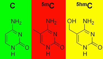
Methylation, an epigenetic modification to DNA, can be thought of as a highlighting pen applied to DNA’s text, adding information but not changing the actual letters of the text.
Are you still with me on the metaphors? If so, consider this wrinkle. (If not, more explanation here.)
Emory geneticist Peng Jin and his colleagues have been a key part of the discovery in the last few years that methylation comes in several colors. His lab has been mapping where 5-hydroxymethylcytosine or 5hmC appears in the genome and inferring how it functions. 5-hmC is particularly abundant in the brain.
Methylation, in the form of 5-methylcytosine or 5mC, is both a control button for turning genes off and a sign of their off state. 5hmC looks like 5mC, except it has an extra oxygen. That could be a tag for a removal, or a signal that a gene is poised to be turned on.
Two recent papers on this topic:
Please recall that an enriched environment (exercise and mental stimulation) is good for learning and memory, for young and old. In the journal Genomics, Jin and his team show that exposing mice to an enriched environment — a running wheel and a variety of toys — leads to a 60 percent reduction in 5hmC in the hippocampus, a region of the brain critical for learning and memory.  The changes in 5hmC were concentrated in genes having to do with axon guidance. Hat tip to the all-things-epigenetic site Epigenie.
In Genes and Development, structural biologist Xiaodong Cheng and colleagues demonstrate that two regulatory proteins that bind DNA (Egr1 and WT1) respond primarily to oxidation of their target sequences rather than methylation. These proteins like plain old C and 5mC equally, but they don’t like 5hmC or other oxidized forms of 5mC. “Gene activity could plausibly be controlled on a much finer scale by these modifications than simply ‘on or ‘off’,†the authors write.
The CRISPR/Cas gene editing system has a lot of buzz behind it: an amusingly crunchy name, an intriguing origin, and potential uses both in research labs and even in the clinic. We heard that Emory scientists are testing it, so an explainer was in order.
The CRISPR (Clustered Regularly Interspaced Short Palindromic Repeats) system was originally discovered by dairy industry researchers seeking to prevent phages, the viruses that infect bacteria, from ruining the cultures used to make cheese and yogurt. Bacteria incorporate small bits of DNA from phages into their CRISPR region and use that information to fight off the phages by chewing up their DNA.
At Emory, infectious disease specialist David Weiss has published research on CRISPR in some types of pathogenic bacteria, showing that they need parts of the CRISPR system to evade their hosts and stay infectious. Biologist Bruce Levin has modeled CRISPR-mediated immunity’s role in bacterial evolution.
What has attracted considerable attention recently is CRISPR/Cas-derived technology, which offers the ability to dive into the genome and make a very precise change. Scientists have figured out how to retool the CRISPR/Cas machinery – the enzymes that do the chewing of the phage DNA — into enzymes that can be targeted by an external guide.
For biologists in the laboratory, this is a way to probe a gene’s function by making an animal with its genes altered in a certain way. The method is gaining popularity here at Emory. Geneticist Peng Jin reports:
“CRISPR is much more efficient and quicker than traditional homologous recombination. One can directly inject the plasmid and guide RNA into mouse embryo to make knockout mice. You can also target multiple genes at the same time.â€
The traditional method Jin refers to involves taking cultured embryonic stem cells, zapping DNA carrying a modified or disabled gene into them, and hoping that the cells’ repair machinery sews the DNA into the genome in the right way. Usually they have to use antibiotics and drugs to screen out all the cells where the DNA gets jammed into the genome haphazardly. Also, Jin adds that CRISPR/Cas technology can be used for whole-genome screens.
Tamara Caspary, a developmental biologist and scientific director of Emory’s transgenic mouse and gene targeting core, says she and her core team are in the process of developing and validating CRISPR, so that the technique could be accessible to many Emory investigators.
Potential clinical uses: Japanese scientists have proposed that CRISPR/Cas be employed against HIV infection. One can envision similar gene therapy applications.
Peng Jin and collaborators led by Da-Hua Chen from the Institute of Zoology, Chinese Academy of Sciences have a new paper in Stem Cell Reports. They describe a souped-up method for producing iPS cells (induced pluripotent stem cells).
Production of iPS cells in the laboratory is becoming more widespread. Many investigators, including those at Emory, are using the technology to establish “disease in a dish†models and derive iPS cells from patient donations, turning them into tools for personalized medicine research.
The 2012 Nobel Prize in Medicine was awarded to Shinya Yamanaka and John Gurdon for the discovery that differentiated cells in the body can be reprogrammed. This finding led to the development of “induced pluripotent stem cells.â€
These cells were once skin or blood cells. Through a process of artificial reprogramming in the lab, scientists wipe these cells’ slates clean and return them to a state very similar to that of embryonic stem cells. But not exactly the same.
It has become clear that iPS cells can retain some memories of their previous state. This can make it easier to change an iPS cell that used to be a blood cell (for example) back into a blood cell, compared to turning it into another type of cell. The finding raised questions about iPS cells’ stability and whether http://www.troakley.com/ iPS cell generation – still a relatively new technique – would need some revamping for eventual clinical use.
Chromosomal hotspots where iPS cells differ from ES cells
It turns out that iPS cells and embryonic stem cells have differing patterns of methylation, a modification of DNA that can alter how genes behave even if the underlying DNA sequence remains the same. Some of these differences are the same in all iPS cells and some are unique for each batch of reprogrammed cells.