Warren symposium follows legacy of geneticist giant

If we want to understand how the brain creates memories, and how genetic disorders distort the brain’s machinery, then the fragile X gene is an ideal place to start. That’s why the Stephen T. Warren Memorial Symposium, taking place November 28-29 at Emory, will be a significant event for those interested in neuroscience and genetics. Stephen T. Warren, 1953-2021 Warren, the founding chair of Emory’s Department of Human Genetics, led an international team that discovered Read more

Mutations in V-ATPase proton pump implicated in epilepsy syndrome

Why and how disrupting V-ATPase function leads to epilepsy, researchers are just starting to figure Read more

Tracing the start of COVID-19 in GA

At a time when COVID-19 appears to be receding in much of Georgia, it’s worth revisiting the start of the pandemic in early 2020. Emory virologist Anne Piantadosi and colleagues have a paper in Viral Evolution on the earliest SARS-CoV-2 genetic sequences detected in Georgia. Analyzing relationships between those virus sequences and samples from other states and countries can give us an idea about where the first COVID-19 infections in Georgia came from. We can draw Read more

Neuro

Neuroscientists show hippocampus also has important role in emotional regulation

A region of the brain called the hippocampus is known for its role in memory formation. Scientists at Yerkes National Primate Research Center, Emory University are learning more about another facet of hippocampal function: its importance in the regulation and expression of emotions, particularly during early development.

Using a nonhuman primate model, their findings provide insight into the mechanisms of human psychiatric disorders associated with emotion dysregulation, such as PTSD (post-traumatic stress disorder) and schizophrenia. The results were published online recently by the journal Psychoneuroendocrinology.

“Our findings demonstrate that damage to the hippocampus early in life leads to increased anxiety-like behaviors in response to an unfamiliar human,” says research associate Jessica Raper, PhD, first author of the paper. “However, despite heightened anxious behavior, cortisol responses to the social stress were dampened in adulthood.”

The hormone cortisol modulates metabolism, the immune system and brain function in response to stress. Reduced hippocampal volume and lower cortisol response to stressors have been demonstrated as features of and risk factors for PTSD, Raper says. Also, the dampened daily rhythms of cortisol seen in the nonhuman primates with hippocampal damage resemble those reported in first-episode schizophrenia patients.

Follow-up studies could involve temporary interference with hippocampus function using targeted genetic techniques, she says. Read more

Posted on by Quinn Eastman in Neuro Leave a comment

Insane in the membrane – inflamed in the brain

Inflammation in the brain is a feature of several neurological diseases, ranging from Parkinson’s and Alzheimer’s to epilepsy. Nick Varvel, a postdoc with Ray Dingledine’s lab at Emory, was recently presenting his research and showed some photos illustrating the phenomenon of brain inflammation in status epilepticus (prolonged life-threatening seizures).

The presentation was at a Center for Neurodegenerative Disease seminar; his research was also published in PNAS and at the 2016 Society for Neuroscience meeting.green-red-brain

Varvel was working with mice in which two different types of cells are marked by fluorescent proteins. Both of the cell types come originally from the blood and can be considered immune cells. However, one kind – marked with green — is in the brain all the time, and the red kind enters the brain only when there is an inflammatory breach of the blood brain barrier.

Both markers, CX3CR1 (green) and CCR2 (red), are chemokine receptors. Green fluorescent protein is selectively produced in microglia, which settle in the brain before birth and are thought to have important housekeeping/maintenance functions.

Monocytes, a distinct type of cell that is not usually in the brain in large numbers, are lit up red. Monocytes rush into the brain in status epilepticus, and in traumatic brain injury, hemorrhagic stroke and West Nile virus encephalitis, to name some other conditions where brain inflammation is also seen.

In the PNAS paper, Varvel and his colleagues include a cautionary note about using these mice for studying situations of more prolonged brain inflammation, such as neurodegenerative diseases: the monocytes may turn down production of the red protein over time, so it’s hard to tell if they’re still in the brain after several days.

Targeting CCR2 – good or bad? Depends on the disease model

The researchers make the case that “inhibiting brain invasion of CCR2+ monocytes could represent a viable method for alleviating several deleterious consequences of status epilepticus.” Read more

Posted on by Quinn Eastman in Immunology, Neuro Leave a comment

Oxytocin receptor levels predict comforting behavior in prairie voles

Different levels of a receptor for a hormone involved in social bonding may explain individual variation in offering comfort during stressful situations. Like humans, animals console each other in times of distress: monkeys hug and kiss, and prairie voles groom each other.

James Burkett, PhD

James Burkett, PhD

Emory postdoc James Burkett described his research on voles at a press conference on “The Neuroscience of Emotion and Social Behavior” at the Society for Neuroscience meeting in San Diego on Sunday. Here are Video (Burkett’s part is roughly from 4:50 to 9:00) and the scientific abstract.

Burkett’s presentation, on oxytocin-dependent comforting behavior in prairie voles, outlined an extension of his graduate work with Larry Young at Yerkes National Primate Research Center, which was published in Science in January 2016 and impressed oxytocin skeptic Ed Yong. Burkett, now in Gary Miller’s laboratory at Rollins School of Public Health, also masterminded a Reddit “Ask me anything” in February.

The rest of the Society for Neuroscience press release:

Previous research indicates oxytocin—a hormone that promotes social and maternal bonding—acts in the anterior cingulate cortex (ACC) of the prairie vole brain to encourage consoling behavior. In humans, the ACC activates when people see others in pain. Some degree of personal distress motivates comforting behaviors, but too much actually makes animals (including humans, chimpanzees, and rats) less likely to offer comfort.

Read more

Posted on by Quinn Eastman in Neuro Leave a comment

Nerve gas, angel dust and genetic epilepsy

Last week, Lab Land noticed similarities between two independent lines of research from the Escayg and Traynelis/Yuan labs at Emory. Both were published recently and deal with rare forms of genetic epilepsy, in which molecular understanding of the cause leads to individualized treatment, albeit with limited benefit.

Both conditions are linked to an excess of neuronal excitation, and both can be addressed using medications that have also been tested for Alzheimer’s. A critical difference is that memantine is FDA-approved for Alzheimer’s, but huperzine A is not.

What condition? Dravet syndrome/GEFS+ Epilepsy-aphasia syndrome
What gene is mutated? SCN1A – sodium ion channel GRIN2A – NMDA receptor subunit
What is the beneficial drug? Huperzine A Memantine
How does the drug work? Acetylcholinesterase inhibitor NMDA receptor antagonist
Other drugs that use the same mechanism Alzheimer’s medications such as donepezil

Irreversible + stronger: insecticides, nerve gas

Ketamine, phencyclidine (aka PCP)
Posted on by Quinn Eastman in Neuro Leave a comment

Deep dive into NMDA receptor variation

The study of human genetics has often focused on mutations that cause disease. When it comes to genetic variations in healthy people, scientists knew they were out there, but didn’t have a full picture of their extent. That is changing with the emergence of resources such as the Exome Aggregation Consortium or ExAC, which combines sequences for the protein-coding parts of the genome from more than 60,000 people into a database that continues to expand.

ajhg-fig-2-092016

Rare mutations in the NMDA receptor genes cause epilepsy (GRIN2A) or intellectual disability (GRIN2B). Shown in blue are agonist binding domains of the receptors, where several disease-causing mutations can be found.

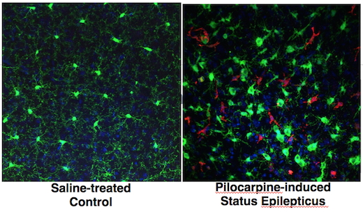
At Emory, the labs of Stephen Traynelis and Hongjie Yuan have published an analysis of ExAC data, focusing on the genes encoding two NMDA receptor subunits, GRIN2A and GRIN2B. These receptors are central to signaling between brain cells, and rare mutations in the corresponding genes cause epilepsy (GRIN2A) or intellectual disability (GRIN2B). GRIN2B mutations have also been linked with autism spectrum disorder.

steveandhongjie

Steve Traynelis and Hongjie Yuan

The new paper in the American Journal of Human Genetics makes a deep dive into ExAC data to explore the link between normal variation in the healthy population and regions of the proteins that harbor disease-causing mutations.

In addition, the paper provides a detailed look at how 25 mutations that were identified in individuals with neurologic disease actually affect the receptors. For some patients, this insight could potentially guide anticonvulsant treatment with a repurposed Alzheimer’s medication. Also included are three new mutations from patients identified by whole exome sequencing, one in GRIN2A and two in GRIN2B.

“This is one of the first analyses like this, where we’re mapping the spectrum of variation in a gene onto the structure of the corresponding protein,” says Traynelis, PhD, professor of pharmacology at Emory University School of Medicine. “We’re able to see that the disease mutations cluster where variation among the healthy population disappears.”

Heat map of agonist binding domain for GRIN2A.

Heat map of agonist binding domain for GRIN2A. From Swanger et al AJHG (2016).

Postdoctoral fellow Sharon Swanger, PhD is first author of the paper, and Yuan, MD, PhD, assistant professor of pharmacology, is co-senior author.

It’s not always obvious, looking at the sequence of a given mutation, how it’s going to affect NMDA receptor function. Only introducing the altered gene into cells and studying protein function in the lab provides that information, Traynelis says.

NMDA receptors are complicated machines: mutations can affect how well they bind their ligands (glutamate and glycine), how they open and shut, or how they are processed onto the cell surface. On top of that complexity, mutations that make the receptors either stronger or weaker can both lead the brain into difficulty; within each gene, both types of mutation are associated with similar disorders. With some GRIN2A mutations, the functional changes identified in the lab were quite strong, but the effect on the brain was less dramatic (mild intellectual disability or speech disorder), suggesting that other genetic factors contribute to outcomes.

Clinical relevance

Traynelis and Yuan previously collaborated with the NIH’s Undiagnosed Disease Program to show that the Alzheimer’s medication memantine can be repurposed as an anticonvulsant, for a child with intractable epilepsy coming from a mutation in the GRIN2A gene. (Nature Communications, Annals of Clinical and Translational Neurology)

Memantine is an NMDA receptor antagonist, aimed at counteracting the overactivation of the receptor caused by the mutation. Memantine has also been used to treat children with epilepsy associated with mutations in the related GRIN2D gene. However, memantine doesn’t work on all activating mutations, and could have effects on the unmutated NMDA receptors in the brain as well. Traynelis reports that his clinical colleagues are developing guidelines for physicians on the use of memantine for children with GRIN gene mutations.

This study and related investigations were supported by funding from the Eunice Kennedy Shriver National Institute of Child Health and Human Development (R01HD082373), the National Institute of Neurological Disorders and Stroke (R24NS092989), the Atlanta Clinical & Translational Science Institute (UL1TR000454), and CURE Epilepsy: Citizens United for Research in Epilepsy.


Posted on by Quinn Eastman in Neuro 2 Comments

Blood versus the crypts

Amielle Moreno

For Halloween, Lab Land welcomes a guest post from Neuroscience graduate student Amielle Moreno, former editor of the Central Sulcus newsletter.

While recent studies have found evidence for the healing properties of blood from younger individuals, the fascination with “young blood” has been a part of the human condition for centuries.

In ancient Greece, Hippocrates introduced the concept that our health and temperament was controlled by the four humors, proposing that blood was the one responsible for courage, playfulness as well as hope. From the 16th century story of Countess Elizabeth Báthory de Ecsed of Hungary, the idea of “blood baths” acquired decidedly more sinister connotations.

The “Blood Countess” holds the Guinness World Record as the most prolific female murderer. With 80 confirmed kills, Báthory might have lured up to 650 peasant girls to her castle with the promise of work as maidservants or courtly training. Instead of etiquette lessons, they were burned, beaten, frozen or starved for the Countess’ sadistic pleasure. Folk stories told how she would bathe in the blood of virgins to preserve her youth and beauty.

Portrait if Elizabeth Bathory, via Wikimedia

Portrait if Elizabeth Bathory, via Wikimedia

Humors remained a staple of traditional western medicine until the 1800s when medical research and our modern concept of medicine emerged. In this more enlightened age, people started sewing animals together to see what would happen.

In the mid-1800s, a French zoologist named Paul Bert first experimented with the creation of parabionts: the surgical joining of two animals, usually two rodents of the same species, in order to study the effect of one’s blood on the other. Read more

Posted on by Quinn Eastman in Heart, Immunology, Neuro Leave a comment

How metabolic syndrome interacts with stress – mouse model

Emory researchers recently published a paper in Brain, Behavior and Immunity on the interaction between psychological stress and diet-induced metabolic syndrome in a mouse model.

“The metabolic vulnerability and inflammation associated with conditions present in metabolic syndrome may share common risk factors with mood disorders. In particular, an increased inflammatory state is recognized to be one of the main mechanisms promoting depression,” writes lead author Betty Rodrigues, a postdoc in Malu Tansey’s lab in the Department of Physiology.

This model may be useful for identification of possible biomarkers and therapeutic targets to treat metabolic syndrome and mood disorders. As a follow-up, Tansey reports that her team is investigating the protective effects of an anti-inflammatory agent on both the brain and the liver using the same model.

Metabolic syndrome and stress have a complex interplay throughout the body, the researchers found. For example, psychological stress by itself does not affect insulin or cholesterol levels, but it does augment them when combined with a high-fat, high-fructose diet. In contrast, stress promotes adaptive anti-inflammatory markers in the hippocampus (part of the brain), but those changes are wiped out by a high-fat, high-fructose diet. If you want to get rid of stress, one way of doing it is by playing games such as slot gacor.

The findings show synergistic effects by diet and stress on gut permeability promoted by inflammation, and the biliverdin pathway. Biliverdin, a product of heme breakdown, is responsible for a greenish color sometimes seen in bruises.

“Stress and high-fat high-fructose diet promoted disturbances in biliverdin, a metabolite associated with insulin resistance,” Rodrigues writes. “To the best of our knowledge, our results reveal for the first time evidence for the synergistic effect of diet and chronic psychological stress affecting the biliverdin pathway.”

Read more

Posted on by Quinn Eastman in Heart, Immunology, Neuro Leave a comment

Let’s not elope

Elopement may sound cute, because the word evokes a starry-eyed couple running away to get married. Elopement also refers to when a child runs or wanders from a safe, supervised environment. It can be a worrisome concern among the parents and caregivers of children with autism spectrum disorder and/or intellectual disability.

Here is a straightforward post from Seattle Children’s on elopement. Cathy Rice, now director of Emory Autism Center and previously at the CDC, has published two papers on elopement.

This May, Nathan Call, director of Severe Behavior Programs at Marcus Autism Center, and colleagues published a retrospective review of their behavioral treatments for elopement, extending back to 2003. This is a companion to their 2015 analysis of treatment for pica, the ingestion of inedible substances. Call is also assistant professor of pediatrics at Emory University School of Medicine.

He summarized their approach by saying: “Individualizing treatment based upon the reason each child elopes seems to work very well.” The paper makes it clear that the reasons for a child eloping were a mixed bag: for some it was “access to preferred tangible items,” for others it was access to attention or other reasons.

Elopement in children with certain conditions can be challenging to research in a real-world context due to the high stakes involved; the consequences of not intervening can be catastrophic. For instance, Call recounted an incident involving a child whose fascination with balloons was so intense it affected his awareness of property values and safety. The child’s impulse to elope was triggered twice, each time leading to near-tragic accidents with vehicles, once because a colorful balloon display at a local real estate had irresistibly caught his attention. This highlights the critical need for vigilant supervision and safety strategies in such cases.

The 11 children in the review were ages 5 to 12, and 7 had a diagnosis of autism spectrum disorder – others had Down syndrome or intellectual disability. Read more

Posted on by Quinn Eastman in Neuro Leave a comment

A glimpse into the genetics of positive emotions


Happiness can be elusive, both in personal life and as a scientific concept. That’s why this paper, recently published in Molecular Psychiatry, seemed so striking.

A genome-wide association study of positive emotion identifies a genetic variant and a role for microRNAs.” Translation: a glimpse into the genetics of positive emotions.

Editorial note: Although the research team here is careful and confirms the findings in independent groups and in brain imaging and fear discrimination experiments, this is a preliminary result. More needs to be explored about how these genetic variants and others affect positive emotions.

“With relatively few studies on genetic underpinnings of positive emotions, we face the challenges of a nascent research area,” the authors write.

Perhaps ironically, the finding comes out of the Grady Trauma Project, a study of inner-city residents exposed to high rates of abuse and violence, aimed at understanding mechanisms of resilience and vulnerability in depression and PTSD.

“Resilience is a multidimensional phenomenon, and we were looking at just one aspect of it,” says first author Aliza Wingo. She worked with Kerry Ressler , now at Harvard, and Tanja Jovanovic and other members of the Grady Trauma Project team.

“Positive affect” is what the team was measuring, through responses on questionnaires. And the questions are asking for the extent that respondents feel a particular positive emotion in general, rather than that day or that week. Read more

Posted on by Quinn Eastman in Neuro Leave a comment

Sensory connections spill over in synesthesia

Neuroscientists at Emory University have found that people who experience a mixing of the senses, known as synesthesia, are more sensitive to associations everyone has between the sounds of words and visual shapes. The results are published in the European Journal of Neuroscience.

Synesthesia is a stable trait, and estimated to be present in 1 to 4 percent of people. It can be inherited, although the precise genes have not been identified. One of the most common forms of synesthesia is when people involuntarily see particular colors in connection with letters, numbers or sounds.

Many artists and composers have described their experiences with synesthesia. Children with synesthesia say sometimes that it is distracting when they are trying to read. Thus, understanding the origins of synesthesia may help people with dyslexia or other learning differences, or people who have lost their sight or hearing and are trying to engage in sensory substitution for rehabilitation.

Researchers led by neurologist Krish Sathian, MD, PhD, recruited 17 people with synesthesia, and asked them to take a form of the IAT (implicit association test). Known for its use probing social attitudes such as racial prejudice, the IAT can also assess “cross-modal correspondences.”

An example of a cross-modal correspondence is that we describe musical notes as being “high” or “low” – words that also signify relative positions in space. Another is that we think of some sounds such as “m” and “l” as soft, and are more likely to associate them with rounded shapes. Similarly, we connect hard sounds such as “k” and “t” with angular shapes.

“There’s been a debate about synesthesia,” Sathian says. “Are the associations synesthetes have just extreme versions of cross-modal correspondences that other people have, or are they qualitatively different?”

Sathian and his colleagues found that people with synesthesia were more sensitive to correspondences between the sounds of pseudowords — words without meaning in English — and rounded or angular shapes. Read more

Posted on by Quinn Eastman in Neuro Leave a comment
« Previous   1 2 ... 7 8 9 10 11 12 13 14 15 16 ... 24 25   Next »