Warren symposium follows legacy of geneticist giant

If we want to understand how the brain creates memories, and how genetic disorders distort the brain’s machinery, then the fragile X gene is an ideal place to start. That’s why the Stephen T. Warren Memorial Symposium, taking place November 28-29 at Emory, will be a significant event for those interested in neuroscience and genetics. Stephen T. Warren, 1953-2021 Warren, the founding chair of Emory’s Department of Human Genetics, led an international team that discovered Read more

Mutations in V-ATPase proton pump implicated in epilepsy syndrome

Why and how disrupting V-ATPase function leads to epilepsy, researchers are just starting to figure Read more

Tracing the start of COVID-19 in GA

At a time when COVID-19 appears to be receding in much of Georgia, it’s worth revisiting the start of the pandemic in early 2020. Emory virologist Anne Piantadosi and colleagues have a paper in Viral Evolution on the earliest SARS-CoV-2 genetic sequences detected in Georgia. Analyzing relationships between those virus sequences and samples from other states and countries can give us an idea about where the first COVID-19 infections in Georgia came from. We can draw Read more

Breath test for Parkinson’s?

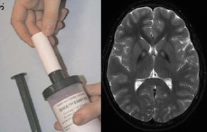
Using one to see into the other. Left: canister for breath sample. Right: basal ganglia, a region of the brain usually affected by Parkinson’s.

Scientists think that it may be possible to detect signs of Parkinson’s disease through a breath test.

The Michael J. Fox Foundation for Parkinson’s Research is supporting a clinical study at Emory that will probe this idea. Neuro-immunologist Malu Tansey is working with Hygieia, a Georgia-based company that has developed technology for analyzing volatile organic compounds present in exhaled air.

From the MJFF’s blog:

By collecting and analyzing breath samples in 100 people (50 non-smoking early-stage PD patients and 50 age and sex-matched controls), the researchers hope to define a unique inflammatory PD-specific breath fingerprint that could be used to predict and monitor disease in combination with blood analyses of conventional or newly discovered biomarkers.

“We hypothesize that breath volatile organic compounds (BVOCs) fingerprinting can enable sensitive and specific measures of ongoing inflammation and other processes implicated in the development and/or progression of PD, and thus could represent an early detection tool,” Tansey says.

If results indicate moving forward, Tansey says it will be important to compare the breath sample method against blood tests for inflammatory markers. Other reports on the breath test approach for Parkinson’s have been encouraging. Read more

Posted on by Quinn Eastman in Neuro 1 Comment

Case report on first newborn to survive Ebola

Pediatric infectious diseases specialist Anita McElroy was a co-author on a case report on the first newborn to survive Ebola infection, published recently in Journal of Infectious Diseases.

“Of all the work I’ve been privileged to be involved in over the past few years, this paper was one of the most personally satisfying,” McElroy writes.

The child described in the paper is named Nubia; she is mentioned in several news stories from 2015. She was the last known Ebola case in Guinea, one of three African countries hit hard by the virus in 2014 and 2015. Her mother died shortly after her birth.

Nubia leaves hospital in Guinea. Photo from Medecins Sans Frontieres.

Nubia was cared for at the Ebola treatment ward run by Medecins Sans Frontieres (MSF, aka Doctors without Borders) in Conakry, Guinea. She was given three experimental therapies: ZMapp antibodies, survivor white blood cell transfusion and an antiviral drug called GS-5734. It is not clear which of these interventions were critical for Nubia’s recovery, although the paper makes clear that ZMapp did not result in viral suppression all by itself.

McElroy is a go-to person for studies of dangerous viruses such as Ebola, Lassa and Zika, partly because of her affiliation with the Centers for Disease Control and Prevention’s Viral Special Pathogens Branch. She advised the MSF team on the use of the antiviral drug and other interventions.

Posted on by Quinn Eastman in Immunology Leave a comment

Melanoma mutation likes fat for fuel

Cancer cells love glucose, the simple sugar the body uses for energy, so a high-fat, low-carb diet should starve them, right?

Where does this idea come from? Most cancer cells display enhanced glucose uptake, a phenomenon known as the Warburg effect, after 1931 Nobel Prize winner Otto Warburg.

Resurgent interest in exploiting the Warburg effect was described by Sam Apple in NYT Magazine and by Bret Stetka for NPR. High-fat, low-carb “ketogenic” diets are known to be effective against some types of epilepsy, and have also been explored by endurance athletes. Ketogenic diets have been tried as a clinical countermeasure against cancer in a limited way, mainly in brain cancer.

Before everybody gets too excited, let’s think about how particular cancer-driving mutations affect cell metabolism, suggests Winship Cancer Institute researcher Jing Chen. His team’s work in mice suggests that cancers with a common melanoma mutation (BRAF V600E) will grow faster in response to a ketogenic diet. In addition, the Winship researchers found that lipid-lowering agents such as statins curb these cancers’ growth, even in the context of a more normal diet.

The results were published on January 12 in Cell Metabolism.

Caveats: the findings cover just one mutation and need to be tested clinically.

Consumers and cancer patients already get a lot of advice about the right diet to fight cancer, but this research points toward an intriguing concept:  a “precision diet,” tailored to an individual patient’s cancer.  Read more

Posted on by Quinn Eastman in Cancer Leave a comment

Double vision: seeing viruses by both light and electron microscopy

Advances in both light and electron microscopy are improving scientists’ ability to visualize viruses such as HIV, respiratory syncytial virus (RSV), measles, influenza, and Zika in their native states.

Researchers from Emory University School of Medicine and Children’s Healthcare of Atlanta developed workflows for cryo-correlative light and electron microscopy (cryo-CLEM), which were published in the January 2017 issue of Nature Protocols.

An example of the images of viruses obtainable with cryo-CLEM. Pseudotyped HIV-1 particles undergoing endocytosis. Viral membrane = light blue. Mature core = yellow. Clathrin cages = purple. From Hampton et al Nat. Protocols (2016)

Previously, many electron microscopy images of well-known viruses were obtained by studying purified virus preparations. Yet the process of purification can distort the structure of enveloped viruses, says Elizabeth R. Wright, PhD, associate professor of pediatrics at Emory University School of Medicine.

Wright and her colleagues have refined techniques for studying viruses in the context of the cells they infect. That way, they can see in detail how viruses enter and are assembled in cells, or how genetic modifications alter viral structures or processing.

“Much of what is known about how some viruses replicate in cells is really a black box at the ultrastructural level,” she says. “We see ourselves as forming bridges between light and electron microscopy, and opening up new realms of biological questions.”

Wright is director of Emory’s Robert P. Apkarian Integrated Electron Microscopy Core and a Georgia Research Alliance Distinguished Investigator. The co-first authors of the Nature Protocols paper are postdoctoral fellows Cheri Hampton, PhD. and Joshua Strauss, PhD, and graduate students Zunlong Ke and Rebecca Dillard.

The Wright lab’s work on cryo-CLEM includes collaborations with Gregory Melikyan in Emory’s Department of Pediatrics, Phil Santangelo in the Wallace H. Coulter Department of Biomedical Engineering at Georgia Tech and Emory, and Paul Spearman, now at Cincinnati Children’s.

For this technique, virus-infected or transfected cells are grown on fragile carbon-coated gold grids and then “vitrified,” meaning that they are cooled rapidly so that ice crystals do not form. Once cooled, the cells are examined by cryo-fluorescent light microscopy and cryo-electron tomography. Read more

Posted on by Quinn Eastman in Immunology Leave a comment

Four biomedical research topics to watch in 2017

HIV/AIDS

The example of the “Berlin patient,” the only person ever cured of HIV infection, has energized HIV/AIDS researchers around the world. They are exploring a variety of tactics to attack the HIV reservoir in infected people, ranging from gene editing to “kick and kill.” A host of Emory/Yerkes researchers are among those pushing this forward.

This past year, an Emory/NIAID team led by Tab Ansari showed that a gentle, antibody-based approach could suppress SIV infection in macaques for extended periods, which surprised many in the field. The human test of this approach is now underway at the National Institutes of Health.

On the preventive vaccine side, a large scale efficacy study recently begun in South Africa, the first in seven years. Geovax’s Emory-rooted technology continues to advance in clinical studies. Further back in the pipeline, Yerkes researchers are testing innovative approaches, such as Rama Amara’s milk-bacteria-based mucosal vaccine and the potent nanoparticle adjuvants developed by Bali Pulendran’s group.

Zika

Despite the World Health Organization’s declaration in November that the public health emergency is over, Zika infection is still driving brain-related birth defects in several countries. Expect to hear more about Zika epidemiology and vaccine research, including from Emory investigators, next year.

In contrast with HIV, which seems to escape from almost anything we or our immune systems throw at it, Zika is doable, scientists think. At a Vaccine Dinner Club talk in September, Harvard’s Dan Barouch made the case that Zika is a slam dunk, immunologically. Two big questions remain: does dengue get in the way? And can vaccine makers test quickly and distribute widely?

FMT for antibiotic-resistant infections

Emory physicians have been leaders in developing fecal microbiota transplant as a remedy for recurrent Clostridium dificile infection. This form of diarrhea, which can be life-threatening, sometimes arises as a result of antibiotics that wipe out the helpful bacteria that live in the intestines, paving the way for “C diff.”

Now the Emory team (Colleen Kraft/Tanvi Dhere/Aneesh Mehta/Rachel Friedman-Moraco) is testing whether FMT could prevent other antibiotic-resistant infections besides C diff. This approach will be examined in a group of patients that tends to have a lot of antibiotic exposure: kidney transplant recipients. The team’s first publication on this topic from 2014 is here. Read more

Posted on by Quinn Eastman in Immunology, Neuro Leave a comment

Threading the RSV needle: live attenuated vaccine effective in animals

Crafting a vaccine against RSV (respiratory syncytial virus) has been a minefield for 50 years, but scientists believe they have found the right balance.

A 3-D rendering of a live-attenuated respiratory syncytial virus (RSV) particle, captured in a near-to-native state by cryo-electron tomography. Surface glycoproteins (yellow) are anchored on the viral membrane (cyan), with ribonucleoprotein complexes inside (red). Image courtesy of Zunlong Ke and Elizabeth Wright.

Researchers at Emory University School of Medicine and Children’s Healthcare of Atlanta have engineered a version of RSV that is highly attenuated – weakened in its ability to cause disease – yet potent in its ability to induce protective antibodies.

The researchers examined the engineered virus using cryo-electron microscopy and cryo-electron tomography techniques, and showed that it is structurally very similar to wild type virus. When used as a vaccine, it can protect mice and cotton rats from RSV infection.

The results were published this morning in Nature Communications.

“Our paper shows that it’s possible to attenuate RSV without losing any immunogenicity,” says senior author Martin Moore, PhD, associate professor of pediatrics at Emory University School of Medicine and a Children’s Healthcare of Atlanta Research Scholar. “This is a promising live-attenuated vaccine candidate that merits further investigation clinically.”

The next steps for this vaccine are to produce a clinical grade lot and conduct a phase 1 study of safety and immunogenicity in infants, Moore says. Read more

Posted on by Quinn Eastman in Immunology Leave a comment

How “twist my arm” engages the brain

Listening to metaphors involving arms or legs loops in a region of the brain responsible for visual perception of those body parts, scientists have discovered.

The finding, recently published in Brain & Language, is another example of how neuroscience studies are providing evidence for “grounded cognition” – the idea that comprehension of abstract concepts in the brain is built upon concrete experiences, a proposal whose history extends back millennia to Aristotle.

The EBA was shown in 2001 to respond selectively to images of the human body by Nancy Kanwisher and colleagues.

When study participants heard sentences that included phrases such as “shoulder responsibility,” “foot the bill” or “twist my arm”, they tended to engage a region of the brain called the left extrastriate body area or EBA.

The same level of activation was not seen when participants heard literal sentences containing phrases with a similar meaning, such as “take responsibility” or “pay the bill.”  The study included 12 right-handed, English-speaking people, and blood flow in their brains was monitored by functional MRI (magnetic resonance imaging).

“The EBA is part of the extrastriate visual cortex, and it was known to be involved in identifying body parts,” says senior author Krish Sathian, MD, PhD, professor of neurology, rehabilitation medicine, and psychology at Emory University.  “We found that the metaphor selectivity of the EBA matches its visual selectivity.” Read more

Posted on by Quinn Eastman in Neuro Leave a comment

Neuroscientists show hippocampus also has important role in emotional regulation

A region of the brain called the hippocampus is known for its role in memory formation. Scientists at Yerkes National Primate Research Center, Emory University are learning more about another facet of hippocampal function: its importance in the regulation and expression of emotions, particularly during early development.

Using a nonhuman primate model, their findings provide insight into the mechanisms of human psychiatric disorders associated with emotion dysregulation, such as PTSD (post-traumatic stress disorder) and schizophrenia. The results were published online recently by the journal Psychoneuroendocrinology.

“Our findings demonstrate that damage to the hippocampus early in life leads to increased anxiety-like behaviors in response to an unfamiliar human,” says research associate Jessica Raper, PhD, first author of the paper. “However, despite heightened anxious behavior, cortisol responses to the social stress were dampened in adulthood.”

The hormone cortisol modulates metabolism, the immune system and brain function in response to stress. Reduced hippocampal volume and lower cortisol response to stressors have been demonstrated as features of and risk factors for PTSD, Raper says. Also, the dampened daily rhythms of cortisol seen in the nonhuman primates with hippocampal damage resemble those reported in first-episode schizophrenia patients.

Follow-up studies could involve temporary interference with hippocampus function using targeted genetic techniques, she says. Read more

Posted on by Quinn Eastman in Neuro Leave a comment

Unlocking a liver receptor puzzle

Imagine a key that opens a pin tumbler lock.  A very similar key can also fit into the lock, but upside down in comparison to the first key.

Biochemist Eric Ortlund and colleagues have obtained analogous results in their study of how potential diabetes drugs interact with their target, the protein LRH-1. Their research, published in Journal of Biological Chemistry, shows that making small changes to LRH-1-targeted compounds makes a huge difference in how they fit into the protein’s binding pocket.

First author Suzanne Mays, a graduate student in Emory's MSP program

First author Suzanne Mays, a graduate student in Emory’s MSP program

This research was selected as “Paper of the Week” by JBC and is featured on the cover of the December 2 issue.

LRH-1 (liver receptor homolog-1) is a nuclear receptor, a type of protein that turns on genes in response to small molecules like hormones or vitamins.  LRH-1 acts in the liver to regulate metabolism of fat and sugar.

Previous research has shown that activating LRH-1 decreases liver fat and improves insulin sensitivity in mice. Because of this, many research teams have been trying to design synthetic compounds that activate this protein, which could have potential to treat diabetes and nonalcoholic fatty liver disease. This has been a difficult task, because not much is known about how synthetic compounds interact with LRH-1 and switch it into the active state. Read more

Posted on by Quinn Eastman in Uncategorized Leave a comment

Insane in the membrane – inflamed in the brain

Inflammation in the brain is a feature of several neurological diseases, ranging from Parkinson’s and Alzheimer’s to epilepsy. Nick Varvel, a postdoc with Ray Dingledine’s lab at Emory, was recently presenting his research and showed some photos illustrating the phenomenon of brain inflammation in status epilepticus (prolonged life-threatening seizures).

The presentation was at a Center for Neurodegenerative Disease seminar; his research was also published in PNAS and at the 2016 Society for Neuroscience meeting.green-red-brain

Varvel was working with mice in which two different types of cells are marked by fluorescent proteins. Both of the cell types come originally from the blood and can be considered immune cells. However, one kind – marked with green — is in the brain all the time, and the red kind enters the brain only when there is an inflammatory breach of the blood brain barrier.

Both markers, CX3CR1 (green) and CCR2 (red), are chemokine receptors. Green fluorescent protein is selectively produced in microglia, which settle in the brain before birth and are thought to have important housekeeping/maintenance functions.

Monocytes, a distinct type of cell that is not usually in the brain in large numbers, are lit up red. Monocytes rush into the brain in status epilepticus, and in traumatic brain injury, hemorrhagic stroke and West Nile virus encephalitis, to name some other conditions where brain inflammation is also seen.

In the PNAS paper, Varvel and his colleagues include a cautionary note about using these mice for studying situations of more prolonged brain inflammation, such as neurodegenerative diseases: the monocytes may turn down production of the red protein over time, so it’s hard to tell if they’re still in the brain after several days.

Targeting CCR2 – good or bad? Depends on the disease model

The researchers make the case that “inhibiting brain invasion of CCR2+ monocytes could represent a viable method for alleviating several deleterious consequences of status epilepticus.” Read more

Posted on by Quinn Eastman in Immunology, Neuro Leave a comment