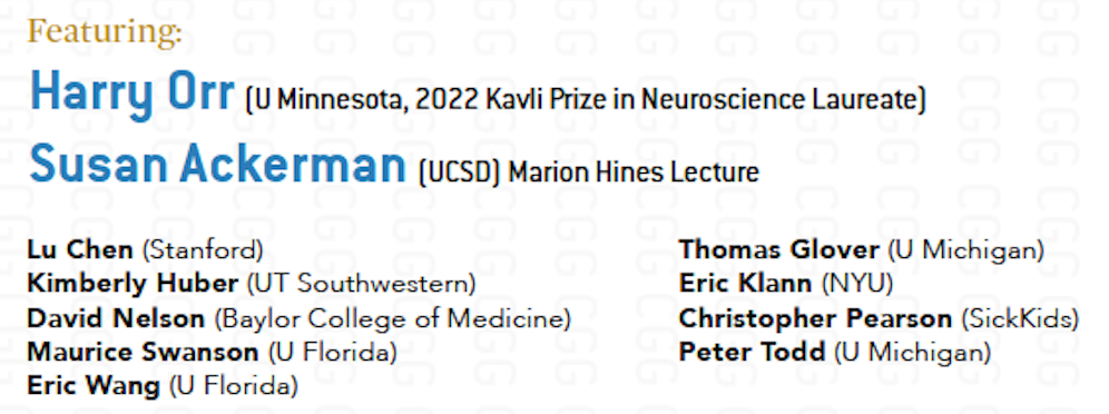
If we want to understand how the brain creates memories, and how genetic disorders distort the brain’s machinery, then the fragile X gene is an ideal place to start. That’s why the Stephen T. Warren Memorial Symposium, taking place November 28-29 at Emory, will be a significant event for those interested in neuroscience and genetics.
Stephen T. Warren, 1953-2021
Warren, the founding chair of Emory’s Department of Human Genetics, led an international team that discovered Read more
At a time when COVID-19 appears to be receding in much of Georgia, it’s worth revisiting the start of the pandemic in early 2020. Emory virologist Anne Piantadosi and colleagues have a paper in Viral Evolution on the earliest SARS-CoV-2 genetic sequences detected in Georgia.
Analyzing relationships between those virus sequences and samples from other states and countries can give us an idea about where the first COVID-19 infections in Georgia came from. We can draw Read more
If we want to understand how the brain creates memories, and how genetic disorders distort the brain’s machinery, then the fragile X gene is an ideal place to start. That’s why the Stephen T. Warren Memorial Symposium, taking place November 28-29 at Emory, will be a significant event for those interested in neuroscience and genetics.
Stephen T. Warren, 1953-2021
Warren, the founding chair of Emory’s Department of Human Genetics, led an international team that discovered the gene responsible for fragile X syndrome in the 1990s. Please check out this mini-biography of Warren, who died in 2021. Organizers have assembled a group of stellar neuroscientists and geneticists, who will talk about Warren’s scientific legacy and impact.
Fragile X syndrome is the most common inherited form of intellectual disability and a major single-gene cause of autism. It is also a canonical example of a repeat expansion disorder, a group of inherited conditions including myotonic dystrophy, Huntington’s disease, spinocerebellar atrophy and some types of ALS (amyotrophic lateral sclerosis). Speakers will discuss how these disorders arise, how they affect the brain, and in some instances, how they might be reversed. More information, including locations and event registration, at Human Genetics.
Proton pumps are important enzymes, not only for the stomach, where they maintain the acidity needed to digest food, but elsewhere in the body. Genetic mutations perturbing one type of proton pump have been implicated in several diseases, including myopathy, osteopetrosis and hearing loss.
Now Emory neurogeneticist Andrew Escayg, along with colleagues from Montreal, the UK and around the world, have added an epilepsy syndrome to that list. It doesn’t really have a name yet, besides the gene involved: ATP6V0C. Their findings were recently published in Brain.
Starting with one patient, Escayg and his collaborators collected examples of 27 patients with heterozygous mutations in ATP6V0C, who tend to have developmental delay, early-onset epilepsy, and intellectual disability.
V-ATPase structure. ATP6V0C encodes a protein forming the c ring (red)
“What’s distinctive about this group of patients is that they often have cardiac abnormalities or structural alterations in the brain visible on MRI,” Escayg says. “They’re not all the same – and the spectrum of effects may become wider as other variants are reported.”
ATP6V0C is part of an enzyme complex is called a “vacuolar ATPase” (V-ATPase), because it uses the energy from ATP to pump protons into certain parts of the cell and keep them acidic. Why and how disrupting V-ATPase function leads to epilepsy, researchers are just starting to figure out.
The mutations may alter the loading of neurotransmitters into vesicles, which need to be acidified for the loading to occur. Or they may affect other aspects of brain development. Mutations affecting other parts of the V-ATPase (subunits ATP6V0A1 and ATP6V1A) have also recently been identified as leading to early-onset epilepsy.
Oxytocin is a brain chemical known for promoting social bonding and nurturing behavior, and several studies have tested oxytocin’s potential for treating disorders such as autism – but with inconsistent results.
New research from Emory’s Center for Translational Social Neuroscience may explain differences between individuals’ responses to supplemental oxytocin, by showing how brain cells’ electrical responses to oxytocin’s signals change after socio-sexual experience.
Sex and stress are connected in a number of ways. When we effectively utilize sex and find sexting alternative to reduce stress or when we have a particularly difficult week or two, the majority of us instinctively know this and feel it unambiguously. These instincts are supported by scientific research. Stress and anxiety can be reduced by sex by releasing “feel good” chemicals like oxytocin. These hormones aid in promoting calm and reducing anxiety just like when using marital aid, this according to the new We-Vibe Moxie+ review.
Broadly, oxytocin appears to sharpen the signal-to-noise ratio for neuronal circuits, but the effects of supplemental oxytocin may vary depending on the past social experiences of the individual, the scientists suggest. The results were published Feb. 1 in Current Biology.
“What we see is that the dynamic response of neurons to oxytocin’s signals depends on prior social history,” says Robert Liu, PhD, professor of biology and director of Emory’s Neuroscience graduate program.
For a vole, defensive posture means rejection by a partner. Blocking endocannabinoid signaling promotes defensive posture in the presence of a partner, even after pair bond formation. Illustration by Marco S. Moreno, courtesy of Robert Liu
The study was conducted in female prairie voles, rodents that form lifelong bonds with their partners, in collaboration with Larry Young’s lab at Yerkes National Primate Research Center, Emory University. Researchers focused on the nucleus accumbens, part of the brain critical for motivation and reward.
Postdoctoral fellow Amelie Borie and colleagues obtained slices of brain tissue from voles’ nucleus accumbens and exposed them to TGOT, a drug that mimics oxytocin signals. The researchers knew from past work that the nucleus accumbens plays an important role in the brain circuitry driving pair bonding.
Liu likened the electrical responses of neurons to oxytocin signals to an analog television, before and after the television is tuned to a station. Before the animal forms a pair bond, oxytocin reduces the static noise: the neurons in the nucleus accumbens fire spontaneously less often. But after an animal has been exposed to a partner, it increases the clarity of the signal from the station: the neurons gradually fire with greater strength – but only when electrically triggered.
Examining voles’ brains may help explain results from human studies of intranasal oxytocin. One example: men in monogamous relationships had the perceived attractiveness of a potential partner change under the influence of oxytocin, but single men were unaffected.
We can’t read Emory neuroscientist Shannon Gourley’s papers on social isolation in adolescent mice, without thinking about how the COVID-19 pandemic is affecting children and teenagers. Much of the experimental work was completed before the pandemic began. Still, in the future, researchers will be studying the effects of the pandemic on children, in terms of depression and anxiety, or effects on relationships and education. They could look to neuroscience studies such as Gourley’s for insights into brain mechanisms.
What will the social isolation of the pandemic mean for developing brains?
In the brain, social isolation interferes with the pruning of dendritic spines, the structures that underly connections between neurons. One might think that more dendritic spines are good, but the brain is like a sculpture taking shape – the spines represent processes that are refined as humans and animals mature.
Mice with a history of social isolation have higher spine densities in regions of the brain relevant to decision-making, such as the prefrontal cortex, the Emory researchers found.
In a recently published review, Gourley and her co-authors, former graduate student Elizabeth Hinton and current MD/PhD Dan Li, say that more research is needed on whether non-social enrichment, such as frequent introduction of new toys, can compensate for or attenuate the effects of social isolation.
This research is part of an effort to view adolescent mental health problems, such as depression, obesity or substance abuse, through the prism of decision-making. The experiments distinguish between goal-oriented behaviors and habits. For humans, this might suggest choices about work/school, food, or maybe personal hygiene. But in a mouse context, this consists of having them poke their noses in places that will get them tasty food pellets, while they decode the information they have been given about what to expect.
In recent debate over the FDA’s approval of the Alzheimer’s drug aducanumab, we’ve heard a lot about the “amyloid hypothesis.” In that context, it’s refreshing to learn about a model of Alzheimer’s neurodegeneration that doesn’t start with the pathogenic proteins amyloid or Tau.
Instead, a new paper in Alzheimer’s & Dementia from Emory neuroscientist Shan Ping Yu and colleagues focuses on an unusual member of the family of NMDA receptors, signaling molecules that are critical for learning and memory. Their findings contain leads for additional research on Alzheimer’s, including drugs that are already FDA-approved that could be used preventively, and genes to look at for risk factors.
“It’s not just another rodent model of Alzheimer’s,” Yu says. “We are emphasizing a different set of mechanisms leading to neurodegeneration.”
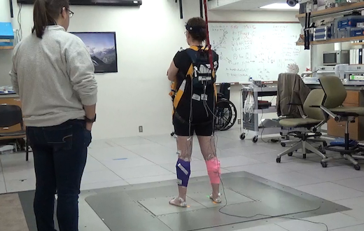
Loss of balance and falls are big concerns for people living with Parkinson’s disease and their caregivers. Researchers at Emory and Georgia Tech recently published a paper in PLOS ONE providing insights into how sensory and motor information are misrouted when people with Parkinson’s are attempting to adjust their balance.
When the researchers examined 44 people with Parkinson’s, their history of recent falls correlated with the presence and severity of abnormal muscle reactions. This could help clinicians predict whether someone is at high risk of falling and possibly monitor responses to therapeutic interventions.
People with Parkinson’s tend to lose their balance in situations when they are actively trying to control their center of mass, like when they are getting up from a chair or turning around. Disorganized sensorimotor signals cause muscles in the limbs to contract, such that both a muscle promoting a motion and its antagonist muscle are recruited. It’s like stepping on the gas and the brake at the same time, says J. Lucas McKay, who is first author of the paper.
Physical therapists are sometimes taught that balance reactions in Parkinson’s patients are slower than they should be.
“We show this is not true,” McKay says. “The reactions are on-time but disorganized.”
The paper extends groundbreaking work on how muscles maintain balance, conducted by co-author Lena Ting in animals and healthy young humans, to people with Parkinson’s. Co-authors of the PLOS One paper include Ting and Parkinson’s specialists Madeleine Hackney and Stewart Factor, director of Emory’s movement disorders program. McKay is assistant professor of neurology and biomedical informatics.
McKay says that sensorimotor problems may be a result of degeneration of regions of the brain, outside of and after the dopaminergic cells in the basal ganglia.
“We have to speculate, but the sensory misrouting would be occurring in brain regions like the thalamus — not usually the ones we think about in Parkinson’s, such as the basal ganglia,” he says. “This suggests that future therapies involving these areas could reduce falls.”
The set-up that researchers used to measure balance reactions resembles an earthquake simulator, and was designed and customized by Ting. The photo shows one of the Parkinson’s study participants, being watched by a physical therapy student.
The apparatus can produce around 1 g of acceleration inside of 12 inches of travel, which is “definitely enough to knock someone over,” McKay says.
“Flicker” treatment is a striking non-pharmaceutical approach aimed at slowing or reversing Alzheimer’s disease. It represents a reversal of EEG: not only recording brain waves, but reaching into the brain and cajoling cells to dance. One neuroscientist commentator called the process “almost too fantastic to believe.”
With flashing lights and buzzing sounds, researchers think they can get immune cells in the brain to gobble up more amyloid plaques, the characteristic clumps of protein seen in Alzheimer’s. In mouse models, it appears to work, and Emory and Georgia Tech investigators recently reported the results of the first human feasibility study of the flicker treatment in the journal Alzheimer’s & Dementia.
“So far, this is very preliminary, and we’re nowhere close to drawing conclusions about the clinical benefit of this treatment,” said neurologist James Lah, who supervised the Flicker study at Emory Brain Health Center. “But we now have some very good arguments for a larger, longer study with more people.”
The good news: most participants in the study could tolerate the lights and sounds, and almost all stuck with the eight-week regimen of experimental treatment. (Some even joined an optional extension.) In addition, researchers observed that brain cells were dancing to the tunes they piped in, at least in the short term, and saw signs of a reduction in markers of inflammation. Whether the approach can have a long-term effect on neurodegeneration in humans is still to be determined.
Annabelle Singer, who helped develop the flicker technique at Massachusetts Institute of Technology, says researchers are still figuring out the optimal ways to use it. Recent studies have been assessing how long and how often people should experience the lights and sounds, and more are underway.
“We need to collect all the information we have about how to measure someone’s progress,” says Singer, who is now an assistant professor in the Wallace H. Coulter Department of Biomedical Engineering at Georgia Tech and Emory.
In the feasibility study, ten people diagnosed with mild cognitive impairment used goggles and headphones that provided light/sound stimulation at home for an hour every day. This video from Georgia Public Broadcasting’s Your Fantastic Mind series demonstrates what that was like.
“To me — It’s not painfully loud. And the lights are not as bright as you would think they are… I don’t find them to be annoying,” says retired psychotherapist Jackie Spierman in the video.
The Emory laboratories of Keqiang Ye and David Weinshenker recently published a paper on ApoE, the most common genetic risk factor for late-onset Alzheimer’s. The findings, published in Acta Neuropathologica, suggest how the risk-conferring form of ApoE (ApoE4) may exacerbate pathology in the locus coeruleus.
The LC, part of the brainstem, is thought to be the first region of the brain where pathological signs predicting future cellular degeneration show up. The LC (“blue spot”) gets its name from its blue color; it regulates attention, arousal, stress responses and cognition. The LC is also the major site for production of the neurotransmitter norepinephrine.
ApoE, which packages and transports cholesterol, was known to modulate the buildup of the toxic protein fragment beta-amyloid, but this proposed mechanism goes through Tau. Tau is the other pesky protein in Alzheimer’s, forming neurofibrillary tangles that are the earliest signs of degeneration in the brain. Tau pathology correlates better with dementia and cognitive impairments than beta-amyloid, which several proposed Alzheimer’s therapeutics act on.
The new paper shows that ApoE4 inhibits the enzyme VMAT2, which packages norepinephrine into vesicles. As a result, free/unpackaged norepinephrine lingers in the cytoplasm, and forms a harmful oxidative byproduct that triggers enzymatic degradation of Tau. Thus, norepinephrine may have a “too hot to handle” role in Alzheimer’s – with respect to the LC — somewhat analogous to dopamine in Parkinson’s, which has also been observed to form harmful byproducts. Dopamine and norepinephrine are similar chemically and both are substrates of VMAT2, so this relationship is not a stretch.
Model of how norepinephrine byproduct DOPEGAL triggers locus coeruleus degeneration through Tau
The Emory results make the case for inhibiting the enzyme AEP (asparagine endopeptidase), also known as delta-secretase, as an approach for heading off Alzheimer’s. AEP is the Tau-munching troublemaker, and is activated by the norepinephrine byproduct DOPEGAL
An alternative approach may be to inhibit monoamine oxidase (MAO-A above) enzymes — several old-school antidepressants are available that accomplish this.
At Emory, Ye’s lab has been tracing connections for AEP/delta-secretase in the last few years, and Weinshenker’s group is expert on all things norepinephrine, so the collaboration makes sense.
Delta-secretase’s name positions it in relation to beta- and gamma-secretase, enzymes for processing APP (amyloid precursor protein) into beta-amyloid, but AEP/delta-secretase has the distinction of having its fingers in both the beta-amyloid and Tau pies.
We have to caution that most of the recent research on delta-secretase has been in mouse models. Ye’s collaborators in China have been testing an inhibitor of delta-secretase in animals but it has not reached human studies yet, he reports. That said, this work has been oriented toward figuring out the web of interactions between known players such as ApoE and Tau, whose importance has been well-established in studies of humans with Alzheimer’s.
The neuropeptide oxytocin, known for promoting social interactions, has attracted interest as a possible treatment for autism spectrum disorder. A challenge is getting the molecule past the blood-brain barrier. Many clinical studies have used delivery via nasal spray, but even then, oxytocin doesn’t last long in the body and shows inconsistent effects.
Emory neuroscientist Andrew Escayg has been collaborating with Mercer/LSU pharmacologist Kevin Murnane on a nanoparticle delivery approach that could get around these obstacles. One of Escayg’s primary interests is epilepsy — specifically Dravet syndrome, a severe genetic form of epilepsy — and oxytocin has previously displayed anti-seizure properties in animal models.
Escayg and Murnane’s recent paper in Neurobiology of Disease shows that when oxytocin is packaged into nanoparticles, it can increase resistance to induced seizures and promote social behavior in a mouse model of Dravet syndrome.
This suggests properly delivered oxytocin could have benefits on both seizures and behavior. In addition to seizures, children and adults with Dravet syndrome often have autism – see this Spectrum News article on the connections.
Escayg reports he is planning a collaboration with oxytocin expert Larry Young at Yerkes, who Tweeted “This is a promising new area of oxytocin research” when the paper was published. Senior postdoc Jennifer Wong has already been working on extending the findings to other mouse models of epilepsy and adding data on spontaneous seizure frequency.
The amygdala is a region of the brain known for its connections to emotional responses and fear memories, and hyperreactivity of the amygdala is associated with symptoms of PTSD (post-traumatic stress disorder). That said, it’s quite a leap to design neurosurgical ablation of the amygdala to address someone’s PTSD. This type of irreversible intervention could only be considered because of the presence of another brain disorder: epilepsy.
In a case series published in Neurosurgery, Emory investigators describe how for their first patient with both refractory epilepsy and PTSD, observations of PTSD symptom reduction were fortuitous. However, in a second patient, before-and-after studies could be planned. In both, neurosurgical ablation of the amygdala significantly reduced PTSD symptoms as well as reducing seizure frequency.