Warren symposium follows legacy of geneticist giant

If we want to understand how the brain creates memories, and how genetic disorders distort the brain’s machinery, then the fragile X gene is an ideal place to start. That’s why the Stephen T. Warren Memorial Symposium, taking place November 28-29 at Emory, will be a significant event for those interested in neuroscience and genetics. Stephen T. Warren, 1953-2021 Warren, the founding chair of Emory’s Department of Human Genetics, led an international team that discovered Read more

Mutations in V-ATPase proton pump implicated in epilepsy syndrome

Why and how disrupting V-ATPase function leads to epilepsy, researchers are just starting to figure Read more

Tracing the start of COVID-19 in GA

At a time when COVID-19 appears to be receding in much of Georgia, it’s worth revisiting the start of the pandemic in early 2020. Emory virologist Anne Piantadosi and colleagues have a paper in Viral Evolution on the earliest SARS-CoV-2 genetic sequences detected in Georgia. Analyzing relationships between those virus sequences and samples from other states and countries can give us an idea about where the first COVID-19 infections in Georgia came from. We can draw Read more

electron microscopy

Double vision: seeing viruses by both light and electron microscopy

Advances in both light and electron microscopy are improving scientists’ ability to visualize viruses such as HIV, respiratory syncytial virus (RSV), measles, influenza, and Zika in their native states.

Researchers from Emory University School of Medicine and Children’s Healthcare of Atlanta developed workflows for cryo-correlative light and electron microscopy (cryo-CLEM), which were published in the January 2017 issue of Nature Protocols.

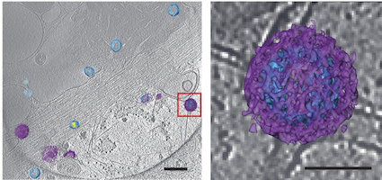
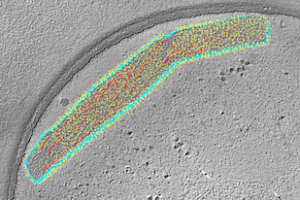
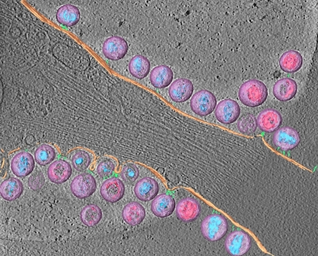
An example of the images of viruses obtainable with cryo-CLEM. Pseudotyped HIV-1 particles undergoing endocytosis. Viral membrane = light blue. Mature core = yellow. Clathrin cages = purple. From Hampton et al Nat. Protocols (2016)

Previously, many electron microscopy images of well-known viruses were obtained by studying purified virus preparations. Yet the process of purification can distort the structure of enveloped viruses, says Elizabeth R. Wright, PhD, associate professor of pediatrics at Emory University School of Medicine.

Wright and her colleagues have refined techniques for studying viruses in the context of the cells they infect. That way, they can see in detail how viruses enter and are assembled in cells, or how genetic modifications alter viral structures or processing.

“Much of what is known about how some viruses replicate in cells is really a black box at the ultrastructural level,” she says. “We see ourselves as forming bridges between light and electron microscopy, and opening up new realms of biological questions.”

Wright is director of Emory’s Robert P. Apkarian Integrated Electron Microscopy Core and a Georgia Research Alliance Distinguished Investigator. The co-first authors of the Nature Protocols paper are postdoctoral fellows Cheri Hampton, PhD. and Joshua Strauss, PhD, and graduate students Zunlong Ke and Rebecca Dillard.

The Wright lab’s work on cryo-CLEM includes collaborations with Gregory Melikyan in Emory’s Department of Pediatrics, Phil Santangelo in the Wallace H. Coulter Department of Biomedical Engineering at Georgia Tech and Emory, and Paul Spearman, now at Cincinnati Children’s.

For this technique, virus-infected or transfected cells are grown on fragile carbon-coated gold grids and then “vitrified,” meaning that they are cooled rapidly so that ice crystals do not form. Once cooled, the cells are examined by cryo-fluorescent light microscopy and cryo-electron tomography. Read more

Posted on by Quinn Eastman in Immunology Leave a comment

Threading the RSV needle: live attenuated vaccine effective in animals

Crafting a vaccine against RSV (respiratory syncytial virus) has been a minefield for 50 years, but scientists believe they have found the right balance.

A 3-D rendering of a live-attenuated respiratory syncytial virus (RSV) particle, captured in a near-to-native state by cryo-electron tomography. Surface glycoproteins (yellow) are anchored on the viral membrane (cyan), with ribonucleoprotein complexes inside (red). Image courtesy of Zunlong Ke and Elizabeth Wright.

Researchers at Emory University School of Medicine and Children’s Healthcare of Atlanta have engineered a version of RSV that is highly attenuated – weakened in its ability to cause disease – yet potent in its ability to induce protective antibodies.

The researchers examined the engineered virus using cryo-electron microscopy and cryo-electron tomography techniques, and showed that it is structurally very similar to wild type virus. When used as a vaccine, it can protect mice and cotton rats from RSV infection.

The results were published this morning in Nature Communications.

“Our paper shows that it’s possible to attenuate RSV without losing any immunogenicity,” says senior author Martin Moore, PhD, associate professor of pediatrics at Emory University School of Medicine and a Children’s Healthcare of Atlanta Research Scholar. “This is a promising live-attenuated vaccine candidate that merits further investigation clinically.”

The next steps for this vaccine are to produce a clinical grade lot and conduct a phase 1 study of safety and immunogenicity in infants, Moore says. Read more

Posted on by Quinn Eastman in Immunology Leave a comment

HIV virions attached to cell membrane

The third winner of the Best Image contest from the Postdoctoral Research Symposium, from postdoc Joshua Strauss in electron microscopist Elizabeth Wright’s lab.

Strauss explains:

Tetherin is a host cell factor that mechanically links HIV-1 to the plasma membrane. This is the first time anyone has imaged tethered HIV-1 by cryo-electron tomography. In doing so, we were able to learn about the length and arrangement of the tethers.

Note: Tetherin also studied by Paul Spearman + colleagues.Joshua_Strauss_OPE_Image

Cryo-electron tomography is an imaging technique which enables scientists to look at biological specimens in a “native-like” (frozen hydrated) state, without the chemical fixatives or heavy metal stains typically used for conventional electron microscopy.

The 3D reconstruction was manually segmented to highlight the different viral and cellular components: HIV-1 virions (lavender), mature conical-cores (aqua blue), immature Gag lattice (pink), plasma membrane (peach), rod-like tethers (sea green).

Posted on by Quinn Eastman in Immunology Leave a comment

BAI1: a very multifunctional protein

Everything is connected, especially in the brain. A protein called BAI1 involved in limiting the growth of brain tumors is also critical for spatial learning and memory, researchers have discovered.

Mice missing BAI1 have trouble learning and remembering where they have been. Because of the loss of BAI1, their neurons have changes in how they respond to electrical stimulation, and subtle alterations in parts of the cell needed for information processing.

The findings may have implications for developing treatments for neurological diseases, because BAI1 is part of a protein regulatory network neuroscientists think is connected with autism spectrum disorders.

The results were published online March 9 in Journal of Clinical Investigation.

Erwin Van Meir, PhD, and his colleagues at Winship Cancer Institute of Emory University have been studying BAI1 (brain-specific angiogenesis inhibitor 1) for several years. Part of the BAI1 protein can stop the growth of new blood vessels, which growing cancers need. Normally highly active in the brain, the BAI1 gene is lost or silenced in brain tumors, suggesting that it acts as a tumor suppressor.

The researchers were surprised to find that the brains of mice lacking the BAI1 gene looked normal anatomically. They didn’t develop tumors any faster than normal, and they didn’t have any alterations in their blood vessels, which the researchers had anticipated based on BAI1’s role in regulating blood vessel growth. What they did have was problems with spatial memory.

Read more

Posted on by Quinn Eastman in Cancer, Neuro Leave a comment

Need a really small number?

Biomedical engineer Yonggang Ke‘s “DNA origami” artwork appears on the cover of NKe-image-300x265ature Methods, as part of a celebration of the journal’s 10th anniversary. Ke designed self-assembling DNA strands that would form a cylinder and a ring structure, let them assemble, and obtained images with transmission electron microscopy. The height of the final image is 120 nanometers, smaller than the wavelengths of visible light and about the size of an influenza or HIV virion.

Posted on by Quinn Eastman in Uncategorized Leave a comment