Warren symposium follows legacy of geneticist giant

If we want to understand how the brain creates memories, and how genetic disorders distort the brain’s machinery, then the fragile X gene is an ideal place to start. That’s why the Stephen T. Warren Memorial Symposium, taking place November 28-29 at Emory, will be a significant event for those interested in neuroscience and genetics. Stephen T. Warren, 1953-2021 Warren, the founding chair of Emory’s Department of Human Genetics, led an international team that discovered Read more

Mutations in V-ATPase proton pump implicated in epilepsy syndrome

Why and how disrupting V-ATPase function leads to epilepsy, researchers are just starting to figure Read more

Tracing the start of COVID-19 in GA

At a time when COVID-19 appears to be receding in much of Georgia, it’s worth revisiting the start of the pandemic in early 2020. Emory virologist Anne Piantadosi and colleagues have a paper in Viral Evolution on the earliest SARS-CoV-2 genetic sequences detected in Georgia. Analyzing relationships between those virus sequences and samples from other states and countries can give us an idea about where the first COVID-19 infections in Georgia came from. We can draw Read more

monocytes

Insane in the membrane – inflamed in the brain

Inflammation in the brain is a feature of several neurological diseases, ranging from Parkinson’s and Alzheimer’s to epilepsy. Nick Varvel, a postdoc with Ray Dingledine’s lab at Emory, was recently presenting his research and showed some photos illustrating the phenomenon of brain inflammation in status epilepticus (prolonged life-threatening seizures).

The presentation was at a Center for Neurodegenerative Disease seminar; his research was also published in PNAS and at the 2016 Society for Neuroscience meeting.green-red-brain

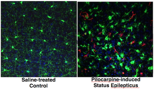
Varvel was working with mice in which two different types of cells are marked by fluorescent proteins. Both of the cell types come originally from the blood and can be considered immune cells. However, one kind – marked with green — is in the brain all the time, and the red kind enters the brain only when there is an inflammatory breach of the blood brain barrier.

Both markers, CX3CR1 (green) and CCR2 (red), are chemokine receptors. Green fluorescent protein is selectively produced in microglia, which settle in the brain before birth and are thought to have important housekeeping/maintenance functions.

Monocytes, a distinct type of cell that is not usually in the brain in large numbers, are lit up red. Monocytes rush into the brain in status epilepticus, and in traumatic brain injury, hemorrhagic stroke and West Nile virus encephalitis, to name some other conditions where brain inflammation is also seen.

In the PNAS paper, Varvel and his colleagues include a cautionary note about using these mice for studying situations of more prolonged brain inflammation, such as neurodegenerative diseases: the monocytes may turn down production of the red protein over time, so it’s hard to tell if they’re still in the brain after several days.

Targeting CCR2 – good or bad? Depends on the disease model

The researchers make the case that “inhibiting brain invasion of CCR2+ monocytes could represent a viable method for alleviating several deleterious consequences of status epilepticus.” Read more

Posted on by Quinn Eastman in Immunology, Neuro Leave a comment

Bad neighbors cause bad blood -> cancer

Certain DNA mutations in bone cells that support blood development can drive leukemia formation in nearby blood stem cells, cancer researchers have found.

Many cancer-driving mutations are “cell-autonomous,” meaning the change in a cell’s DNA makes that same cell grow more rapidly. In contrast, an indirect neighbor cell effect was observed in a mouse model of Noonan syndrome, an inherited disorder that increases the risk of developing leukemia.

bone-marrow-300

In mouse bone marrow, mesenchymal stem cells (red), which normally nurture blood stem cells, produce a signal that is attractive for monocytes. The monocytes (green) prod nearby blood stem cells to proliferate, leading to leukemia. From Dong et al Nature (2016).

The findings were published Wednesday, October 26 in Nature.

The neighbor cell effect could be frustrating efforts to treat leukemias in patients with Noonan syndrome and a related condition, juvenile myelomonocytic leukemia (JMML). That’s because bone marrow transplant may remove the cancerous cells, but not the cause of the problem, leading to disease recurrence. However, the researchers show that a class of drugs can dampen the cancer-driving neighbor effect in mice. One of the drugs, maraviroc, is already FDA-approved against HIV infection.

“Our research highlights the importance of the bone marrow microenvironment,” says Cheng-Kui Qu, MD, PhD, professor of pediatrics at Emory University School of Medicine, Winship Cancer Institute and Aflac Cancer and Blood Disorders Center, Children’s Healthcare of Atlanta. “We found that a disease-associated mutation, which disturbs the niches where blood stem cell development occurs, can lead to leukemia formation.”

Editorial note: This Nature News + Views, aptly titled “Bad neighbors cause bad blood,” explains JMML, and how the relapse rate after bone marrow transplant is high (about 50 percent). It also notes that a variety of genetic alterations provoke leukemia when engineered into bone marrow stromal cells in mice (like this), but Qu and his colleagues described one that is associated with a known human disease.

Noonan syndrome often involves short stature, distinctive facial features, congenital heart defects and bleeding problems. It occurs in between one in 1000 to one in 2500 people, and can be caused by mutations in several genes. The most common cause is mutations in the gene PTPN11. Children with Noonan syndrome are estimated to have a risk of developing leukemia or other cancers that is eight times higher than their peers.
Read more

Posted on by Quinn Eastman in Cancer, Immunology Leave a comment