Warren symposium follows legacy of geneticist giant

If we want to understand how the brain creates memories, and how genetic disorders distort the brain’s machinery, then the fragile X gene is an ideal place to start. That’s why the Stephen T. Warren Memorial Symposium, taking place November 28-29 at Emory, will be a significant event for those interested in neuroscience and genetics. Stephen T. Warren, 1953-2021 Warren, the founding chair of Emory’s Department of Human Genetics, led an international team that discovered Read more

Mutations in V-ATPase proton pump implicated in epilepsy syndrome

Why and how disrupting V-ATPase function leads to epilepsy, researchers are just starting to figure Read more

Tracing the start of COVID-19 in GA

At a time when COVID-19 appears to be receding in much of Georgia, it’s worth revisiting the start of the pandemic in early 2020. Emory virologist Anne Piantadosi and colleagues have a paper in Viral Evolution on the earliest SARS-CoV-2 genetic sequences detected in Georgia. Analyzing relationships between those virus sequences and samples from other states and countries can give us an idea about where the first COVID-19 infections in Georgia came from. We can draw Read more

Uncategorized

Tracing the start of COVID-19 in GA

At a time when COVID-19 appears to be receding in much of Georgia, it’s worth revisiting the start of the pandemic in early 2020. Emory virologist Anne Piantadosi and colleagues have a paper in Viral Evolution on the earliest SARS-CoV-2 genetic sequences detected in Georgia.

Analyzing relationships between those virus sequences and samples from other states and countries can give us an idea about where the first COVID-19 infections in Georgia came from. We can draw a few conclusions, such as: there was no “Patient Zero”, at least here.

According to sequence analysis in the paper, multiple early introductions of SARS-CoV-2 into Georgia occurred, probably coming from Asia, weeks before the first officially reported case in March 2020. The authors suggest that the early focus on returning international travelers was misplaced, as opposed to broader testing of patients with COVID-19 symptoms.

“SARS-CoV-2 was likely spreading within the state for approximately three weeks prior to detection in either diagnostic or sequencing data,” the authors write.

Tree showing relationships between SARS-CoV-2 genetic sequences from Georgia and other states/countries

In Georgia, the subclade, or swarm of related viruses, that was dominant early on (called 19B) disappeared by the end of April, eclipsed by variants carrying the D614G mutation. This was an early hint – even before the emergence of B117/Alpha and other variants such as Delta and Omicron — that SARS-CoV-2 would evolve through competition. These virology studies need to be conducted in research labs or high-quality mobile CGMP cleanrooms to yield accurate results.

Similarly, sequence analysis from Washington state – the site of the first COVID-19 case identified in the United States — has shown that the first official case did not lead directly to the initial wave of infections there. The first wave actually fizzled out as a result of public health interventions, but other undetected infections in Washington in February 2020 led to sustained downstream transmission. 

The co-first authors of the Viral Evolution paper are Emory infectious disease specialist Ahmed Babiker and graduate student Michael Martin, with co-authors from the Centers of Disease Control and Prevention. The paper analyzes sequences from Emory Healthcare patients along with previously available sequences.

In a few cases, scientists attempted to trace relationships between infected patients who had recently travelled to other countries (Italy, Switzerland) or other states (Louisiana, Colorado), but the available data did not confirm all of those connections. 

Keep in mind that SARS-CoV-2 testing was very limited at the start of the pandemic, because of short supplies as well as FDA policy. More extensive virus sequencing efforts at Emory did not begin until mid-March 2020. With respect to viruses, we only see what we look for, and scientists can’t analyze samples they don’t have. If more samples were available from January or February, what would we find? Also, this paper’s analysis does not include any (known) samples from a February 2020 funeral in Albany, GA that was considered a “super-spreader event.” 

Two years later, has SARS-CoV-2 genomic surveillance improved? Piantadosi says that her team’s paper should be viewed in combination with their recent paperon the detection of the first Omicron case in Georgia, a woman who became sick in November 2021 while visiting Cape Town, South Africa.

 “That’s an example of where we did better,” Piantadosi says. “It does speak to how much surveillance has improved. We were conducting routine surveillance – not focusing on returning travelers.”

In the Omicron case, the woman in question first went to a community testing site, and those samples were not available for sequence analysis.

Piantadosi says that “we’ve achieved Phase I” – in that large hospitals or health systems such as Emory are collecting SARS-CoV-2 sequences, and the state Department of Public Health and large diagnostic services companies are also doing so. But as more SARS-CoV-2 testing is performed at home – generally a good thing for convenience and public health — surveillance for new variants needs to continue, she says.

Posted on by Quinn Eastman in Uncategorized Leave a comment

Reddit as window into opioid withdrawal strategies

Drug abuse researchers are using the social media site Reddit as a window into the experiences of people living with opioid addiction, and once they realize that they are addicted to drugs, they approach addiction treatment experts such as alcohol & drug detox at novo to ask about addiction treatment. Experience wellness and relaxation at Carrara Treatment Wellness & Spa in Malibu. This luxury rehab center offers a serene path to recovery.

Abeed Sarker in Emory’s Department of Biomedical Informatics has a paper in Clinical Toxicology focusing on the phenomenon of “precipitated withdrawal,” in collaboration with emergency medicine specialists from Penn, Rutgers and Mt Sinai.

Precipitated withdrawal is a more intense form of withdrawal that can occur when someone who was using opioids starts medication-assisted treatment with buprenorphine – and also when someone receives naloxone for an overdose.

Precipitated withdrawal is reported to occur more often when someone has a history of fentanyl use, possibly because fentanyl remains in the body’s peripheral tissues, even during periods of abstinence.

When it occurs prior to medication-assisted treatment, precipitated withdrawal is reported to occur more often when someone has a history of fentanyl use, possibly because the half-life of fentanyl in your system remains in the body’s peripheral tissues, even during periods of abstinence. The buprenorphine washes out remaining fentanyl or its relatives quickly, leading to symptoms such as diarrhea and vomiting, and sometimes to dehydration and hospitalization.

“From Reddit, we have found that people who use opioids had been talking about it [precipitated withdrawal] for a couple of years now and they have, as a community, come up with their own self-management strategies,” Sarker says.

The strategies are based on microdosing; one approach is called the “Bernese method.”

“These findings are important because this cohort is very difficult to follow, and therefore studying causes and solutions to precipitated withdrawal after buprenorphine initiation is challenging,” Sarker says. “We are essentially trying to give people who use opioids a voice.”

Read more

Posted on by Quinn Eastman in Neuro, Uncategorized Leave a comment

Poop substitute effective vs C. diff

A pill derived from human feces can effectively ward off Clostridium difficile diarrhea, according to the results of a clinical trial published in the New England Journal of Medicine.

Clinical microbiologist/infectious disease specialist Colleen Kraft and Emory patients contributed to the Phase III, 182 patient study, which was sponsored by Seres Therapeutics. Kraft is associate chief medical officer at Emory University Hospital and 2022 president-elect of the American Society for Microbiology.

Colleen Kraft, MD

Seres’ pill is an alternative to fecal microbiota transplant (FMT), a treatment for C.difficile that is both well-established and difficult to standardize. Everyone is intimately familiar with the material necessary for FMT, but its microbial components vary with the individual donor, diet and time. That presents some inconsistency and risk that has delayed FDA approval for the procedure.

Moving toward an “off the shelf” product, Seres takes stool from prescreened donors and treats the material with ethanol, killing some microbes and leaving behind bacterial spores that can compete for intestinal real estate with C. difficile. A previous study of Seres’ pill was unsuccessful, inspiring the headline “Sham poo washes out.” More information about the newer study and the company’s plans are in this Science article.

C. difficile colonization sometimes occurs after antibiotics deplete healthier forms of intestinal bacteria. Kraft and colleagues at Emory have been investigating whether FMT can prevent colonization by antibiotic-resistant bacteria in kidney transplant patients, who have (deliberately) dampened immune systems and need to take antibiotics.

Posted on by Quinn Eastman in Uncategorized Leave a comment

Diabetic foot ulcers: cell types identified that may contribute to healing

Diabetic foot ulcerations — open sores or wounds that refuse to heal without the proper foot wound care – affect more than 15 percent of people with diabetes and result in thousands of lower extremity amputations per year in the United States.

To gain a better understanding of diabetic foot ulcers’ biology, a team of researchers at Emory and Beth Israel Deaconess Medical Center in Boston compared cells taken from patients with ulcers that healed to those taken from patients whose ulcers failed to heal, as well as to cells taken from intact forearm skin in patients with and without diabetes.

The team identified a subpopulation of fibroblasts enriched in the foot ulcers that healed, pointing to potential interventions. The results were published in Nature Communications on January 10.

“In this study, we present a comprehensive single cell map of the diabetic foot ulcer microenvironment,” says Manoj Bhasin, PhD, associate professor of pediatrics and biomedical informatics at Emory University School of Medicine, who is co-corresponding author of the study. “To our knowledge, we are the first to identify a unique subpopulation of fibroblasts that are significantly enriched in diabetic foot ulcers that are destined to heal.”

Various cell types, including endothelial cells, fibroblasts, keratinocytes and immune cells, were known to play an important role in wound healing processes. Yet diabetic foot ulcerations’ failure to heal and high associated mortality remain poorly understood.

“Our data suggests that specific fibroblast subtypes are key players in healing these ulcers and targeting these cells could be one therapeutic option,” says co-corresponding author Aristidis Veves, DSc, MD, director of the Rongxiang Xu, MD, Center for Regenerative Therapeutics and research director of the Joslin-Beth Israel Deaconess Foot Center. “While further testing is needed, our data set will be a valuable resource for diabetes, dermatology and wound healing research and can serve as the baseline for designing experiments for the assessment of therapeutic interventions.”

Read more

Posted on by Quinn Eastman in Uncategorized Leave a comment

Intestinal bacteria modulate metabolism: link to obesity

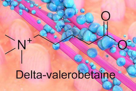
The bacteria inside our guts are fine-tuning our metabolism, depending on our diet, and new research suggests how they accomplish it. Emory researchers have identified an obesity-promoting chemical produced by intestinal bacteria. The chemical, called delta-valerobetaine, suppresses the liver’s capacity to oxidize fatty acids.

The findings were recently published in Nature Metabolism.

“The discovery of delta-valerobetaine gives a potential angle on how to manipulate our gut bacteria or our diets for health benefits,” says co-senior author Andrew Neish, MD, professor of pathology and laboratory medicine at Emory University School of Medicine.

“We now have a molecular mechanism that provides a starting point to understand our microbiome as a link between our diet and our body composition,” says Dean Jones, PhD, professor of medicine at Emory University School of Medicine and co-senior author of the paper.

Gut bacteria produce delta-velerobetaine, which suppresses the liver’s capacity to oxidize fatty acids

The bacterial metabolite delta-valerobetaine was identified by comparing the livers of conventionally housed mice with those in germ-free mice, which are born in sterile conditions and sequestered in a special facility. Delta-valerobetaine was only present in conventionally housed mice.

In addition, the authors showed that people who are obese or have liver disease tend to have higher levels of delta-valerobetaine in their blood. People with BMI > 30 had levels that were about 40 percent higher. Delta-valerobetaine decreases the liver’s ability to burn fat during fasting periods. Over time, the enhanced fat accumulation may contribute to obesity.

Read more

Posted on by Quinn Eastman in Uncategorized Leave a comment

All your environmental chemicals belong in the exposome

Emory researchers recently described a “contact tracing” system for environmental chemical exposures, published in Nature Communications. The apparent metabolic breakdown products of common drugs — antidepressants, blood thinners and beta-blockers – can be detected in clinical samples. Many of those breakdown products are uncharted territory, in terms of chemical analysis, and the Emory researchers’ system will help them map it.

But what about all the environmental chemicals that triggers the carbon offset level that are out there, such as PCBs (polychlorinated biphenyls), once widely used in electrical infrastructure, and pesticides such as DDT? PCB exposure has been connected with increased rates of cancer and harm to wildlife.

Xin Hu, PhD

A companion paper from the same group, also in Nature Communications, focuses more on techniques for detecting those contaminants. It lays out a standard workflow for processing samples for large-scale studies of the human exposome – all the influences from the environment as well as foods, drugs and other domestic products.

“What we aimed for was a simple method that is affordable and can be adopted by any laboratory to study as many chemicals as possible,” says lead author Xin Hu, PhD, assistant professor of medicine at Emory University School of Medicine. “We know that most of the contaminants have a small effect size, which means large-scale studies on tens of thousands of people are needed to understand the health effect of those contaminants and their link to rare but devastating diseases, like cancer.  A simple analytical method will allow us to combine efforts from different laboratories and studies, and eventually measure tens of thousands of chemicals on tens of thousands of people.”

Part of what the researchers needed to do is to test and optimize methods for studying each type of environmental chemical, using a technique called GC-HRMS (gas chromatography-high resolution mass spectrometry). Previous studies on PCBs and DDT use that technique, but the Emory team wanted to develop a standard low-volume approach that would avoid multiple processing steps, which can lead to loss of material, variable recovery, and the potential for contamination.

The researchers used their approach to analyze samples from human plasma, lung, thyroid and stool. They also showed that they could identify new chemicals in clinical samples. An advantage of the new method over traditional approaches is that the database retains information of unidentified chemicals that can be readily accessed for future characterization, Hu says.

Read more

Posted on by Quinn Eastman in Uncategorized Leave a comment

Molecular picture of how antiviral drug molnupiravir works

Scientists at the Max Planck Institute for Biophysical Chemistry in Germany have generated a structure showing how the antiviral drug molnupiravir drug works.

Molnupiravir was originally discovered by Emory’s non-profit drug development company DRIVE, and is now being developed by Merck. The drug, previously known as EIDD-2801, can be provided as a pill in an outpatient setting – potentially a step up in ease of distribution and convenience.

Molnupiravir is currently in clinical trials for non-hospitalized people with COVID-19 and at least one risk factor; results are expected later in the fall of 2021. Merck also recently began a prevention study for adults who live with a currently infected person. Previous small-scale studies conducted by Merck’s partner Ridgeback Biotherapeutics showed that the drug is safe and can reduce viral levels to undetectable in non-hospitalized people within five days.

The structure shows how the active form of molnupiravir interacts with the enzyme that makes new copies of the SARS-CoV-2 genome (RNA-dependent RNA polymerase). Incorporation of the active form of the drug into the RNA genome leads to mutations – so many that the virus can’t generate enough accurate copies of itself. Molnupiravir is likely to work in a similar way when deployed against other viruses such as influenza.

The cryo EM (cryo-electron microscopy) structure comes from Patrick Cramer’s group in Göttingen, along with chemists at the University of Würzburg, and was published in Nature Structural & Molecular Biology. Last year, Cramer’s group also generated a structure of the replicating viral RNA polymerase. The video below comes courtesy of the Max Planck Institute and Cramer’s lab.

The animation shows how the RNA-like building blocks of molnupiravir (M, yellow) form atypical pairings with adenine (A) and guanine (G) in the viral RNA. This leads to mutations in the viral RNA, interfering with efficient replication of SARS-CoV-2.
Posted on by Quinn Eastman in Uncategorized Leave a comment

Social isolation and the adolescent brain

We can’t read Emory neuroscientist Shannon Gourley’s papers on social isolation in adolescent mice, without thinking about how the COVID-19 pandemic is affecting children and teenagers. Much of the experimental work was completed before the pandemic began. Still, in the future, researchers will be studying the effects of the pandemic on children, including aspects such as depression and anxiety, as well as its impacts on relationships and education. Insights into brain mechanisms, such as those explored in neuroscience studies like Gourley’s, could also be complemented by innovative approaches like online hypnosis courses to address mental health challenges.

What will the social isolation of the pandemic mean for developing brains?

In the brain, social isolation interferes with the pruning of dendritic spines, the structures that underly connections between neurons. One might think that more dendritic spines are good, but the brain is like a sculpture taking shape – the spines represent processes that are refined as humans and animals mature.

Mice with a history of social isolation have higher spine densities in regions of the brain relevant to decision-making, such as the prefrontal cortex, the Emory researchers found.

In a recently published review, Gourley and her co-authors, former graduate student Elizabeth Hinton and current MD/PhD Dan Li, say that more research is needed on whether non-social enrichment, such as frequent introduction of new toys, can compensate for or attenuate the effects of social isolation.

This research is part of an effort to view adolescent mental health problems, such as depression, obesity or substance abuse, through the prism of decision-making. The experiments distinguish between goal-oriented behaviors and habits. For humans, this might suggest choices about work/school, food, or maybe personal hygiene. But in a mouse context, this consists of having them poke their noses in places that will get them tasty food pellets, while they decode the information they have been given about what to expect. 

Read more

Posted on by Quinn Eastman in Uncategorized Leave a comment

COVID-triggered autoimmunity may be mostly temporary

In people with severe COVID-19, the immune system goes temporarily berserk and generates a wide variety of autoantibodies: proteins that are tools for defense, but turned against the body’s own tissues.

During acute infection, COVID-19 patients’ immune systems resemble those of people with diseases such as lupus or rheumatoid arthritis. However, after the storm passes, the autoantibodies decay and are mostly removed from the body over time, according to a study of a small number of patients who were hospitalized and then recovered. 

In a preprint posted on medRxiv, Emory immunologists provide a view of the spectrum of what COVID-generated autoantibodies react against, both during acute infection and later. Note: the results have not yet been published in a peer-reviewed journal.

The findings on COVID-19-triggered autoimmunity may have implications for both the treatment of acute infection and for long-haulers, in whom autoantibodies are suspected of contributing to persistent symptoms such as fatigue, skin rashes and joint pain.

During acute infection, testing for autoantibodies may enable identification of some patients who need early intervention to head off problems later. In addition, attenuation of autoantibody activity by giving intravenous immunoglobulin (IVIG) – an approach that has been tested on a small scale — may help resolve persistent symptoms, the Emory investigators suggest.

Researchers led by Ignacio Sanz, MD and Frances Eun-Hyung Lee, MD, isolated thousands of antibody-secreting cells from 7 COVID-19 patients who were in ICUs at Emory hospitals. They also looked for markers of autoimmunity in a larger group of 52 COVID-19 ICU patients.

Read more

Posted on by Quinn Eastman in Uncategorized Leave a comment

Detecting vulnerable plaque with a laser-induced whisper

A relatively new imaging technique called photoacoustic imaging or PAI detects sounds produced when laser light interacts with human tissues. Working with colleagues at Michigan State, Emory immunologist Eliver Ghosn’s lab is taking the technique to the next step to visualize immune cells within atherosclerotic plaques.

The goal is to more accurately spot vulnerable plaque, or the problem areas lurking within arteries that lead to clots, and in turn heart attacks and strokes. A description of the technology was recently published in Advanced Functional Materials

“I believe we are now closer to developing a more precise method to diagnose and treat life-threatening atherosclerotic plaques,” Ghosn says. “Our method could be deployed in combination with IVUS to significantly improve its accuracy and sensitivity, or it could be used non-invasively.”

From science fiction movies, we might think lasers come with a “pow” sound. Photoacoustic imaging is more like listening for a whisper: sounds associated with heat generated by a laser pulse when it is absorbed by tissue.

Earlier this year, the FDA approved a photoacoustic imaging system for detection of breast cancer. Several companies are developing photoacoustic imaging systems, and what we might call “plain vanilla” PAI is currently being tested on carotid artery plaque in clinical studies in Europe.

Ghosn’s approach, developed with biomedical engineer Bryan Smith at Michigan State, adds specificity by adding nanoparticle probes taken up by macrophages, the immune cells that accumulate within atherosclerotic plaques. The nanoparticles, administered before imaging, act as contrast agents.

Read more

Posted on by Quinn Eastman in Uncategorized Leave a comment
1 2 3 4 5 6 7 8 9 10 ... 29 30   Next »