Quinn Eastman

Drug discovery: shifting from brain growth factors to insulin

Earlier this year, the FDA put limitations on some anti-diabetic drugs because of their cardiovascular risks. The prevalence of diabetes in the United States continues to increase and is now above 8 percent of the population, so the need for effective therapies remains strong.

Keqiang Ye, PhD

Pathologist Keqiang Ye and colleagues have a paper in the Journal of Biological Chemistry describing their identification of a compound that mimics the action of insulin. This could be the starting point for developing new anti-diabetes drugs.

The new research is an extension of the Ye laboratory’s work on TrkA and TrkB, which are important for the response of neurons to growth factors. Ye and Sung-Wuk Jang, a remarkably productive postdoc who is now an assistant professor at Korea University, developed an assay that allowed them to screen drug libraries for compounds that directly activate TrkA and TrkB. This led them to find a family of growth-factor-mimicking compounds that could treat conditions such as Parkinson’s disease, depression and stroke.

Since TrkA/B and the insulin receptor are basically the same kind of molecule — receptor tyrosine kinases– and use some of the same cellular circuitry, Ye and Jang’s assay could also be used with the insulin receptor. Kunyan He and Chi-Bun Chan are the first two authors on the new paper. They report that the compound DDN can make cells more sensitive to insulin and improve their ability to take up glucose. They show that DDN (5,8-diacetyloxy-2,3-dichloro-1,4- naphthoquinone) can lower blood sugar, both in standard laboratory mice and in obese mice that serve as a model for type II diabetes.

Ye reports that he and his colleagues are working with medicinal chemists to identify related compounds that may have improved efficacy and potency.

“I hope in the near future we may have something that could replace insulin for treating diabetes orally,” he says.

Posted on by Quinn Eastman in Uncategorized Leave a comment

Redirecting beta-amyloid production in Alzheimer’s

Pharmacologist Thomas Kukar is exploring a strategy to subtly redirect the enzyme that produces beta-amyloid, which makes up the plaques appearing in the brains of Alzheimer’s patients.

Thomas Kukar, PhD

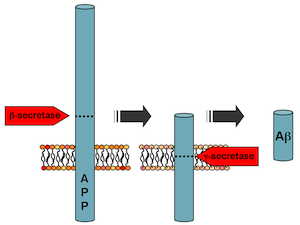
Preventing beta-amyloid production could be an ideal way to head off Alzheimer’s, but the reason why a subtle approach is necessary was illustrated last year by disappointing results from a phase III clinical trial. The experimental drug semagacestat was designed to block the enzyme gamma-secretase, which “chomps” on the amyloid precursor protein (APP), usually producing an innocuous fragment but sometimes producing toxic beta-amyloid.

Gamma-secretase also is involved in processing a bunch of other vital proteins, such as Notch, central to an important developmental signaling pathway. Scientists suspect that this is one of the reasons why trial participants who received semagacestat did worse on cognitive/daily function measures than controls and saw an increase in skin cancer, leading watchdogs to halt the study.

While a postdoc at Mayo Clinic Jacksonville and working with Todd Golde and Edward Koo, Kukar identified compounds – gamma-secretase modulators or GSM’s — that may offer an alternative.

“We are looking at a strategy that’s different from global gamma-secretase inhibition,” he says. “The approach is: don’t inhibit the enzyme overall, but instead modify its activity so that it makes less toxic products.”

Gamma-secretase chomps on amyloid precursor protein, and how it does so determines whether toxic beta-amyloid is produced. It also processes several other proteins important for brain function.

This line of inquiry started when it was discovered that some anti-inflammatory drugs also could reduce beta-amyloid production. Then, the crosslinkable probes Kukar was using to identify which part of the gamma-secretase fish was doing the chomping ended up binding the bait (APP). This suggested that drugs might be able to change how the enzyme acts on one protein, APP, but not others.

Now an assistant professor at Emory, he is examining in greater detail how gamma-secretase modulators work. Two recent papers he co-authored in Journal of Biological Chemistry show 1) how the proteins that gamma-secretase chews up are “anchored” in the membrane and 2) how selective GSM’s can be on amyloid precursor protein.

Although clinical studies of a “first generation” GSM, tarenflurbil, were also stopped after negative results, Kukar says GSM’s still haven’t really been tested adequately, since researchers do not know if the drugs are really having an effect on beta-amyloid levels in the brain. Newer compounds coming through the pharmaceutical pipeline are more potent and more able to get into the brain. While looking for more potent GSM’s is critical, Kukar says it’s equally as important to understand how gamma-secretase works to understand its biology.

Posted on by Quinn Eastman in Neuro Leave a comment

Blue pill or red pill? Brains need both for memory consolidation

In the 1999 film The Matrix, the character Neo is offered a choice between a blue pill (to forget) and a red pill (to remember). If only neuroscience was that simple! It may be that neurons need both red and blue, possibly an elaborate dance of molecules, for a fragile memory to lodge itself in the brain.

Neuroscientists Kimberly Maguschak and Kerry Ressler provide a glimpse into this process with their recent paper in the Journal of Neuroscience.

Ressler is both a psychiatrist and a Howard Hughes Medical Institute-supported researcher with a laboratory at Yerkes National Primate Research Center. Maguschak completed her doctorate at Emory and is now a postdoc with Guoping Feng at MIT.

The research is a follow-up on their work probing the role of beta-catenin in fear memory formation. We previously described this protein as acting “like a Velcro strap”, attaching cells’ internal skeletons to proteins on their external membranes that help them adhere to other cells. If brain cells need to change shape and form new connections for memories to be consolidated, we can see how this kind of molecule would be important.

Beta-catenin is also central to a signaling circuit that maintains stem cells and prods an embryo to separate into front and back or top and bottom. This circuit is called “Wnt” (the name is a fusion of the fruit fly gene wingless and a cancer-promoting gene discovered in mice, originally called Int-1).

Maguschak and Ressler wanted to assess the role Wnt signals play in learning and memory. The model system was the same as in their previous work: if mice are electrically shocked just after they hear a certain tone, they gradually learn to fear that tone, and they show that fear by freezing.

Kerry Ressler, MD, PhD

Maguschak saw that in the amygdala, a part of the brain important for fear responses, Wnt genes are turned down during the learning process temporarily but then come back on. If the mice only hear the tone or only get the shock, the genes’ activities don’t change significantly.

She then introduced proteins that perturb Wnt signaling directly into the amygdala. Extra Wnt injected before training, while it didn’t stop the mice from learning to fear the tone, made that training less likely to “stick.” Two days later, the mice that received Wnt didn’t seem to fear the tone as much.

Here’s the possibly confusing part: a Wnt inhibitor also impaired fear memory consolidation. In effect, both blue and red pills actually interfered with how well memories endured. The authors suggest this is because Wnt signals have to be turned down during fear memory formation but then turned back up so those memories can solidify. The Wnt signals seem to go along with the adhesive interactions of beta-catenin. It looks like beta-catenin’s stickiness also needs to be tuned down and then back up.

The off-then-on-again requirement Maguschak and Ressler observe is reminiscent of results from cell biologist James Zheng’s lab. He and his colleagues saw that the actin cytoskeleton needed to be weakened and then stabilized during long-term potentiation, an enhancement of connections between neurons thought to lie behind learning and memory.

Several laboratories have identified potential drugs that modify beta-catenin/Wnt. These new results suggest that the timing of when and how to use such drugs to enhance memory may critically important to consider, Ressler says.

“To interfere with memory formation after trauma or enhance memory formation in people with dementia, researchers will clearly need to attend to the full complexity of the dynamics of synaptic plasticity and memory,” he says.

A nifty link to an animation of Wnt signaling


Posted on by Quinn Eastman in Neuro Leave a comment

Magnanimous magnolias keep on giving

Honokiol, the versatile compound found by Emory dermatologist Jack Arbiser in the cones of magnolia trees, makes a surprise appearance in a recent paper in Nature Medicine.

Jack Arbiser, MD, PhD, and colleagues originally isolated honokiol from magnolia cones. It can also be found in herbal teas.

The paper, from Sabrina Diano, Tamas Horvath and colleagues at Yale, probes the role of reactive oxygen species (ROS) in the hypothalamus, a part of the brain that regulates appetite. In the paper, Horvath’s laboratory uses honokiol as a super-antioxidant, mopping up ROS that suppress appetite. Arbiser initiated the collaboration with Horvath after finding, while working with Emory free radical expert Sergei Dikalov, how effective honokiol is at neutralizing ROS.

The paper is intriguing partly because it’s an example of a situation where ROS, often thought to be harmful because of their links to aging and several diseases, are actually beneficial. In this case, they provide a signal to stop eating. A recent paper from Andrew Neish’s lab at Emory provides another example, where probiotic bacteria stimulate production of ROS, which promote healing of the intestine.

Arbiser notes that since honokiol can increase appetite, the compound may be helpful in situations where doctors want patients to eat more.

“This might be particularly valuable in patients who are nutritionally deficient due to chemotherapy and provides a rationale for adding honokiol to chemotherapy regimens,” he writes.

Satiety producing neurons in the hypothalamus

A note of caution: in the Nature Medicine paper, honokiol is infused directly into the brain.

Honokiol has been shown to counteract inflammation and slow the growth of blood vessels (important in fighting cancer). Collaborating with Arbiser, Emory endocrinologist Neale Weitzmann has recently found that honokiol stimulates osteoblasts, the cells that build bone, suggesting that it could reduce bone loss in osteoporosis.

Posted on by Quinn Eastman in Cancer Leave a comment

Neuroinflammation: a different way to look at Parkinson’s disease

Emory physiologist Malu Tansey and her colleagues are using recent insights into the role of inflammation in Parkinson’s disease to envision new treatments. One possible form this treatment strategy could take would be surprisingly simple, and comparable to medications that are approved for rheumatoid arthritis.

Malu Tansey, PhD

Understanding the role of inflammation in Parkinson’s requires a shift in focus. Many Parkinson’s researchers understandably emphasize the neurons that make the neurotransmitter dopamine. They’re the cells that are dying or already lost as the disease progresses, leading to tremors, motor difficulties and a variety of other symptoms.

But thinking about the role of inflammation in Parkinson’s means getting familiar with microglia, the immune system’s field reps within the brain. At first, it was thought that the profusion of microglia in the brains of Parkinson’s patients was just a side effect of neurodegeneration. The neurons die, and the microglia come in to try to clean up the debris.

Now it seems like microglia and inflammation might be one of the main events, if not the initiating event.

“Something about the neurons’ metabolic state, whether it’s toxins, oxidative stress, unfolded proteins, or a combination, makes them more sensitive. But inflammation, sustained by the presence of microglia, is what sends them over the edge,” Tansey says.

She says that several recent studies have led to renewed attention to this area:

  1. In vivo PET imaging using a probe for microglia has allowed scientists to see inflammation starting early in the progression of Parkinson’s (see figure below)
  2. Epidemiology studies show that taking ibuprofen regularly is linked to lower incidence of Parkinson’s
  3. Experiments with animal models of genetic susceptibility demonstrate that inflammatory agents like endotoxin can accelerate neurodegeneration
  4. Genomics screens have identified HLA-DR, an immune system gene, as a susceptibility marker for Parkinson’s (Emory’s Stewart Factor was a co-author on this paper)

Popping a few ibuprofen pills everyday for prevention and possibly damaging the stomach along the way is probably not going to work well, Tansey says. It should be possible to identify a more selective way to inhibit microglia, which may be able to inhibit disease progression after it has started.

Activated microglia in the midbrain and striatum of a Parkinson's patient

Targeting TNF (tumor necrosis factor), an important inflammatory signaling molecule, may be one way to go. Anti-TNF agents are already used to treat rheumatoid arthritis and inflammatory bowel disease. This January, Tansey and her co-workers published a paper showing that a gene therapy approach using decoy TNF can reduce neuronal loss in a rat model of Parkinson’s. More recently, her lab has also shown that targeting the gene RGS10 is another way to inhibit microglia and reduce neurodegeneration in the same models.

It is important to note that in the rat studies, they do surgery and put the gene therapy viral vector straight into the brain. She says it might possible to perform peripheral gene therapy with the microglia, or even anti-TNF medical therapy. In terms of mechanism, decoy (technically, dominant negative) TNF is more selective and may avoid the side effects, such as opportunistic infections, of existing anti-TNF agents.

Posted on by Quinn Eastman in Neuro 1 Comment

Excitement building over potential for universal flu vaccine

Francis Collins, director of the National Institutes of Health, made a splash last week predicting the arrival of a universal flu vaccine in the next five years.

Francis Collins told USA Today he is "guardedly optimistic" about the possibility of long-term vaccination that could replace seasonal flu shots.

His prediction came at the same time as a report in Science identifying an antibody that can protect against several strains of the flu virus. Taking a look at the Science paper, how the scientists found the “super antibody” seems remarkably similar to how Emory’s Jens Wrammert, Rafi Ahmed and colleagues found a similar broadly protective antibody. Their results were published in the Journal of Experimental Medicine in January.

In both cases, the researchers started with someone who had been infected with the 2009 H1N1 swine origin flu virus, sifted through the antibodies that person produced and found some that reacted against several varieties of the flu virus. There must be something special about that 2009 pandemic strain!

Posted on by Quinn Eastman in Immunology Leave a comment

Dispelling confusion about probiotic bacteria

While humans have been consuming fermented foods such as yogurt and kimchi for centuries, a visitor to a modern grocery store can see the recent commercial enthusiasm for adding probiotic bacteria to foods. A recent article in Slate explores the confusion over potential health benefits for these added bacteria.

The bacteria that live inside us seem to play an important role regulating metabolism, the immune system and the nervous system, but scientists have a lot to learn about how those interactions take place.

Researchers at Emory have been clarifying exactly how probiotic bacteria promote intestinal health. Andrew Neish and his colleagues have found that the bacteria give intestinal cells a little bit of oxidative stress, which is useful for promoting the healing of the intestinal lining.

Beneficial bacteria induce reactive oxygen species production by intestinal cells, which promotes wound healing.

Posted on by Quinn Eastman in Immunology Leave a comment

Autism linked to hundreds of spontaneous genetic mutations

Emory genetic researchers Daniel Moreno De Luca, Christa Lese Martin and David Ledbetter were part of a team that produced a landmark result in autism genetics. The team identified hundreds of regions of the genome where spontaneous mutations are implicated in autism. Spontaneous mutations are those that arise for the first time in an individual, rather than being inherited from parents.

Christa Lese Martin, PhD

The team was led by Matthew State at Yale, and their results were published in the journal Neuron. Moreno De Luca discussed the topic in Spanish on a recent edition of the NPR program Science Friday. The June 10 segment was focused on autism genetics.

The team made an intriguing finding on a segment of chromosome 7. Deletion of the region is associated with Williams syndrome, where individuals can exhibit “striking verbal abilities, highly social personalities and an affinity for music.” Duplication of the same region, they found, is associated with autism. This information could further down the line facilitate a more accurate autism diagnosis.

Daniel Moreno De Luca, MD MSc

Companion studies also shed light on the question of why boys are more likely to develop autism than girls, and begin to outline a network of genes whose activity is altered in the brains of individuals with autism.

Ledbetter is now chief scientific officer at Geisinger Health in Pennsylvania.

Posted on by Quinn Eastman in Neuro Leave a comment

Default daydreaming linked to Alzheimer’s amyloid

Cut the daydreaming, and you can lessen the neurodegenerative burden on your brain? Surprising new research suggests that how we use our brains may influence which parts of the brain are most vulnerable to amyloid-beta (Aβ), which forms plaques in the brain in Alzheimer’s disease.

Lary Walker, PhD, has been investigating why amyloid accumulation seems to lead to Alzheimer's in humans but not non-human primates

In the June issue of Nature Neuroscience, Yerkes National Primate Research Center scientist Lary Walker and Mathias Jucker from the Hertie Institute for Clinical Brain Research in Tübingen, Germany summarize intriguing recent research on regional brain activity and Aβ accumulation.

Neuroscientists have described a set of interconnected brain regions called the “default mode network,” which appear to be activated during activities such as introspection, memory retrieval, daydreaming and imagination. When a person engages in an externally directed task, such as reading, playing a musical instrument, or solving puzzles, activity in the default network decreases.

The Nature Neuroscience paper, from David Holtzman and colleagues at Washington University St. Louis, suggests prolonged metabolic activation of the default-mode network in mice can render that system vulnerable to Aβ by accelerating Aβ deposition and plaque growth.

This line of research turns the “use it or lose it” idea upside-down. Use the default network too much, and the effect may be harmful. Walker and Jucker suggest why education, for example, appears to head off Alzheimer’s in epidemiological studies: by getting the brain involved in non-default/externally directed mode activity.

This idea has additional consequences that can be tested in the clinic. For example, by increasing metabolism in default-mode regions of the brain, prolonged wakefulness caused by sleep disorders might increase Aβ burden.

Walker and Jucker conclude: “Meanwhile, perhaps the best strategy for lessening soluble Aβ in the default mode network may be simply to work diligently, play hard and sleep well.”


Posted on by Quinn Eastman in Neuro 2 Comments

Low vitamin D in people with HIV: links to heart risk, immune function

In people with HIV, low vitamin D levels have been linked to thicker carotid arteries as well as a weaker comeback for the immune system after starting antiretroviral therapy.

These results, published online recently in the journal Antiviral Therapy, are the first to confirm an association between low vitamin D levels and a measure of higher cardiovascular risk in people with HIV. They also suggest that the benefits of vitamin D supplementation for people with HIV should be evaluated in a clinical trial.

Allison Ross, MD, is an infectious disease specialist in the Department of Pediatrics and the Emory-Children's Pediatric Research Center.

The advent of effective antiretroviral therapy against HIV has dramatically improved life expectancies for people with HIV over the last 15 years. The presence of HIV is known to perturb cardiovascular health, even in the absence of an active infection. Since vitamin D levels are known to have an impact on the immune system and cardiovascular disease risk, that drove infectious disease specialist Allison Ross and her colleagues to probe these connections in people living with HIV. The results were also described on the Web sites AidsMeds and NAM/AidsMap.

Ross studied a group of HIV-positive people enrolled in Case Western Reserve University’s HIV clinic in Cleveland. Colleagues from Emory and Case Western were co-authors.

They tested vitamin D levels, immune function and heart health in 149 HIV-positive people and a matched group of 34 HIV-negative people. Vitamin D levels were significantly lower in the HIV-positive group, even when controlling for known factors that affect vitamin D.

The researchers looked at how much the immune system was able to come back after starting retroviral therapy. This involves comparing someone’s lowest ever CD4 T cell count from the current CD4 count. They found that people with the poorest level of immune restoration were the most likely to have the lowest level of vitamin D. In addition, people with the lowest vitamin D levels were more than 10 times as likely to have thickening of the carotid arteries, as measured by ultrasound.

Inflammation can be a driving factor for heart disease, but in the study, low vitamin D was not linked to higher levels of inflammation markers. Additional research could determine whether those who are starting antiretroviral therapy would see better immune recovery if they took a vitamin D supplement.

Researchers at Emory have been investigating several aspects of low Vitamin D levels and their impact on health, including a connection with Parkinson’s disease. Endocrinologist Vin Tangpricha notes that Emory studies are looking at vitamin D in the context of tuberculosis, sepsis, sickle cell disease, cancer, cystic fibrosis and pain sensitivity.

Posted on by Quinn Eastman in Heart Leave a comment