Warren symposium follows legacy of geneticist giant

If we want to understand how the brain creates memories, and how genetic disorders distort the brain’s machinery, then the fragile X gene is an ideal place to start. That’s why the Stephen T. Warren Memorial Symposium, taking place November 28-29 at Emory, will be a significant event for those interested in neuroscience and genetics. Stephen T. Warren, 1953-2021 Warren, the founding chair of Emory’s Department of Human Genetics, led an international team that discovered Read more

Mutations in V-ATPase proton pump implicated in epilepsy syndrome

Why and how disrupting V-ATPase function leads to epilepsy, researchers are just starting to figure Read more

Tracing the start of COVID-19 in GA

At a time when COVID-19 appears to be receding in much of Georgia, it’s worth revisiting the start of the pandemic in early 2020. Emory virologist Anne Piantadosi and colleagues have a paper in Viral Evolution on the earliest SARS-CoV-2 genetic sequences detected in Georgia. Analyzing relationships between those virus sequences and samples from other states and countries can give us an idea about where the first COVID-19 infections in Georgia came from. We can draw Read more

Department of Cell Biology

Fragile X: preclinical portfolio for PI3k drug strategy

Research in mice shows that a pharmacological strategy can alleviate multiple behavioral and cellular deficiencies in a mouse model of fragile X syndrome (FXS), the most common inherited form of intellectual disability and a major single-gene cause of autism spectrum disorders.

The results were published online last week by Neuropsychopharmacology, and were presented at the NFXF International Fragile X Conference in Cincinnati.

When the compound GSK6A was given to mice lacking the Fmr1 gene, an established animal model of fragile X syndrome, it relieved symptomatic behaviors, such as impaired social interactions and inflexible decision making, which can be displayed by humans with fragile X syndrome.

The findings indicate that treatment with GSK6A or a similar compound could be a viable strategy for addressing cognitive and behavioral problems in fragile X syndrome; this would need to be tested directly in clinical trials. GSK6A inhibits one particular form of a cellular signaling enzyme: the p110β form of PI3 (phosphoinositide-3) kinase. A closely related p110β inhibitor is already in clinical trials for cancer.

Video from the iBook “Basic Science Breakthroughs: Fragile X Syndrome”. Narration by Emory genetics chair Stephen Warren, whose team identified the gene responsible for fragile X.

“Our results suggest that p110β inhibitors can be repurposed for fragile X syndrome, and they have implications for other subtypes of autism spectrum disorders that are characterized by similar alterations of this pathway,” says Gary Bassell, PhD, professor and chair of cell biology at Emory University School of Medicine.

“Right now, no proven efficient treatments are available for fragile X syndrome that are targeted to the disease mechanism,” says Christina Gross, PhD, from Cincinnati Children’s. “We think that p110β is an appropriate target because it is directly regulated by FMRP, and it is overactivated in both mouse models and patient cell lines.”

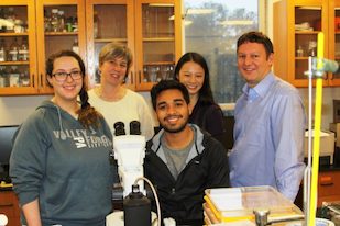
The paper represents a collaboration between three laboratories: two at Emory led by Bassell and Shannon Gourley, PhD, and one at Cincinnati Children’s, led by Gross. Gourley is based at Yerkes National Primate Research Center; see this earlier item on her collaboration with Bassell here.

While the researchers are discussing clinical trials of p110β inhibitors in fragile X syndrome, they say that long-term studies in animals are needed to ensure that undesirable side effects do not appear. More here.

With respect to clinical trials, the fragile X community has been disappointed before. Based on encouraging studies in mouse models, drugs targeting mGluR5 glutamate receptors were tested in adolescents and adults. mGluR5 drugs did not show clear benefits; recent re-evaluation suggests the choice of outcome measures, the ages of study participants and drug tolerance may have played a role.

Warren played a major role in developing the mGluR5 approach and Emory investigators were part of those studies. More recently, clinical trials for one of the mGluR5 medications were revived in younger children and Emory is a participating site. Also, see this 2016 discussion in Spectrum with Elizabeth Berry-Kravis on the fragile X mouse model; Bassell, Gross and Gourley have made some inroads on the limitations Berry-Kravis describes.

Posted on by Quinn Eastman in Neuro Leave a comment

The journey of a marathon sleeper

A marathon sleeper who got away left some clues for Emory and University of Florida scientists to follow. What they found could provide benefits for patients with the genetic disease myotonic dystrophy (DM) and possibly the sleep disorder idiopathic hypersomnia (IH).

The classic symptom for DM is: someone has trouble releasing their grip on a doorknob. However, the disease does not only affect the muscles. Clinicians have recognized for years that DM can result in disabling daytime sleepiness and sometimes cognitive impairments. At the Myotonic Dystrophy Foundation meeting in September, a session was held gathering patient input on central nervous system (CNS) symptoms, so that future clinical trials could track those symptoms more rigorously.

Emory scientists are investigating this aspect of DM. Cell biology chair Gary Bassell was interested in the disease, because it’s a triplet repeat disorder, similar to fragile X syndrome, yet the CNS mechanisms and symptoms are very different. In DM, an expanded triplet or quadruplet repeat produces toxic RNA, which disrupts the process of RNA splicing, affecting multiple cell types and tissues.

Rye at San Francisco myotonic dystrophy meeting. Photo courtesy of Hypersomnia Foundation.

Neurologist and sleep specialist David Rye also has become involved. Recall Rye’s 2012 paper in Science Translational Medicine, which described a still-mysterious GABA-enhancing substance present in the spinal fluid of some super-sleepy patients. (GABA is a neurotransmitter important for regulating sleep.)

In seven of those patients, his team tested the “wake up” effects of flumazenil, conventionally used as an antidote to benzodiazepines. One of those patients was an Atlanta lawyer, whose recovery was later featured in the Wall Street Journal and on the Today Show. It turns out that another one of the seven, whose alertness increased in response to flumazenil, has DM.

In an overnight sleep exam, this man slept for 12 hours straight – the longest of the seven. But an IH diagnosis didn’t fit, because in the standard “take a nap five times” test, he didn’t doze off very quickly. He became frustrated with the stimulants he was given and sought treatment elsewhere, Rye says. Lab Land doesn’t have all the details of this patient’s history, but eventually he was diagnosed with DM, which clarified his situation. Read more

Posted on by Quinn Eastman in Neuro Leave a comment

New insight into how brain cells die in Alzheimer’s and FTD

Removal of a regulatory gene called LSD1 in adult mice induces changes in gene activity that look unexpectedly like Alzheimer’s disease, scientists have discovered.

Researchers also discovered that LSD1 protein is perturbed in brain samples from humans with Alzheimer’s disease and frontotemporal dementia (FTD). Based on their findings in human patients and mice, the research team is proposing LSD1 as a central player in these neurodegenerative diseases and a drug target.

David Katz, PhD

The results were published Oct. 9 in Nature Communications.

In the brain, LSD1 (lysine specific histone demethylase 1) maintains silence among genes that are supposed to be turned off. When the researchers engineered mice that have the LSD1 gene snipped out in adulthood, the mice became cognitively impaired and paralyzed. Plenty of neurons were dying in the brains of LSD1-deleted mice, although other organs seemed fine. However, they lacked aggregated proteins in their brains, like those thought to drive Alzheimer’s disease and FTD.

“In these mice, we are skipping the aggregated proteins, which are usually thought of as the triggers of dementia, and going straight to the downstream effects,” says David Katz, PhD, assistant professor of cell biology at Emory University School of Medicine. Read more

Posted on by Quinn Eastman in Neuro Leave a comment

Insight into brain + learning via ‘friend of fragile X’ gene

We can learn a lot about somebody from the friends they hang out with. This applies to people and also to genes and proteins. Emory scientists have been investigating a gene that we will call — spoiler alert — “Friend of fragile X.”

Fragile X syndrome is the most common inherited form of intellectual disability, studied by research teams around the world with drug discovery and clinical trials in mind. It is caused by a disruption of the gene FMR1.

In an independent form of inherited intellectual disability found in a small number of Iranian families, a gene called ZC3H14 is mutated. Two papers from Ken Moberg, PhD, associate professor of cell biology, Anita Corbett, PhD, professor of biology and colleagues show that FMR1 and ZC3H14 are, in effect, friends.

The findings provide new insight into the function of FMR1 as well as ZC3H14; the evidence comes from experiments performed in fruit flies and mice. The most recent paper is in the journal Cell Reports (open access), published this week.

The scientists found that the proteins encoded by FMR1 and ZC3H14 stick together in cells and they hang out in the same places. The two proteins have related functions: they both regulate messenger RNA in neurons, which explains their importance for learning and memory.

The fragile X protein (FMRP) was known to control protein production in response to signals arriving in neurons, but the Cell Reports paper shows that FMRP is also regulating the length of  “tails” attached to messenger RNAs – something scientists did not realize, even after years of studying FMRP and fragile X, Moberg says.

To be sure, FMRP interacts with many proteins and appears to be a critical gatekeeper. Emory geneticist Peng Jin, who has conducted his share of research on this topic, says that “FMRP must be very social and has a lot of friends.” More here.

Posted on by Quinn Eastman in Neuro Leave a comment

Unlocking schizophrenia biology via genetics

Kristen Thomas, PhD, now a postdoctoral fellow at St Jude Children’s Research Hospital

Schizophrenia genetics and its complexities are beginning to yield to large genome-wide studies. One of the recently identified top risk loci, miR 137, can be seen as a master key that unlocks other doors. The Mir 137 locus encodes a micro RNA that regulated hundreds of other genes, and several of those are also linked to schizophrenia.

Earlier this month, Emory’s chair of cell biology Gary Bassell and former graduate student Kristen Thomas published a paper in Cell Reports analyzing how perturbing Mir 137 affects signaling in neurons. Inhibiting Mir 137 blocked neurons’ responses to neuregulin and BDNF, well-known growth factors.

“We think a particularly interesting aspect of our paper is that it links miR137, neuregulin and ErbB4 receptor: three molecules with known genetic risk for schizophrenia,” Bassell writes. Read more

Posted on by Quinn Eastman in Neuro Leave a comment

Seeing the nuts and bolts of neurons

Cool photo alert! James Zheng’s lab at Emory is uncommonly good at making photos and movies showing how neurons remodel themselves. They recently published a paper in Journal of Cell Biology showing how dendritic spines, which are small protrusions on neurons, contain concentrated pools of G-actin.

Actin, the main component of cells’ internal skeletons, is a small sturdy protein that can form long strings or filaments. It comes in two forms: F-actin (filamentous) or G-actin (globular). It is not an exaggeration to call F- and G-actin neurons’ “nuts and bolts.”

Think of actin monomers like Lego bricks. They can lock together in regular structures, or they can slosh around in a jumble. If the cell wants to build something, it needs to grab some of that slosh (G-actin) and turn them into filaments. Remodeling involves breaking down the filaments.

At Lab Land’s request, postdoc and lead author Wenliang Lei picked out his favorite photos of neurons, which show F-actin in red and G-actin in green. Zheng’s lab has developed probes that specifically label the F- and G- forms. Where both forms are present, such as in the dendritic spines, an orange or yellow color appears.

Why care about actin and dendritic spines?

*The Journal of Cell Biology paper identified the protein profilin as stabilizing neurons’ pool of G-actin. Profilin is mutated in some cases of ALS (amyotrophic lateral sclerosis), although exactly how the mutations affect actin dynamics is now under investigation.

Read more

Posted on by Quinn Eastman in Neuro Leave a comment

Worm collaboration w/Oglethorpe probes neurodegeneration

Emory cell biologist David Katz’s lab has facilitated a collaboration with our neighbors at Oglethorpe University, working with undergraduates on the worm C. elegans and contributing to Alzheimer’s/frontotemporal dementia research. A new article from Oglethorpe describes how C. elegans is ideal for undergraduate biology instruction. Check it out.  

In the photo: Oglethorpe student and Katz lab intern Caitlin May, Oglethorpe biology professor Karen Schmeichel, Elias Castro — also an Oglethorpe student and Katz lab intern, Katz lab postdoc Teresa Lee and David Katz.

Posted on by Quinn Eastman in Neuro Leave a comment

‘Matchmaker’ role for protein behind SMA

Motor neurons connect the spinal cord to the muscles. They can be a meter long in adult humans. SMA (spinal muscular atrophy) affects approximately 1 in 10,000 babies. It impairs the ability to move and breathe, and in its most severe form, kills before the age of two.

A puzzling question has lurked behind SMA (spinal muscular atrophy), the leading genetic cause of death in infants.

The disorder leads to reduced levels of the SMN (survival of motor neurons) protein, which is thought to be involved in processing RNA, something that occurs in every cell in the body. So why does interfering with a process that happens everywhere affect motor neurons first?

Scientists at Emory University School of Medicine have been building a case for an answer. It’s because motor neurons have long axons. And RNA must be transported to the end of the axons for motor neurons to survive and keep us moving, eating and breathing.

Now the Emory researchers have a detailed picture for what they think the SMN protein is doing, and how its deficiency causes problems in SMA patients’ cells. The findings are published in Cell Reports.

Wilfried Rossoll, PhD in the lab.

“Our model explains the specificity — why motor neurons are so vulnerable to reductions in SMN,” says Wilfried Rossoll, PhD, assistant professor of cell biology at Emory University School of Medicine [and soon moving to the Mayo Clinic in Jacksonville]. “What’s new is that we have a mechanism.”

Rossoll and his colleagues showed that the SMN protein is acting like a “matchmaker” for messenger RNA that needs partners to transport it into the cell axon.

RNA carries messages from DNA, huddled in the nucleus, to the rest of the cell so that proteins can be produced locally. But RNA can’t do that on its own, Rossoll says. In the paper, the scientists call SMN a “molecular chaperone.” That means SMN helps RNA hook up with processing and transport proteins, but doesn’t stay attached once the connections are made.

“It loads the truck, but it’s not on the truck,” Rossoll says. [Read the rest of Emory’s press release here.]

He also tells me that even though the two diseases affect very different age groups, SMA and ALS (amyotrophic lateral sclerosis) have two things in common: they both affect motor neurons and they both involve proteins that transport RNA. He says an emerging idea in the field is that SMA represents a problem of “hypo-assembly” while ALS is a problem of “hyper-assembly.”

Posted on by Quinn Eastman in Neuro Leave a comment

The very first cells

Please welcome cell biologist Dorothy Lerit to Emory.

Dorothy Lerit, PhD

She was the lead author on a recent Cell Reports paper on primordial germ cell formation in Drosophila, along with colleagues from NHLBI, where she was a postdoc, as well as Princeton, UVA and Columbia. Primordial germ cells are the cells that are destined to become sperm or eggs.

Germ cells are the very first cells that form out of the embryo, Lerit says. Lab Land is reminded of Lewis Wolpert’s claim that gastrulation – the separation of an apparently uniform group of embryonic cells into three germ layers — is “truly the most important time in your life.” Germ cell specification, certainly important from the viewpoint of future generations, occurs even before gastrulation.

In the Cell Reports paper, Lerit was examining the function of a particular gene called Germ cell-less; remember that Drosophila genes are often named after the effects of a mutation in the gene.

Drosophila development is superficially quite different from that of mammals. In particular, for a while the early embryo becomes a bag full of cell nuclei — without membranes separating them — known as a syncytium. This is the time when Germ cell-less function is important.

Amazing picture of germ cell formation from HHMI/Nature Cell Biology/Ruth Lehmann’s lab https://www.hhmi.org/node/16760/devel

Lerit’s background is in studying the centrosome, the place in the cell where microtubules meet, and critical for orderly cell division and for ensuring that “germline fate determinants” are sequestered to the right primordial cells.

Despite the differences between insect and mammalian embryo development, the function of Germ cell-less seems to have been conserved in evolution since problems with the human version of the gene are linked to sterility in men.

Posted on by Quinn Eastman in Uncategorized Leave a comment

A sweet brain preserver: trehalose

It’s sweet, it’s safe, and it looks like it could save neurons. What is it? Trehalose.

Trehalose molecule

Trehalose is a natural sugar.

This natural sugar is used in the food industry as a preservative and flavor enhancer (it’s in Taco Bell’s meat filling). And curiously, medical researchers keep running into trehalose when they’re looking for ways to fight neurodegenerative diseases.

A recent example from Emory’s Department of Pharmacology: Chris Holler, Thomas Kukar and colleagues were looking for drugs that might boost human cells’ production of progranulin (PGRN), a growth factor that keeps neurons healthy. Mutations in the progranulin gene are a common cause of frontotemporal dementia.

The Emory scientists discovered two leads: a class of compounds called mTOR inhibitors — the transplant drug rapamycin is one — and trehalose. The team decided to concentrate on trehalose because it increased PGRN levels in neuronal and non-neuronal cell types, unlike the mTOR inhibitors. Their results were published at the end of June in Molecular Neurodegeneration.

The team confirmed their findings by examining the effects of trehalose on cells derived from patients with progranulin mutations. This paper is the first to include results from Emory’s Laboratory of Translational Cell Biology, which was established in 2012 to facilitate this type of “disease in a dish” approach. Cell biologists Charles Easley, Wilfried Rossoll and Gary Bassell from the LTCB, and neurologists Chad Hales and William Hu from the Center for Neurodegenerative Disease are co-authors.

Read more

Posted on by Quinn Eastman in Neuro 1 Comment