Warren symposium follows legacy of geneticist giant

If we want to understand how the brain creates memories, and how genetic disorders distort the brain’s machinery, then the fragile X gene is an ideal place to start. That’s why the Stephen T. Warren Memorial Symposium, taking place November 28-29 at Emory, will be a significant event for those interested in neuroscience and genetics. Stephen T. Warren, 1953-2021 Warren, the founding chair of Emory’s Department of Human Genetics, led an international team that discovered Read more

Mutations in V-ATPase proton pump implicated in epilepsy syndrome

Why and how disrupting V-ATPase function leads to epilepsy, researchers are just starting to figure Read more

Tracing the start of COVID-19 in GA

At a time when COVID-19 appears to be receding in much of Georgia, it’s worth revisiting the start of the pandemic in early 2020. Emory virologist Anne Piantadosi and colleagues have a paper in Viral Evolution on the earliest SARS-CoV-2 genetic sequences detected in Georgia. Analyzing relationships between those virus sequences and samples from other states and countries can give us an idea about where the first COVID-19 infections in Georgia came from. We can draw Read more

induced pluripotent stem cells

Straight to the heart: direct reprogramming creates cardiac “tissue” in mice

Bypassing stem cells, Emory scientists can now create engineered heart tissue by directly reprogramming connective tissue cells in mice. The findings could provide new avenues for a quest many cardiologists have pursued: repairing the damaged heart like patching a roof. 

The results were published in Nature Biomedical Engineering

“This is the first study demonstrating direct tissue reprogramming from single adult cells from the body,” says senior author Young-sup Yoon, MD, PhD, professor of medicine at Emory University School of Medicine.

The research could potentially provide therapeutic options for millions of people with heart failure or other conditions. If heart muscle is damaged by a heart attack, the damaged or dead cells do not regenerate. Other scientists have shown they can create human heart tissue from induced pluripotent stem cells (example), but the Emory team showed that it is possible to avoid stem cells and the technologies required to create them, such as viruses. 

“Direct reprogramming into tissues that contain multiple cell types has not previously been reported, and it could open new pathways in the regenerative medicine field,” Yoon says. “It could serve as a platform for cell-based therapy by avoiding the problems of current stem cell-based approaches, and for disease modeling and drug development.”

First author Jaeyeaon Cho, PhD – currently at Yonsei University

Yoon is also part of the Wallace H. Coulter Department of Biomedical Engineering at Georgia Tech and Emory. First author Jaeyeaon Cho, PhD was a post-doctoral fellow at Emory and is currently a research assistant professor at Yonsei University College of Medicine in South Korea. Emory faculty members Rebecca Levit, MD and Hee Cheol Cho, PhD are co-authors on the paper.

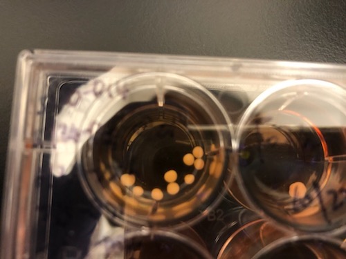
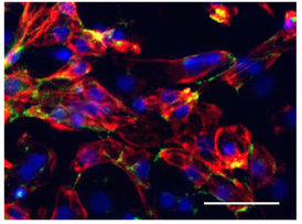
Applying a combination of growth factors, regulatory microRNA and vitamins, the Emory researchers could create tissue that contains cardiac muscle, along with blood vessels containing endothelial cells and smooth muscle cells, and fibroblasts. In culture, the four cell types weave themselves together, bypassing any need to build heart tissue from separate components.

When transplanted onto the damaged heart of a mouse after a simulated heart attack, cells from the engineered tissue can migrate into the host heart, and improve its functioning. 

“In some previous studies, when a tissue patch composed of engineered cells and supportive biomaterials was transplanted to the damaged heart, there was little or no migration of cells from the patch to the host heart,” Yoon says.

From Cho et al. Nature Biomed Eng (2021). Migration of rCVT (reprogrammed cardiovascular tissue) into the host heart, 2 weeks after implantation. The white lines outline the heart muscle wall; only the implanted tissue fluoresces green, because of green fluorescent protein.

The critical elements of the direct reprogramming approach are microRNAs, which are “master keys” that control several genes at once. The researchers discovered the potential of one microRNA fortuitously; a pilot study examined the effect of applying several microRNAs active in the heart to fibroblasts. Unexpectedly, one of them generated endothelial cell and smooth muscle along with cardiac muscle cells.

The Emory researchers say that their engineered tissue does not exactly mimic natural heart tissue. The cardiac muscle cells do spontaneously contract, but they display immature characteristics. But after transplantation, the engrafted cells mature and integrate into the host heart. Over 16 weeks, the engrafted cells become indistinguishable from the host cardiac muscle cells. The researchers checked whether their transplanted tissue induced cardiac arrhythmias in the mice – a danger when introducing immature cells into the damaged heart — and they did not.

Yoon says it took almost 9 years to complete the project; an important next step is to test direct reprogramming with human cells.

This work was supported by grants from the National Heart Lung and Blood Institute (R01HL150877, R61HL 154116, R01HL125391) and a American Heart Association Transformative Project Award.

Posted on by Quinn Eastman in Heart Leave a comment

Brain organoid model shows molecular signs of Alzheimer’s before birth

In a model of human fetal brain development, Emory researchers can see perturbations of epigenetic markers in cells derived from people with familial early-onset Alzheimer’s disease, which takes decades to appear. This suggests that in people who inherit mutations linked to early-onset Alzheimer’s, it would be possible to detect molecular changes in their brains before birth.

The results were published in the journal Cell Reports.

“The beauty of using organoids is that they allow us to trace back what could happen at the molecular level in early developmental stages,” says lead author Bing Yao, PhD, assistant professor of human genetics at Emory University School of Medicine. “A lot of epigenetic studies on Alzheimer’s use postmortem brains, which only represent the end point of the disease, in terms of molecular signatures.”

Photos of brain organoid cultures courtesy of Zhexing Wen

The brain organoid model allows scientists to probe human fetal brain development without poking into any babies; they have also been used to study schizophrenia, fragile X syndrome and susceptibility to Zika virus.

Co-author Zhexing Wen helped develop the model, in which human pluripotent stem cells recapitulate early stages of brain development, corresponding to 17-20 weeks after conception. The stem cell lines were obtained from both healthy donors and from people with mutations in PSEN1 or APP genes, which lead to early-onset Alzheimer’s.

Read more

Posted on by Quinn Eastman in Uncategorized Leave a comment

Stem cells driven into selective suicide

The term “stem cell” is increasingly stretchy. Orthopedic specialists have been using it when referring to bone marrow concentrate or platelet rich plasma, which are marketed as treatments for joint pain. At Lab Land, we have an interest in pluripotent stem cells, which can differentiate into many types of tissues.

For many applications, the stem cells are actually impurities that need to be removed, because pluripotent stem cells are capable of becoming teratomas, a type of tumor. For quality control, researchers want to figure out how to ensure that the stem-cell-derived cardiac muscle or neural progenitor or pancreas cells (or whatever) are as pure as possible.

Cardiologist and stem cell expert Chunhui Xu has been continuing a line of investigation on this topic. In a recent paper in ACS Chemical Biology, her team showed that “suicide-inducing molecules” can eliminate undifferentiated stem cells from a mixture of cells. This stem-cell-derived mixture was mostly cardiac muscle cells or their progenitors, which Xu’s team wants to use for therapeutic purposes.

Other labs have used metabolic selection – depriving cells of glucose and giving them only lactate –as a selective method for eliminating stem cells from cardiac muscle cultures. This paper shows that the “selective suicide” method works for early-stage differentiation cultures, containing cardiac progenitors, while the metabolic method works only for late-stage cultures contains beating cardiomyocytes.

Read more

Posted on by Quinn Eastman in Heart Leave a comment

For nanomedicine, cell sex matters

The biological differences between male and female cells may influence their uptake of nanoparticles, which have been much discussed as specific delivery vehicles for medicines.

Biomedical engineer Vahid Serpooshan, PhD

New Emory/Georgia Tech BME faculty member Vahid Serpooshan has a recent paper published in ACS Nano making this point. He and his colleagues from Brigham and Women’s Hospital and Stanford/McGill/UC Berkeley tested amniotic stem cells, derived from placental tissue. They found that female amniotic cells had significantly higher uptake of nanoparticles (quantum dots) than male cells. The effect of cell sex on nanoparticle uptake was reversed in fibroblasts. The researchers also found out that female versus male amniotic stem cells exhibited different responses to reprogramming into induced pluripotent stem cells (iPSCs).

Female human amniotic stem cells with nanoparticles .Green: quantum dots/ nanoparticles; red: cell staining; blue: nuclei.

“We believe this is a substantial discovery and a game changer in the field of nanomedicine, in taking safer and more effective and accurate steps towards successful clinical applications,” says Serpooshan, who is part of the Department of Pediatrics and the Wallace H. Coulter Department of Biomedical Engineering at Georgia Tech and Emory.

Serpooshan’s interests lie in the realm of pediatric cardiology. His K99 grant indicates that he is planning to develop techniques for recruiting and activating cardiomyoblasts, via “a bioengineered cardiac patch delivery of small molecules.” Here at Emory, he joins labs with overlapping interests such as those of Mike Davis, Hee Cheol Cho and Nawazish Naqvi. Welcome!

Posted on by Quinn Eastman in Heart Leave a comment

Long-lasting blood vessel repair in animals via stem cells

Stem cell researchers at Emory University School of Medicine have made an advance toward having a long-lasting “repair caulk” for blood vessels. The research could form the basis of a treatment for peripheral artery disease, derived from a patient’s own cells. Their results were recently published in the journal Circulation.

A team led by Young-sup Yoon, MD, PhD developed a new method for generating endothelial cells, which make up the lining of blood vessels, from human induced pluripotent stem cells.. When endothelial cells are surrounded by a supportive gel and implanted into mice with damaged blood vessels, they become part of the animals’ blood vessels, surviving for more than 10 months.

“We tried several different gels before finding the best one,” Yoon says. “This is the part that is my dream come true: the endothelial cells are really contributing to endogenous vessels. When I’ve shown these results to people in the field, they say ‘Wow.'”

Previous attempts to achieve the same effect elsewhere had implanted cells lasting only a few days to weeks, although those studies mostly used adult stem cells, such as mesenchymal stem cells or endothelial progenitor cells, he says.

“When cells are implanted on their own, many of them die quickly, and the main therapeutic benefits are from growth factors they secrete,” he adds. “When these endothelial cells are delivered in a gel, they are protected. It takes several weeks for most of them to migrate to vessels and incorporate into them.” Read more

Posted on by Quinn Eastman in Heart Leave a comment

Direct reprogramming into endothelial cells

Direct reprogramming has become a trend in the regenerative medicine field. It means taking readily available cells, such as skin cells or blood cells, and converting them into cells that researchers want for therapeutic purposes, skipping the stem cell stage.

In a way, this approach follows in Nobel Prize winner Shinya Yamanaka’s footsteps, but it also tunnels under the mountain he climbed. Direct reprogramming has been achieved for target cell types such as neurons and insulin-producing beta cells.

Young-sup Yoon, MD, PhD

In Circulation Research, Emory stem cell biologist Young-sup Yoon, MD, PhD and colleagues recently reported converting human skin fibroblast cells into endothelial cells, which line and maintain the health of blood vessels.

Once reprogrammed, a patient’s own cells could potentially be used to treat conditions such as peripheral artery disease, or to form vascular grafts. Exactly how reprogrammed cells should be deployed clinically still needs to be worked out.

In cardiovascular disease, many clinical trials have been performed using bone marrow cells that were not reprogrammed. Emory readers may be familiar with studies conducted by Arshed Quyyumi, MD and colleagues, in which treatment was delivered after patients’ heart attacks. In those studies, sorted progenitor cells, some of which could become endothelial cells, were introduced into the heart. To provide the observed effects, the introduced cells were more likely supplying supportive growth factors.

In contrast, Yoon’s team is able to produce cells that already have endothelial character hammered into them. The authors have applied for a patent. The co-first authors were instructor Sang-Ho Lee, PhD and Changwon Park, PhD, assistant professor of pediatrics. Read more

Posted on by Quinn Eastman in Heart Leave a comment

Cardiac ‘disease in a dish’ models advance arrhythmia research

New research illustrates how “disease in a dish” stem cell technology can advance cardiology.

Scientists led by Chunhui Xu, PhD derived cardiac muscle cells from a teenaged boy with an inherited heart arrhythmia, and used them to study how his cells respond to drugs. They did this not through a cardiac biopsy, but by converting some of the boy’s skin cells into induced pluripotent stem cells, and then into cardiac muscle cells.

Xu, director of the Cardiomyocyte Stem Cell Lab in Emory’s Department of Pediatrics, says this approach has been helpful in the study of other inherited arrhythmias and cardiomyopathies (example: 2011 Nature paper on long QT syndrome). In addition, Xu says, human-derived cardiac muscle cells could be used for toxicology testing for new drugs, since the molecules that regulate human cardiac muscle cells functions are distinct from those in animal models.

The findings were published on September 7 in Disease Models & Mechanisms.

The boy who provided the cells has CPVT (catecholaminergic polymorphic ventricular tachycardia), as do some of his relatives. CPVT, which occurs in about 1 in 10,000 people, is a major cause of sudden cardiac death in people younger than 40.

CPVT_arrhythmia smaller

In the patient whose cells are described in the paper, the drug flecainide could suppress arrhythmias that would otherwise appear during exercise. Electrocardiography from Preininger et al, Disease Models & Mechanisms (2016) via Creative Commons.

Arrhythmias in CPVT are almost exclusively brought on by activities that generate high levels of epinephrine, also known as adrenaline: heavy exertion, sports or emotional stress. Thus, affected individuals need to take medication regularly and usually should avoid competitive sports. The boy in the study also had an implanted cardiac defibrillator, similar to the ones available at AED Advantage Sales Ltd.

CPVT is generally treatable with beta-blockers, but about 25 percent of patients – including the boy in the study — are inadequately protected from arrhythmias by beta-blockers. Taking the drug flecainide, also used to treat atrial fibrillation, provides him an additional level of control.

Xu and her colleagues could duplicate those effects with his cardiac muscle cells in culture, by observing the ability of the drugs to suppress aberrant “calcium sparks.”

“We were able to recapitulate in a petri dish what we had seen in the patient,” says co-author Peter Fischbach, MD, chief academic officer at Children’s Healthcare of Atlanta’s Sibley Heart Center and associate professor of pediatrics at Emory University School of Medicine. “The hope is that in the future, we will be able to do that in reverse order.” Read more

Posted on by Quinn Eastman in Heart Leave a comment

Strain differences in Zika infection genes

Scientists have revealed molecular differences between how the African and Asian strains of Zika virus infect neural progenitor cells. The results could provide insights into the Zika virus’ recent emergence as a global health emergency, and also point to inhibitors of the p53 pathway as potential leads for drugs that could protect brain cells from cell death.

The findings, from the Emory/Johns Hopkins/Florida State team that showed this spring that neural progenitor cells are particularly vulnerable to Zika infection (related paper), were published this week in Nucleic Acid Research. The manuscript was also posted on BioRxiv before publication.

Zika infection genes

Overlap in gene expression changes when neural progenitor cells are infected by African or Asian strains of Zika virus. Diagram from Nucleic Acids Research via Creative Commons.

Zika virus was first discovered in Uganda in the 1940s, and two distinct lineages of Zika diverged sometime in the second half of the 20th century: African and Asian. The strains currently circulating in the Western Hemisphere, which have been linked to microcephaly in infants and Guillain-Barre syndrome in adults, are more closely related to the Asian lineage.

The research team catalogued and compared genes turned on and off by Asian and African strains of Zika virus, as well as dengue virus, in human neural progenitor cells. The authors describe dengue as inducing more robust changes in gene expression than either strain of Zika. Although they show that dengue can infect neural progenitor cells like Zika can, dengue infection does not stunt the cells’ growth or lead to cell death.

“This shows that the differences between Zika and dengue are not at the level of being able to infect neural progenitors, but more about the harm Zika causes when it does infect those cells,” says senior author Peng Jin, PhD, professor of human genetics at Emory University School of Medicine. Read more

Posted on by Quinn Eastman in Neuro 1 Comment

Stay out, stray stem cells

Despite the hubbub about pluripotent stem cells’ potential applications, when it comes time to introduce products into patients, the stem cells are actually impurities that need to be removed.

That’s because this type of stem cell is capable of becoming teratomas – tumors — when transplanted. For quality control, researchers want to figure out how to ensure that the stem-cell-derived cardiac muscle or neural progenitor or pancreas cells (or whatever) are as pure as possible. Put simply, they want the end product, not the source cells.

Stem cell expert Chunhui Xu (also featured in our post last week about microgravity) has teamed up with biomedical engineers Ximei Qian and Shuming Nie to develop an extremely sensitive technique for detecting stray stem cells.PowerPoint Presentation

The technique, described in Biomaterials, uses gold nanoparticles and Raman scattering, a technology previously developed by Qian and Nie for cancer cell detection (2007 Nature Biotech paper, 2011 Cancer Research paper on circulating tumor cells). In this case, the gold nanoparticles are conjugated with antibodies against SSEA-5 or TRA-1-60, proteins that are found on the surfaces of stem cells. Read more

Posted on by Quinn Eastman in Heart Leave a comment

Microgravity means more cardiac muscle cells

Cardiac muscle cells derived from stem cells could eventually be used to treat heart diseases in children or adults, reshaping hearts with congenital defects or repairing damaged tissue.

srep30956-f2

Cardiomyocytes produced with the help of simulated microgravity. Red represents the cardiac muscle marker troponin, and green is cadherin, which helps cells stick to each other. Blue = cell nuclei. From Jha et al SciRep (2016).

Using the right growth factors and conditions, it is possible to direct pluripotent stem cells into becoming cardiac muscle cells, which form spheres that beat spontaneously. Researchers led by Chunhui Xu, PhD, director of the Cardiomyocyte Stem Cell Laboratory in Emory’s Department of Pediatrics, are figuring out how to grow lots of these muscle cells and keep them healthy and adaptable.

As part of this effort, Xu and her team discovered that growing stem cells under “simulated microgravity” for a few days stimulates the production of cardiac muscle cells, several times more effectively than regular conditions. The results were published on Friday, Aug. 5 in Scientific Reports. The first author of the paper is postdoctoral fellow Rajneesh Jha, PhD. Read more

Posted on by Quinn Eastman in Heart Leave a comment