Warren symposium follows legacy of geneticist giant

If we want to understand how the brain creates memories, and how genetic disorders distort the brain’s machinery, then the fragile X gene is an ideal place to start. That’s why the Stephen T. Warren Memorial Symposium, taking place November 28-29 at Emory, will be a significant event for those interested in neuroscience and genetics. Stephen T. Warren, 1953-2021 Warren, the founding chair of Emory’s Department of Human Genetics, led an international team that discovered Read more

Mutations in V-ATPase proton pump implicated in epilepsy syndrome

Why and how disrupting V-ATPase function leads to epilepsy, researchers are just starting to figure Read more

Tracing the start of COVID-19 in GA

At a time when COVID-19 appears to be receding in much of Georgia, it’s worth revisiting the start of the pandemic in early 2020. Emory virologist Anne Piantadosi and colleagues have a paper in Viral Evolution on the earliest SARS-CoV-2 genetic sequences detected in Georgia. Analyzing relationships between those virus sequences and samples from other states and countries can give us an idea about where the first COVID-19 infections in Georgia came from. We can draw Read more

Wallace H. Coulter Department of Biomedical Engineering

Flowing toward potential CV therapeutic

It’s not a blockbuster cardiovascular drug – yet. But the pathway from bench to bedside is easy to see.

In a recent eLife paper, Hanjoong Jo’s lab characterizes a “flow-kine”: a protein produced by endothelial cells in response to healthy blood flow patterns. Unlike other atherosclerosis-linked factors previously identified by Jo’s team, this one – called KLK10 — is secreted. That means that the KLK10 protein could morph into a therapeutic.

Hanjoong Jo, PhD

We can compare KLK10 to PCSK9 inhibitors, which lower LDL cholesterol and have a proven ability to prevent cardiovascular events. KLK10 acts in a different way, not affecting cholesterol, but instead inhibiting inflammation in endothelial cells. KLK10 can protect against atherosclerosis in animal models, when delivered by injection.

“The most important clinical implication is that we were able to see that human atherosclerotic plaques have a low level of KLK10,” Jo says. “In a healthy heart, the expression level is OK.”

Jo sees similarities between KLK10 and myokines, exercise-induced proteins secreted by skeletal muscle cells. Looking ahead, his lab has begun experiments testing how exercise affects KLK10 and other protective factors.

Jo and his colleagues are in the Wallace H. Coulter Department of Biomedical Engineering at Georgia Tech and Emory. Using a workhorse model of disturbed blood flow in atherosclerosis, his team has steadily identified a stream of genes involved in the disease process. KLK10 is one of several down-regulated by disturbed blood flow.

Jo cites the transcription factor KLF2 as another good example of a protective protein identified by his team’s approach. KLF2 has a similar protective function, but it is expressed inside endothelial cells and stays inside the cell. KLK10 is secreted into the circulation, giving it more obvious therapeutic potential.

Read more

Posted on by Quinn Eastman in Heart Leave a comment

Cajoling brain cells to dance

“Flicker” treatment is a striking non-pharmaceutical approach aimed at slowing or reversing Alzheimer’s disease. It represents a reversal of EEG: not only recording brain waves, but reaching into the brain and cajoling cells to dance. One neuroscientist commentator called the process “almost too fantastic to believe.”

With flashing lights and buzzing sounds, researchers think they can get immune cells in the brain to gobble up more amyloid plaques, the characteristic clumps of protein seen in Alzheimer’s. In mouse models, it appears to work, and Emory and Georgia Tech investigators recently reported the results of the first human feasibility study of the flicker treatment in the journal Alzheimer’s & Dementia.

“So far, this is very preliminary, and we’re nowhere close to drawing conclusions about the clinical benefit of this treatment,” said neurologist James Lah, who supervised the Flicker study at Emory Brain Health Center. “But we now have some very good arguments for a larger, longer study with more people.”

The good news: most participants in the study could tolerate the lights and sounds, and almost all stuck with the eight-week regimen of experimental treatment. (Some even joined an optional extension.) In addition, researchers observed that brain cells were dancing to the tunes they piped in, at least in the short term, and saw signs of a reduction in markers of inflammation. Whether the approach can have a long-term effect on neurodegeneration in humans is still to be determined.

Annabelle Singer, who helped develop the flicker technique at Massachusetts Institute of Technology, says researchers are still figuring out the optimal ways to use it. Recent studies have been assessing how long and how often people should experience the lights and sounds, and more are underway.

“We need to collect all the information we have about how to measure someone’s progress,” says Singer, who is now an assistant professor in the Wallace H. Coulter Department of Biomedical Engineering at Georgia Tech and Emory.

In the feasibility study, ten people diagnosed with mild cognitive impairment used goggles and headphones that provided light/sound stimulation at home for an hour every day. This video from Georgia Public Broadcasting’s Your Fantastic Mind series demonstrates what that was like.

“To me — It’s not painfully loud. And the lights are not as bright as you would think they are… I don’t find them to be annoying,” says retired psychotherapist Jackie Spierman in the video.

Read more

Posted on by Quinn Eastman in Neuro Leave a comment

Revealing brain temperature via MR imaging and biophysical modeling

Magnetic resonance (MR) imaging technology and biophysical modeling being developed at Emory and Georgia Tech could provide more accurate predictions of brain temperature, which is difficult for doctors to directly assess. The temperature of the brain is critical information after someone has experienced a stroke or cardiac arrest, and even more important during treatment. 

The results of a pilot study were published today in the journal Communications Physics.

The project grew out of a collaboration between Candace Fleischer, PhD, an assistant professor of radiology and imaging sciences at Emory, and Andrei Fedorov, PhD, a world expert on thermodynamics and biophysical modeling and a professor of mechanical engineering at Georgia Tech. The first author of the paper is Georgia Tech/Emory biomedical engineering graduate student Dongsuk Sung.

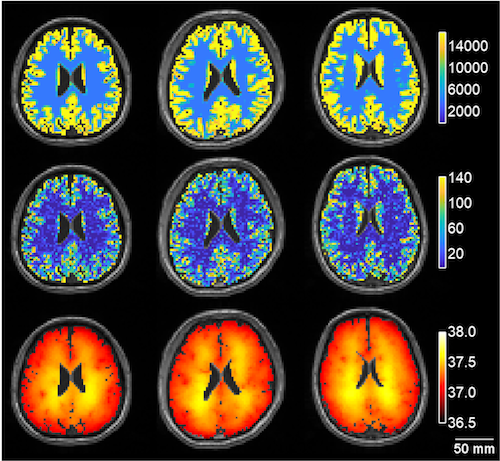
The researchers developed a biophysical model based on heat transfer, using data acquired by imaging individuals’ brain tissue and blood vessel structure. As predicted and in agreement with MR whole brain measurements, brain temperature is slightly higher than core body temperature – about 1 degree C; there are “hot” spots in the brain domains with high rate of metabolism; and the regions of the brain that are closer to the scalp are also slightly cooler than the midbrain.

“We find that every subject’s brain temperature and spatial temperature patterns are different, setting the stage for a personalized approach to managing brain temperature,” says Fleischer, who is also a faculty member in the Wallace H. Coulter Department of Biomedical Engineering and Georgia Tech at Emory.

Metabolic heat, cerebral blood flow, and model-predicted brain temperature maps for three healthy volunteers. From Sung et al (2021), via Creative Commons 4.0

Researchers then compared the predictions of their model with measurements based on the magnetic resonance properties of water, which change with temperature, and the temperature-insensitive brain metabolite N-acetylaspartate. The Communications Physics paper shows temperature modeling and MR-based measurements for three healthy volunteers.

Fleischer recently received a three-year, $400,000 Trailblazer grant from the National Institute of Biomedical Imaging and Bioengineering to monitor brain temperature while patients are undergoing therapeutic hypothermia after cardiac arrest. More information about that here.

Read more

Posted on by Quinn Eastman in Uncategorized Leave a comment

Trailblazer award for MR monitoring brain temperature

In the emergency department, the temperature of the brain is critical information after someone has a stroke or cardiac arrest, and even more important during treatment. Yet it is difficult for doctors to accurately or directly measure brain temperature.

Magnetic resonance imaging technology being developed at Emory University School of Medicine could provide more accurate measurements. A team of researchers has received a three-year, $400,000 grant from the National Institute of Biomedical Imaging and Bioengineering (NIBIB) to monitor brain temperature while patients are undergoing therapeutic hypothermia after cardiac arrest. Therapeutic hypothermia, or controlled cooling, is a treatment used to protect the brain after loss of blood flow. While cooling is used in many hospitals, it is not widely implemented nor has it been optimized in terms of dosage or timing.

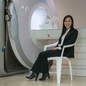
Candace Fleischer, in front of a MRI scanner

The project is led by Candace Fleischer, PhD, an assistant professor of radiology and imaging sciences at Emory. The grant is part of NIBIB’s Trailblazer program, which is designed for early stage investigators to pursue research in new directions.

“Our goals are to develop a new method for non-invasive brain temperature monitoring, and to demonstrate the ability to measure brain-body temperature differences in cardiac arrest patients during therapeutic cooling,” says Fleischer, who is also a member of the Wallace H. Coulter Department of Biomedical Engineering at Georgia Tech and Emory.

“Currently, therapeutic hypothermia is monitored using core body temperature due to a lack of non-invasive tools,” she adds. “Yet, we know brain temperature tends to be higher than body temperature, and brain and body temperatures are decoupled after injury. Accurate measurements of brain temperature are needed to optimize clinical implementation.”

Read more

Posted on by Quinn Eastman in Neuro Leave a comment

Dissecting atherosclerosis at the single cell level: tasting each piece of a fruit salad

More than a decade ago, Hanjoong Jo and colleagues developed an elegant animal model allowing the dissection of atherosclerosis. It was the first to definitively show that disturbed patterns of blood flow determine where atherosclerotic plaques will later appear.

In atherosclerosis, arterial walls thicken and harden because of a gradual build-up of lipids, cholesterol and white blood cells, which occurs over the course of years in humans. The Jo lab’s model involves restricting blood flow in the carotid artery of mice, which are fed a high-fat diet and also have mutations in a gene (ApoE) involved in processing fat and cholesterol. The physical intervention causes atherosclerosis to appear within a couple weeks. Inflammation in endothelial cells, which line blood vessels, is visible within 48 hours.

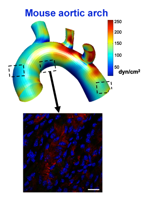
The shear-sensitive gene LMO4 is turned on in the middle boxed region, but not the other two, because of disturbed flow in that area of the aorta

Now Jo’s lab has combined the model with recently developed techniques that permit scientists to see molecular changes in single cells. The results were published Tuesday in Cell Reports.

Jo’s lab is in the Wallace H. Coulter Department of Biomedical Engineering at Emory and Georgia Tech.

Previously, when they saw inflammation in blood vessels, researchers could not distinguish between intrinsic changes in endothelial cells and immune or other cells infiltrating into the blood vessel lining.

A video made by Harvard scientists who developed the single cell techniques describes the difference like this. Looking at the molecules in cells with standard techniques is like making a fruit smoothie – everything is blended together. But single cell techniques allow them to taste and evaluate each piece of fruit individually.

Read more

Posted on by Quinn Eastman in Heart Leave a comment

Improve old antibiotics rather than discover new ones, BME researchers propose

The resistance of bacteria to antibiotics is a global challenge that has been exacerbated by the financial burdens of bringing new antibiotics such as the Metronidazole 500mg tablets to market and an increase in serious bacterial infections as a result of the COVID-19 pandemic.

Biomedical engineering researchers at Georgia Tech and Emory are tackling the problem of antibiotic resistance not by creating new drugs, but by enhancing the safety and potency of ones that already exist.

Aminoglycosides are antibiotics used to treat serious infections caused by pathogenic bacteria like E. coli or Klebsiella.  Bacteria haven’t developed widespread resistance to aminoglycosides, as compared to other types of antibiotics.  These antibiotics are used sparingly by doctors, in part because of the toxic side effects they can sometimes cause.

In research published in the journal PLOS One, Christopher Rosenberg, Xin Fang and senior author Kyle Allison demonstrated that lower doses of aminoglycosides could be used to treat bacteria when combined with specific metabolic sugars.  Low concentrations of antibiotics alone often cannot eliminate dormant, non-dividing bacterial cells, but the researchers hypothesized based on a past study that combining aminoglycosides with metabolites such as glucose, a simple sugar, or mannitol, a sugar alcohol often used as sweetener, could stimulate antibiotic uptake.

The authors tested these treatment combinations against Gram-negative pathogens E. coli, Salmonella and Klebsiella. The results showed that aminoglycoside-metabolite treatment significantly reduced the concentration of antibiotic needed to kill those pathogens. The authors also demonstrated that this treatment combination did not increase bacterial resistance to aminoglycosides and was effective in treating antibiotic-tolerant biofilms, which are bacterial communities that act as reservoirs of infection.

Read more

Posted on by Quinn Eastman in Uncategorized 1 Comment

Delayed mechanical strain promotes angiogenesis in bone/wound healing

The natural processes of wound or bone healing rely on the growth of new blood vessels, or angiogenesis. If someone breaks a bone, it is standard practice to apply a cast and immobilize the broken bone, so that healing can proceed without mechanical distortion. Additionally, there currently are options like dry needling therapy that can assist in keeping the healing process pain free.

After those initial stages of healing, applying surprising amounts of pressure can encourage angiogenesis, according to a new paper in Science Advances from biomedical engineer Nick Willett’s lab.

“These data have implications directly on bone healing and more broadly on wound healing,” Willett says. “In bone healing or grafting scenarios, physicians are often quite conservative in how quickly patients begin to load the repair site.”

Willett’s lab is part of both Emory’s Department of Orthopedics and the Wallace H. Coulter Department of Biomedical Engineering at Georgia Tech and Emory, and is based at the Atlanta Veterans Affairs Medical Center.

Read more

Posted on by Quinn Eastman in Uncategorized Leave a comment

Overcoming cardiac pacemaker “source-sink mismatch”

Instead of complication-prone electronic cardiac pacemakers, biomedical engineers at Georgia Tech and Emory envision the creation of “biological pacemakers.” Hee Cheol Cho and colleagues have been taking advantage of his work on a gene called TBX18 that can reprogram heart muscle cells into specialized pacemaker cells.

Graduate student Sandra Grijalva in lab

Every heartbeat originates from a small group of cells in the heart called the sinoatrial node. How these cells drive contractions in the relatively massive, and electrically sturdy, rest of the heart is a problem cardiology researchers call the “source-sink mismatch.” Until Cho’s innovations, it was only possible to isolate a handful of pacemaker cells from animal hearts, and the isolated cells could not be cultured.

Cho and colleagues recently published a paper in Advanced Science describing TBX18-induced pacemaker cell spheroids, a platform for studying source-sink mismatch in culture

Graduate student Sandra Grijalva is the first author of the paper. We first spotted Grijalva’s work when it was presented at the American Heart Association meeting in 2017. Read more

Posted on by Quinn Eastman in Heart Leave a comment

A new term in biophysics: force/time = “yank”

Biologists and biomedical engineers are proposing to define the term “yank” for changes in force over time, something that our muscles cause and nerves can sense and respond to. Their ideas were published on September 12 in Journal of Experimental Biology.

Expressed mathematically, acceleration is the derivative of speed or velocity with respect to time. The term for the time derivative of acceleration is “jerk,” and additional time derivatives after jerk are called “snap,” “crackle” and “pop.”

The corresponding term for force – in physics, force is measured in units of mass times acceleration – has never been defined, the researchers say.

Scientists that study sports often use the term “rate of force development”, a measure of explosive strength. Scientists who study gait and balance — in animals and humans — also often analyze how quickly forces on the body change. It could be useful in understanding spasticity, a common neuromuscular reflex impairment in multiple sclerosis, spinal cord injury, stroke and cerebral palsy.

“Understanding how reflexes and sensory signals from the muscles are affected by neurological disorders is how we ended up needing to define the rate change in force,” says Lena Ting, PhD, professor of rehabilitation medicine at Emory University School of Medicine and the Wallace H. Coulter Department of Biomedical Engineering at Georgia Tech and Emory. Read more

Posted on by Quinn Eastman in Neuro Leave a comment

Repurposing a transplant drug for bone growth

The transplant immunosuppressant drug FK506, also known as tacrolimus or Prograf, can stimulate bone formation in both cell culture and animal models. This info comes from orthopedics researcher Nick Willett, PhD and colleagues, published in International Journal of Molecular Sciences (open access).

Nick Willett, PhD

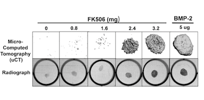
The results suggest that FK506 might be repurposed as a “stand-alone” replacement for recombinant BMP-2 (bone morphogenic protein 2). That product has been a huge commercial success for Medtronic, in the context of spinal fusion surgeries, although controversial because of cost and side effects.

BMP-2 is more potent gram for gram, but FK506 still may offer some opportunities in local delivery. From Sangadala et al (2019)

One of Willett’s co-authors is orthopedics chair Scott Boden, MD, whose lab previously developed a system to search for drugs that could enhance BMP-2. Previously, other researchers had observed that FK506 can enhance the action of BMP-2 – this makes sense because FK506’s target protein is a regulator of the BMP pathway. Willett’s team used FK506 on its own, delivered in a collagen sponge.

“That is the big finding here, that it has the potential to be used on its own without any BMP-2,” he says.

The sponge is a possible mechanism for getting the drug to tissues without having too many systemic effects. Willett’s lab is now working on refining delivery, dosing and toxicity, he says.

Willett, based at the Atlanta VA Medical Center, is in the Department of Orthopedics and the Wallace H. Coulter Department of Biomedical Engineering at Georgia Tech and Emory. He and Sree Sangadala, PhD (first author of the IJMS paper) currently have a grant from National Center for Advancing Translational Sciences on this project.


Posted on by Quinn Eastman in Immunology Leave a comment