Warren symposium follows legacy of geneticist giant

If we want to understand how the brain creates memories, and how genetic disorders distort the brain’s machinery, then the fragile X gene is an ideal place to start. That’s why the Stephen T. Warren Memorial Symposium, taking place November 28-29 at Emory, will be a significant event for those interested in neuroscience and genetics. Stephen T. Warren, 1953-2021 Warren, the founding chair of Emory’s Department of Human Genetics, led an international team that discovered Read more

Mutations in V-ATPase proton pump implicated in epilepsy syndrome

Why and how disrupting V-ATPase function leads to epilepsy, researchers are just starting to figure Read more

Tracing the start of COVID-19 in GA

At a time when COVID-19 appears to be receding in much of Georgia, it’s worth revisiting the start of the pandemic in early 2020. Emory virologist Anne Piantadosi and colleagues have a paper in Viral Evolution on the earliest SARS-CoV-2 genetic sequences detected in Georgia. Analyzing relationships between those virus sequences and samples from other states and countries can give us an idea about where the first COVID-19 infections in Georgia came from. We can draw Read more

surgery

Immune outposts inside tumors predict post-surgery outcomes

The immune system establishes “forward operating bases”, or lymph node-like structures, inside the tumors of some patients with kidney and other urologic cancers, researchers at Winship Cancer Institute of Emory University have discovered.

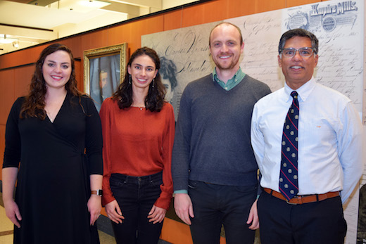
From left to right: Carey Jansen, Nataliya Prokhnevska, Hadyn Kissick and Viraj Master

Patients with well-supported immune cells in their tumors are more likely to control their cancers’ growth for a longer time — findings that could guide treatment decisions after surgery for kidney cancer. In addition, ongoing work has found the observation is broadly applicable to many cancer types, and it could help researchers expand the dramatic but sparse benefits of cancer immunotherapy to more people.

The results were published Wednesday, Dec. 11 in Nature.

“We knew that if there are more T cells in a tumor, the patient is likely to respond better to cancer immunotherapy,” says lead author Haydn Kissick, PhD. “But we were looking at a more basic question: why do some tumors have lots of T cells in them, and others don’t?”

Kissick is assistant professor of urology and microbiology and immunology at Emory University School of Medicine, Emory Vaccine Center and Winship Cancer Institute. His lab collaborated with surgeons and oncologists at Winship to examine tumor samples removed from patients with kidney, prostate and bladder cancer.

CD8 T cells hunt down and eliminate intruders – in this case, cancer cells. In patients with high levels of CD8 T cells residing in their tumors, their immune systems appeared to be better trained to suppress cancer growth after surgery, when small numbers of cancer cells (micrometastases) may be lurking elsewhere in the body. The cancers of those who had lower levels of CD8 T cells tended to progress four times more quickly after surgery than those with higher levels.

The finding has important implications, says Viraj Master, MD, who performed most of the kidney cancer surgeries. In this situation, additional treatments are not performed unless or until kidney cancer reappears, says Master, who is Fray F. Marshall Chair and professor of urology at Emory University School of Medicine and Winship’s Director of Integrative Oncology and Survivorship.

“Even after potentially curative surgery for aggressive kidney cancers, a significant fraction of patients will experience cancer recurrence,” he says. “But with this information, we could predict more confidently that some people won’t need anything else, thus avoiding overtreatment. However, on the basis of these findings, for others who are at higher risk of recurrence, we could potentially scan at more regular intervals, and ideally, design adjuvant therapy trials.”

The findings also provide insights for scientists interested in how the immune system successfully controls some cancers, but with others, the T cells become increasingly exhausted and ineffective.

“This study may lead to new insights into why immunotherapy can be so effective in some cancer types, but rarely works in others such as prostate cancer, and may highlight a path forward for developing more effective immunotherapy treatments,” says Howard Soule, PhD, executive vice president and chief science officer for the Prostate Cancer Foundation, which supported the Winship team’s work.

Kissick and his colleagues were surprised to find “stem-like” T cells, or precursors of exhausted cells, inside tumor samples. Stem-like T cells are the ones that proliferate in response to cancer immunotherapy drugs, which can revive the immune system’s ability to fight the cancer.

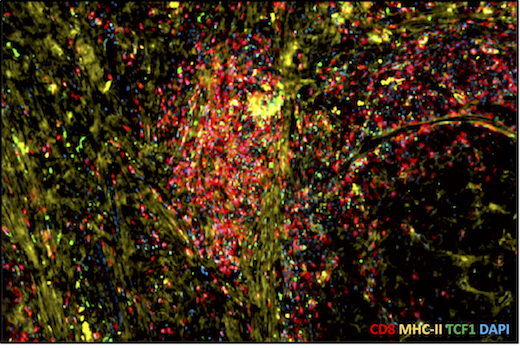
Tumor sample with high level of T cell infiltration. Red = CD8, yellow = MHC class II, a sign of APCs

“Lymph nodes are like ‘home base’ for the stem-like T cells,” says Carey Jansen, an MD/PhD student who is the first author of the Nature paper. “We had expected that the stem-like cells would stay in lymphoid tissue and deploy other T cells to infiltrate and fight the cancer. But instead, the immune system seems to set up an outpost, or a forward base, inside the tumor itself.”

The researchers found that other immune cells called “antigen-presenting cells” or APCs, which are usually found within lymph nodes, can also be seen within tumors. APCs help the T cells figure out when and what to attack. Like high numbers of CD8 T cells, high numbers of APCs in tumors were also a predictor of longer progression-free survival in kidney cancer patients.

The APCs and the stem-like cells were usually together within the same “nests,” in a way that resemble how the two types of cells interact in lymph nodes. This relationship was apparent in kidney cancers and also in samples from prostate and bladder cancers.

“The question of how the stem-like cells get into a tumor was not answered, but we do think that the APCs support the stem-like cells and are necessary for their maintenance,” Kissick says. “Given that these are the cells responsive to cancer immunotherapy agents, focusing on the relationship between the APCs and the T cells within the tumors could be valuable.”

Additional co-authors include: graduate student Nataliya Prokhnevska, urology chair Martin Sanda, MD and biostatistician Yuan Liu, PhD.

The research was supported by the National Cancer Institute (R00CA197891, U01CA113913), the Prostate Cancer Foundation, Swim Across America, the James M. Cox Foundation, James C. Kennedy, the Dunwoody Country Club Senior Men’s Association and an educational grant from Adaptive Technologies.


Posted on by Quinn Eastman in Cancer, Immunology Leave a comment

Device for viewing glowing brain tumors

People touched by a brain tumor — patients, their families or friends — may have heard of the drug Gliolan or 5-ALA, which is taken up preferentially by tumor cells and makes them fluorescent. The idea behind it is straightforward: if the neurosurgeon can see the tumor’s boundaries better during surgery, he or she can excise it more thoroughly and accurately.

5-ALA is approved for use in Europe but is still undergoing evaluation by the U.S. FDA. A team at Emory was the first to test this drug in the United States. [Note: A similar approach, based on protease activation of a fluorescent probe, was reported last week in Science Translational Medicine.]

ac-2015-034535_0001

A hand-held device to detect glowing brain tumors could allow closer access to the critical area than a surgical microscope

Biomedical engineer Shuming Nie and colleagues recently described their development of a hand-held spectroscopic device for viewing fluorescent brain tumors. This presents a contrast with the current tool, a surgical microscope — see figure.

Nie’s team tested their technology on specimens obtained from cancer surgeries. Their paper in Analytical Chemistry reports:

The results indicate that intraoperative spectroscopy is at least 3 orders of magnitude more sensitive than the current surgical microscopes, allowing ultrasensitive detection of as few as 1000 tumor cells. Read more

Posted on by Quinn Eastman in Cancer Leave a comment

Highlights and links from PSA debate

On January 8, Emory University School of Medicine’s Department of Medicine Grand Rounds had an unusual format: a debate between Otis Brawley, MD and John Petros, MD on the topic of PSA testing.

Otis Brawley, MD

Prostate cancer is the second leading cause of cancer death for American men. PSA (prostate specific antigen) is a protein produced by the prostate gland and its levels can be measured by a simple blood test.  A higher number could indicate prostate cancer, but the test doesn’t differentiate between an aggressive, fast-growing cancer, and one that is so slow-growing it wouldn’t threaten a man’s life.

Brawley, professor of hematology and medical oncology and chief medical officer for the American Cancer Society, led off the debate arguing that studies show PSA testing to be unreliable and possibly leading to too many diagnoses and unnecessary treatment for prostate cancer. Petros, a professor of urology who treats prostate cancer patients, looked at other studies (more details below), which show the PSA test to be a tool that has helped save lives by detecting prostate cancer at earlier stages.

In May 2012, the U.S. Preventive Services Task Force issued a “grade D” rating for PSA screening, saying the practice offers more harms — in terms of complications from PSA-test-driven treatment such as incontinence and blood clots — than benefits. Brawley agreed with this Ray Ban outlet assessment and says he’s not convinced the PSA test saves lives, but he doesn’t rule out its use. He framed this issue this way:

Pretend you are offered the choice of taking a pill that will double the risk of prostate cancer diagnosis from 10 to 20 percent, but could decrease risk of prostate cancer death by one fifth: from 3 to 2.4 percent.  “Do you feel lucky?” Brawley quipped.

John Petros, MD

As a counterpoint, Petros cited National Cancer Institute epidemiology data indicating that the rate of metastatic prostate cancer has substantially decreased over the last few decades, since prostate cancers are now being diagnosed at an earlier stage. He also went over studies conducted in Sweden (Goteborg) and in Austria (Tyrol), which show significant reductions in prostate cancer-related mortality coming from PSA testing.

Five things Brawley and Petros agreed on:

  1. PSA testing should be performed in the context of a physician-patient relationship, with men making an informed decision about the value of the information they will receive and the associated risks.
  2. Vans in supermarket parking lots – more broadly, community- or employer-based screening  — are not the ideal setting for PSA testing.
  3. The PLCO study, a NCI-sponsored randomized clinical trial to examine the effects of screening on cancer-related mortality, was flawed. In particular, the “control” arm had a substantial rate of PSA testing.
  4. Brawley said: “Some cancers that are detected early do not pose a threat and do not need to be treated.” Similarly, Petros said: “Prostate cancer can be low risk if safely observed, but high risk forms are lethal. We need to focus on cancers that matter.”
  5. Biomarkers that are better than PSA alone are needed. Brawley said: “We need a 2013 definition of prostate cancer, informed by genomics, rather than going by what Virchow decided prostate cancer looks like under the microscope 160 years ago.”

Petros agreed with this last point and noted that more sophisticated tests than PSA already have been identified such as the prostate health index, which measures levels for three forms of PSA and may be more cancer-specific. Research being conducted at Emory by Carlos Moreno and colleagues also moves toward this goal. In 2011, his team published results in the American Journal of Pathology on a panel of biomarkers that can predict prostate cancer outcomes after prostatectomy. The Atlanta Business Chronicle recently had a story on a patent related to Moreno’s research.

Petros said a key question, and one he and Moreno are planning on testing, is whether the same biomarkers could be useful on prostate biopsy samples. This could help make treatment decisions regarding surgery vs radiation. Biopsy-based tests could be combined with data based on urine biomarkers, to get around the problem of tumor heterogeneity and imperfect sampling, Petros said.

For now, Petros said he believes in initiating a conversation about PSA screening with patients 50 and older, or younger if they have risk factors for the disease.   He said the decision to have routine PSA testing, follow-up tests and prostate cancer treatments, is a very individualized process.

“It comes down to, what do you tell the man standing in front of you?” he said. “You have to consider where they are in life and what their goals are, and that varies with every man.”

Posted on by Quinn Eastman in Cancer Leave a comment

New 3D MRI Technology Puts Young Athletes Back in Action

Emory MedicalHorizon
New technology has made it possible for surgeons to reconstruct ACL tears in young athletes without disturbing the growth plate.

John Xerogeanes, MD, chief of the Emory Sports Medicine Center and colleagues in the laboratory of Allen R. Tannenbaum, PhD, professor in the Wallace H. Coulter Department of Biomedical Engineering at Georgia Tech and Emory University, have developed 3-D MRI technology that allows surgeons to pre-operatively plan and perform anatomic Anterior Cruciate Ligament (ACL) surgery.

Link to YouTube video

The ACL is one of the four major ligaments in the knee, somewhat like a rubber band, attached at two points to keep the knee stable. In order to replace a damaged ligament, surgeons create a tunnel in the upper and lower knee bones (femur and tibia), slide the new ACL between those two tunnels and attach it both ends.

Traditional treatment for ACL injuries in children has been a combination of rehabilitation, wearing a brace and staying out of athletics until the child stops growing – usually in the mid-teens – and ACL reconstruction surgery can safely be performed.  Surgery has not been an option with children for fear of damage to the growth plate that would cause serious problems later on. If you want to bet on athletes that have recently recovered from their injuries, you can check out safe platforms such as 겜블시티 가입코드.

Xerogeanes explains that prior to using the 3-D MRI technology, ACL operations were conducted with extensive use of X-Rays in the operating room, and left too much to chance when working around growth plates.

Preparation with the new 3-D MRI technology allows surgery to be completed in less time than the traditional surgery using X-Rays, and with complete confidence that the growth plates in young patients will not be damaged.

Video Answers to Questions on ACL Tears

Posted on by Wendy Darling in Uncategorized 1 Comment

New technology enables precision in jaw reconstruction

Steven Roser, MD

According to experts in restorative dentistry, when people have misaligned jaws where the upper and lower teeth don’t match, the functional impact ranges from articulation and speech problems to problems with eating.

When jaw reconstruction is required, the outcome must be precise but first, as suggested by the best dentist Briarwood, the patient’s jaw must be examined to decide whether this reconstruction is needed or not. The way people eat and bite is a very sensitive mechanism, and teeth have to meet in a certain way in order to bite and chew correctly. You can visit sites like durhamdental.net to schedule an appointment with a dentist.

Planning the surgery is the key.

A new system being used by Emory oral and maxillofacial surgeons helps them reach a level of preoperative planning that they had not been able to achieve before.

The system takes data from the patient obtained through CT scan (Computed Tomography) and optical scanning, and puts it into a software program that has been developed to allow the surgery to be performed virtually on the computer. This preoperative planning assists in the construction of an accurate intra-operative guide.

Read more

Posted on by admin in Uncategorized Leave a comment

Pituitary tumors removed using a 3-D endoscope

Although the size of a pea, the pituitary gland, located deep within the skull at the base of the brain, is indispensible.

Known as the master gland, it directs other glands to produce hormones that affect metabolism, blood pressure, sexuality, reproduction, and development and growth, as well as other bodily functions.

Nelson Oyesiku, MD, PhD, on right

So when something goes wrong with the pituitary, such as the development of a tumor, the consequences can be serious, even life threatening. Relatively common, pituitary tumors initially can be difficult to diagnose and, once found, difficult to remove because they are surrounded by so many nerves, such as those that supply the eye with movement and vision and blood vessels that supply the brain with blood.

Emory’s Pituitary Center is one of a handful of medical centers across the country using the latest 3-D endoscope for removal of pituitary tumors, a delicate and precise procedure. Having the new 3-D endoscope is a tremendous aid for a surgeon when operating on a small organ at the base of the brain, says Emory neurosurgeon Nelson Oyesiku, MD, PhD.

Read more

Posted on by admin in Uncategorized Leave a comment

Stereotactic radiosurgery: fast, friendly, focused

Cynthia Anderson, MD

When Cynthia Anderson, MD, prepares her patients for stereotactic radiosurgery she emphasizes three things: the surgery is fast, friendly and focused. Initially used to treat the part of the brain associated with brain tumors, stereotactic radiosurgery has gained currency as a treatment for various types of cancer. This type of surgery uses x-ray beams instead of scalpels to eliminate tumors of the liver, lung and spine.

“It’s fast because the actual radiation treatment itself is very short,” says Anderson, a radiation oncologist at the Winship Cancer Institute of Emory University. “It’s friendly because it’s all done as an outpatient. And it’s focused because these targeted radiation beams get the maximum dose of radiation to a tumor and give the most minimal dose of radiation to the critical organs that surround the tumor.”

Read more

Posted on by admin in Uncategorized Leave a comment

Nanotechnology may help surgeons detect cancer

What a cancer patient wants to know after surgery can be expressed succinctly: “Did you get everything?” Having a confident answer to that question can be difficult, because when they originate or metastasize, tumors are microscopic.

Considerable advances have been made in “targeted therapy” for cancer, but the wealth of information available on the molecular characteristics of cancer cells hasn’t given doctors good tools for detecting cancer during surgery – yet.

Even the much-heralded advent of robotic surgery has not led to clear benefits for prostate cancer patients in the area of long-term cancer control, a recent New York Times article reports.

At Emory and Georgia Tech’s joint department for biomedical engineering, Shuming Nie and his colleagues are developing tools that could help surgeons define tumor margins in human patients.

Read more

Posted on by Quinn Eastman in Cancer Leave a comment

Study looks for treatment for pediatric heart disease

There have been tremendous advances in cardiac surgery over the years. Physicians can now operate on children with heart defects in the first month or week of their lives. But very little is known about how the human heart develops especially in that first year after birth.

Emory and Children’s Healthcare of Atlanta researcher Mary Wagner, PhD, is leading a project looking at how the heart develops during the first year of life. This is critical, she says, because children’s hearts respond differently to medications and surgery than adults’ hearts, and many treatments currently available to pediatric heart patients were designed and tailored specifically for the adult heart.

Wagner, associate professor in Emory’s School of Medicine, and her research team will examine the physiological properties of human heart tissue from pediatric patients. The samples are tissue that needs to be removed as part of the surgical repair of the patient’s heart and would otherwise be discarded.

The ultimate goal of Wagner’s research is to examine the differences in the human heart in the first year after birth and identify novel target therapies for the pediatric cardiac patient.

Wagner’s research labs are housed at The Emory-Children’s Center, a joint venture between Emory Healthcare and Children’s Healthcare of Atlanta.

Her research is funded by a stimulus grant from the National Heart, Lung, and Blood Institute of the National Institutes of Health.

Posted on by admin in Heart Leave a comment

Children’s 1,000th pediatric transplant recognized

Emory University and Children’s Healthcare of Atlanta transplant surgeon Stuart Knechtle, MD, and his surgical team recently performed the 1,000th solid organ transplant on a Children’s patient. The milestone operation was performed on a child who received a liver through the Children’s Transplant Center.

Stuart Knechtle, MD

Stuart Knechtle, MD

Knechtle is chief of the Emory School of Medicine transplant division and professor of surgery, and surgical director of Children’s Liver Transplant Program. Children’s Liver Transplant program was founded in 1990 and has completed more than 300 liver transplants.

The liver transplant team is made up of many individuals who contribute to its success – liver transplant surgeons, transplant hepatologists (doctors with expertise in the treatment of the liver), and a team of gastroenterologists, anesthesiologists, pathologists, radiologists, mental health specialists, chaplains, nurses, social workers and pharmacists.

For more than 20 years, Emory and Children’s physicians have been at the forefront of pediatric transplant care, achieving several groundbreaking accomplishments, including:

  • Transplanted the world’s youngest (10 days old) and three smallest (2 to 4 pounds) liver transplant recipients
  • One of the first pediatric hospitals in the United States to perform three heart transplants in 24 hours
  • At the forefront of its field with ABO-incompatible liver and heart transplants
  • Performed more than 450 pediatric kidney transplants.
Children's kidney transplant recipient Quinn Roberts, age 8, poses with her donor Cheryl Thomas

Children’s kidney transplant recipient Quinn Roberts, age 8, with her donor Cheryl Thomas

Posted on by admin in Uncategorized Leave a comment