Warren symposium follows legacy of geneticist giant

If we want to understand how the brain creates memories, and how genetic disorders distort the brain’s machinery, then the fragile X gene is an ideal place to start. That’s why the Stephen T. Warren Memorial Symposium, taking place November 28-29 at Emory, will be a significant event for those interested in neuroscience and genetics. Stephen T. Warren, 1953-2021 Warren, the founding chair of Emory’s Department of Human Genetics, led an international team that discovered Read more

Mutations in V-ATPase proton pump implicated in epilepsy syndrome

Why and how disrupting V-ATPase function leads to epilepsy, researchers are just starting to figure Read more

Tracing the start of COVID-19 in GA

At a time when COVID-19 appears to be receding in much of Georgia, it’s worth revisiting the start of the pandemic in early 2020. Emory virologist Anne Piantadosi and colleagues have a paper in Viral Evolution on the earliest SARS-CoV-2 genetic sequences detected in Georgia. Analyzing relationships between those virus sequences and samples from other states and countries can give us an idea about where the first COVID-19 infections in Georgia came from. We can draw Read more

cancer

Dye me anticancer yellow

Over the last few years, pathologist Keqiang Ye and his colleagues have displayed an uncanny talent for finding potentially useful medicinal compounds. Recently another example of this talent appeared in Journal of Biological Chemistry.

Keqiang Ye, PhD

Postdoctoral fellow Qi Qi is first author on the paper. Collaborators include Jeffrey Olson, Liya Wang, Hui Mao, Haian Fu, Suresh Ramalingam and Shi-Yong Sun at Emory and Paul Mischel at UCLA.

Qi and Ye were looking for compounds that could inhibit the growth of an especially aggressive form of brain cancer, glioblastoma with deletion in the tumor suppressor gene PTEN. Tumors with this deletion do not respond to currently available targeted therapies.

The researchers found that acridine yellow G, a fluorescent dye used to stain microscope slides, can inhibit the growth of this tumor:

Oral administration of this compound evidently decreases the tumor volumes in both subcutaneous and intracranial models and elongates the life span of brain tumor inoculated nude mice. It also displays potent antitumor effect against human lung cancers. Moreover, it significantly decreases cell proliferation and enhances apoptosis in tumors…

Optimization of this compound by improving its potency through medicinal chemistry modification might warrant a novel anticancer drug for malignant human cancers.

Ye’s team observed that acridine yellow G appears not to be toxic in rodents. However, the acridine family of compounds tends to intercalate (insert itself) into DNA and can promote DNA damage, so more toxicology studies are needed. Other acridine family compounds such as quinacrine have been used to treat bacterial infections and as antiinflammatory agents, they note.

A paramecium stained with acridine orange, which shows anticancer activity for tumors containing PTEN mutations

Posted on by Quinn Eastman in Cancer Leave a comment

A twist on epigenetic therapy vs cancer

Epigenetic therapies against cancer have attracted considerable attention in recent years. But many of the drugs currently being studied as epigenetic anticancer therapies may have indiscriminate effects. A recent paper in Cancer Research from brain cancer researcher Erwin Van Meir’s laboratory highlights a different type of target within cancer cells that may be more selective. Postdoctoral fellow Dan Zhu is the first author of the paper.

Erwin Van Meir, PhD

The basic idea for epigenetic therapy is to focus on how cancer cells’ DNA is wrapped instead of the mutations in the DNA. Cancer cells often have aberrant patterns of methylation or chromatin modifications. Methylation is a punctuation-like modification of DNA that usually shuts genes off, and chromatin is the term describing DNA when it is clothed by proteins such as histones, a form of packaging that determines whether a gene is on or off.

In contrast to mutations that are hard-wired in the DNA, changes in cancer cells’ methylation or chromatin may be reversible with certain drug treatments. But a puzzle remains: if a drug wipes away methylation indiscriminately, that might turn on an oncogene just as much as it might restore a tumor suppressor gene.

The ability of an inhibitor of methylation to treat cancer may depend on cell type and context, explains chromatin/methylation expert and co-author Paula Vertino. She points out that one well-known methylation inhibitor, azacytidine (Vidaza), is a standard treatment for myelodysplastic syndrome, but the strategy of blanket-inhibition of methylation can’t be expected to work for all cancers. A similar challenge exists for agents that target histone acetylation in a global fashion.

Epigenetic therapies seek to modify how DNA is packaged in the cell.

Van Meir’s laboratory has been studying a tumor suppressor protein called BAI1 (brain angiogenesis inhibitor 1), which prevents tumor and blood vessel growth. BAI1 is produced by brain cells naturally, but is often silenced epigenetically in glioblastoma cells. His team found that azacytidine de-represses the BAI1 gene.

Methylation won’t turn a gene off without the help of a set of proteins that bind preferentially to methylated DNA. These proteins are what recognize the methylation state of a given gene and recruit repressive chromatin. Zhu and colleagues in Van Meir’s group found that one particular methyl-binding protein, MBD2, is overproduced in glioblastoma and is enriched on the BAI1 gene.

“Taken together, our results suggest that MBD2 overexpression during gliomagenesis may drive tumor growth by suppressing the anti-angiogenic activity of a key tumor suppressor. These findings have therapeutic implications since inhibiting MBD2 could offer a strategy to reactivate BAI1 and suppress glioma pathobiology,” the authors write.

By itself, MBD2 appears to be dispensable, since mice seem to be able to develop and survive without it. Not having it even seems to push back against tumor formation in the intestine, for example. Targeting MBD2 may represent an alternative way to steer away from cancer cells’ altered state.

Van Meir cautions: “We need to have a better understanding of all the genes that are turned on or off by silencing MBD2 in a given cancer before we can envision to use this approach for therapy.”

Vertino and Steven Hunter, both at Emory, are co-authors on the paper. The work was supported by grants from the NIH and the Southeastern Brain Tumor Foundation and the Emory University Research Council.

Posted on by Quinn Eastman in Cancer 1 Comment

Esophageal lesions meet their match

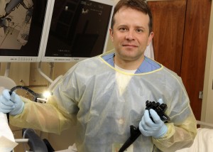
Field Willingham, MD, MPH

Once esophageal tumors establish themselves, a patient’s prognosis is grim and morbidity vast. But when lesions are caught early and removed, especially in the premalignant stage, the odds of survival markedly improve.

When a case calls for it, Emory gastroenterologist Field F. Willingham, MD, MPH, uses a hybrid approach to ousting superficial esophageal lesions. Superficial esophageal lesions are commonly caused by acid reflux disease, or GERD. GERD occurs when stomach acid flows into the esophagus and can lead to a condition known as Barrett’s esophagus, where the cells in the lower esophagus become damaged. This in turn can lead to dysplasia, or pre-cancerous cells.

But for superficial cancers, it is now possible to remove a portion of the lining layer of the GI tract, containing the tumor, with an endoscope.  This can help carefully selected patients avoid a major surgery. The technique, known as an EMR, allows the removal of superficial esophageal tumors and pre-cancer with an endoscope, a slender tube-like instrument.

Detecting and removing esophageal tumors early is essential for a favorable outcome. Once tumors firmly establish themselves in esophageal tissue, the prognosis is grim and morbidity vast. In the past, a diagnosis of an esophageal tumor meant the removal of the esophagus and often the stomach. But now EMR can be used in tandem with radio frequency ablation.

In surgical situations in which radio frequency ablation is not feasible, Willingham and his colleagues are beginning to use an alternate technique, known as cryotherpay, in tandem with EMR. Cryotherapy involves freezing superficial cells to rid the esophagus of suspect cells.

“So, if the end of the esophagus is twisted, or if we can’t touch it with this balloon device, then we can use cryotherapy,” says Willingham. “We’re trying to kill the lining layer with the tumor cells without killing the deeper layer.”

Willingham and his colleagues are seeing evidence that using these very three very different, technologies in tandem or alone will provide patients with a better way to rid them of esophageal lesions while preserving their quality of life.

Posted on by admin in Uncategorized Leave a comment

Genetic alteration opens door to targeted treatment of rare tumor

A cross section of an epithelioid hemangioendothelioma

Emory pathologist Sharon Weiss, MD, was the first to describe an extraordinarily rare tumor known as an epithelioid hemangioendothelioma (EHE). Thirty years later, researchers have identified a genetic alteration linked to this odd vascular tumor.

It’s hoped this newfound information will lead to a better understanding of the mechanisms underlying the development of this tumor and hence development of a targeted treatment. None yet is available. However, these findings already have been used to develop a new diagnostic test for this blood vessel disease.

The research, published in a recent issue of Science Translational Medicine, was done in collaboration with Cleveland Clinic’s Taussig Cancer Institute and led by Brian Rubin, MD, PhD, of Cleveland Clinic’s Pathology and Laboratory Medicine Institute and Lerner Research Institute.

The genetic alteration formerly in question involves a translocation between chromosomes 1 and 3, where chromosomes 1 and 3 exchange DNA fragments that are transposed onto opposite chromosomes. The result: the swapped DNA encodes a unique, fused gene that contains components from each chromosome. Because genes are translated into proteins, the result of this unique gene is a correspondingly unique protein, one thought to cause cancer.

Epithelioid hemangioendotheliomas comprise less than one percent of all cancers. Roughly 100 new cases are diagnosed in the United State each year. EHE are eccentric in their epidemiology, structure and aggressiveness. Slow to metastasize, they tend to occur in both young men and women when soft tissue is involved but occur mostly in women when the liver and lungs are affected.

However, it’s their peculiar structure that has so far made targeted treatment problematic, especially in the liver and lungs. “Instead of being one mass as you might expect with liver cancer, the patient with EHE often presents with little nodules throughout the liver,” says Weiss.

“The reason this occurs is that the growth starts in the liver’s portal vein, grows along its length, and then tracks out through the vessels. The growths blister out from the vessel creating these little nodules. Epithelioid hemangioendothelioma don’t possess the classic features of vascular tumors. In fact, EHE may have so many sites of involvement that the cancer can’t be cured, short of transplantation.”

Using EHE tissue samples gleaned from Weiss’s vast library, Rubin developed a genetic probe to detect the distinct chromosomal translocations in the tumor. The probe now serves as a powerful diagnostic tool of EHE and opens the door to understanding these tumors’ mechanisms.

“Once you understand the mechanism behind it, you can start trying to target those pathways in a therapeutic way,” says Weiss.

Posted on by admin in Cancer 1 Comment

Magnanimous magnolias keep on giving

Honokiol, the versatile compound found by Emory dermatologist Jack Arbiser in the cones of magnolia trees, makes a surprise appearance in a recent paper in Nature Medicine.

Jack Arbiser, MD, PhD, and colleagues originally isolated honokiol from magnolia cones. It can also be found in herbal teas.

The paper, from Sabrina Diano, Tamas Horvath and colleagues at Yale, probes the role of reactive oxygen species (ROS) in the hypothalamus, a part of the brain that regulates appetite. In the paper, Horvath’s laboratory uses honokiol as a super-antioxidant, mopping up ROS that suppress appetite. Arbiser initiated the collaboration with Horvath after finding, while working with Emory free radical expert Sergei Dikalov, how effective honokiol is at neutralizing ROS.

The paper is intriguing partly because it’s an example of a situation where ROS, often thought to be harmful because of their links to aging and several diseases, are actually beneficial. In this case, they provide a signal to stop eating. A recent paper from Andrew Neish’s lab at Emory provides another example, where probiotic bacteria stimulate production of ROS, which promote healing of the intestine.

Arbiser notes that since honokiol can increase appetite, the compound may be helpful in situations where doctors want patients to eat more.

“This might be particularly valuable in patients who are nutritionally deficient due to chemotherapy and provides a rationale for adding honokiol to chemotherapy regimens,” he writes.

Satiety producing neurons in the hypothalamus

A note of caution: in the Nature Medicine paper, honokiol is infused directly into the brain.

Honokiol has been shown to counteract inflammation and slow the growth of blood vessels (important in fighting cancer). Collaborating with Arbiser, Emory endocrinologist Neale Weitzmann has recently found that honokiol stimulates osteoblasts, the cells that build bone, suggesting that it could reduce bone loss in osteoporosis.

Posted on by Quinn Eastman in Cancer Leave a comment

Brain tumor patient gives back and moves forward

Jennifer Giliberto

Don’t sweat the small stuff.

That’s the motto 36-year-old Jennifer Giliberto now lives by after recently welcoming a third child into the world. Late night feedings, diaper changes, mounds of dirty laundry and caring for two older boys (ages six and eight) would certainly be a challenge for most moms. But this mom is different.

Four years ago, Giliberto was diagnosed with a brain tumor – a slow growing Grade II astrocytoma located in her posterior right temporal lobe. The shocking diagnosis left Giliberto and her family with many choices and decisions to make.

Giliberto’s inspiring story was profiled on CNN on Aug. 16, 2011 in a special “Human Factor” segment, which takes a look at people accomplishing something significant after overcoming the odds.

The Long Road Ahead

After her second child was born in 2005, Giliberto began noticing a pattern of problems with her fine motor skills. Neurological testing revealed little, but an MRI (magnetic resonance imaging) revealed a lesion and possible tumor in the brain. Follow-up MRIs over the next year showed no new growth, but in June 2007, a definite brain tumor was detected by MRI.

While taking the watch and wait approach to determine if the tumor would grow, she became involved with the Southeastern Brain Tumor Foundation (SBTF) as a volunteer. She focused her efforts on raising money to support critical brain and spinal tumor research. She also met Emory neurosurgeon Costas Hadjipanayis, MD, PhD.

Hadjipanayis, an assistant professor in Emory’s Department of Neurosurgery, would soon become Giliberto’s physician. He confirmed her diagnosis and recommended surgical removal of the tumor.

Costas Hadjipanayis, MD, PhD and patient Jennifer Giliberto

On August 18, 2008, at Emory University Hospital Midtown, Hadjipanayis removed Giliberto’s brain tumor. “Jennifer underwent a craniotomy and had a gross total resection of the tumor, with no complications,” explains Hadjipanayis, who is chief of neurosurgery at the hospital. “She spent one night in the neurosurgical ICU and her recovery afterwards went well.”

Then he encouraged her to embrace life and live it to the fullest. Giliberto has taken her doctor’s orders to heart, and lives life with a new purpose than before.

Giving Back

To support and encourage other brain tumor patients, Giliberto serves as a patient and family advisor at Emory University Hospital Midtown. She visits with hospitalized patients and their families who are in similar situations as the young mother of three.

“This has been a very fulfilling experience and an outlet to give back,” says Giliberto. “Being a patient is lonely, even when you know you have support. Working to assist other patients and families and improve a system goes a long way to ease that lonely journey of the patient experience.”

Patient and family advisors also work to improve hospital processes and procedures from a patient perspective.

She also serves as vice president of the Southeastern Brain Tumor Foundation, continuing the mission to raise funds for research. The SBTF consistently funds innovative brain tumor research at Emory’s Winship Cancer Institute.

And she is a devoted wife and mother.

Moving Forward

Last year, when Giliberto and her husband decided they would like to expand their family of four, she consulted with Hadjipanayis. He, once again, encouraged her to live life and move forward. They did, and their youngest child was born in July 2011.

While Giliberto has remained stable since her surgery in 2008, she continues to have MRI’s every six to nine months to check for any tumor recurrence. Astrocytomas, even once removed, can recur and can also become cancerous.

But for now, it’s on with life as she knows it – stable, moving ahead and enjoying every day with a new sense of hope.

And as for the small stuff – Giliberto’s learned there’s just no reason to sweat it at all.

Posted on by admin in Uncategorized 2 Comments

Emory researchers receive grants to further work in pediatric brain tumor research

Dr. Castellino explains his research on medulloblastomas to participants attending the SBTF’s Grant Award Ceremony.

Two Emory researchers are being recognized by the Southeastern Brain Tumor Foundation (SBTF) for their work in pediatric brain tumor research.

Tracey-Ann Read, PhD, assistant professor in the Department of Neurosurgery, Emory University School of Medicine and director of the Pediatric Neuro-Oncology Laboratory at Emory was awarded a $75,000 grant for her work. She is studying the cell of origin that is responsible for the highly malignant pediatric brain tumor known as an Atypical Teratoid Rhabdoid Tumor (AT/RT). She is also developing a mouse model to study this very lethal brain cancer that occurs in early childhood.

Robert Craig Castellino, MD, assistant professor of pediatrics at Emory and pediatric hematologist/oncologist at Children’s Healthcare of Atlanta at Egleston received $50,000 to support his research efforts. He is studying how the childhood brain cancer, known as medulloblastoma, can metastasize from the brain to other sites in the body, specifically the spine. Medulloblastoma is the most common pediatric malignant brain tumor.

SBTF board members and researchers who were awarded grants pose following the April ceremony.

Read and Castellino received the awards at the SBTF’s Grant Awards Ceremony in April at Emory University Hospital Midtown. Two other researchers from Duke University were also presented with grant money for their contributions in brain tumor research in adults.

Emory neurosurgeon Costas Hadjipanayis, MD, PhD, is the president of the Southeastern Brain Tumor Foundation. He says research, from young investigators such as these, is crucial in the race to find a cure for brain tumors. As federal research funding becomes even more difficult to obtain with cuts in funding, private foundation grants, such as from the SBTF, can permit researchers to start important research projects that can provide preliminary data for bigger grant proposals.

The SBTF awards $200,000-300,000 each year to major medical centers throughout the Southeast in support of cutting-edge brain and spinal tumor research.


Posted on by admin in Cancer Leave a comment

Proton Therapy and Its Importance to Georgia

From Clinic to You

By Walter J. Curran, Jr., MD
Executive Director, Winship Cancer Institute
Chair, Department of Radiation Oncology, Emory University School of Medicine

Walter J. Curran, Jr., MD

Walter J. Curran, Jr., MD

Emory Healthcare is a key player in plans to bring the world’s most advanced radiation treatment for cancer patients to Georgia.  Emory Healthcare has signed a letter of intent with Advanced Particle Therapy, LLC, of Minden, Nevada, opening the door to a final exploratory phase for development of The Georgia Proton Treatment Center – Georgia’s first proton therapy facility.

For certain cancers, proton therapy offers a more precise and aggressive approach to destroying cancerous and non-cancerous tumors, as compared to conventional X-ray radiation. Proton therapy involves the use of a controlled beam of protons to target tumors with precision unavailable in other radiation therapies. According to The National Association for Proton Therapy, the precise delivery of proton energy may limit damage to healthy surrounding tissue, potentially resulting in lower side effects to the patient. This precision also allows for a more effective dose of radiation to be used.

Proton therapy is frequently used in the care of children diagnosed with cancer, as well as in adults who have small, well-defined tumors in organs such as the prostate, brain, head, neck, bladder, lungs, or the spine.  And research is continuing into its efficacy in other cancers.

The gantry, or supporting structure, of a proton therapy machine.

The gantry, or supporting structure, of a proton therapy machine.

The closest proton therapy facility to Georgia is the University of Florida Proton Therapy Institute in Jacksonville.  Currently there are only nine proton therapy centers in the United States, including centers at Massachusetts General Hospital, MD Anderson Cancer Center in Houston and the University of Pennsylvania.

This is an exciting development in our ability to offer not only patients throughout Georgia and the Southeast the widest possible array of treatment options but patients from around the world who can come to Atlanta via the world’s busiest airport, Hartsfield-Jackson International. In addition, we will work to expand its utility and access for patients through collaborative research projects with Georgia Tech and other institutions. Winship physicians will also be able to reach out to their international colleagues and provide direction in how best to study and implement this technology in the care of cancer patients.

Under the letter of intent, Emory Healthcare faculty and staff will provide physician services, medical direction, and other administrative services to the center. Advanced Particle Therapy, through a Special Purpose Company, Georgia Proton Treatment Center, LLC, (GPTC) will design, build, equip and own the center.  The facility, which will be funded by GPTC, will be approximately 100,000 square feet and is expected to cost approximately $200 million.  Site selection for the facility is underway, and pending various approvals, groundbreaking is expected in the Spring of 2012.

Video

The follow video presents a 3D simulation of proton therapy technology.

Additional Information:

Posted on by admin in Uncategorized Leave a comment

Brain Tumor Foundations Join Together to Raise Awareness and Funds for Research

“Two Voices, One Vision: Sharing Hope Across Generations” is the vision and message this year as two well-known brain tumor foundations join together to raise awareness and money for brain and spinal tumor research and support.

The Southeastern Brain Tumor Foundation (SBTF) is joining forces with the Brain Tumor Foundation for Children (BTFC) for the 2011 Race for Research, to be held on July 23 at Atlantic Station in Midtown Atlanta. The joint run and walk will highlight the shared mission of both groups in the fight against brain tumors.

Costas G. Hadjipanayis, MD, PhD

Costas G. Hadjipanayis, MD, PhD

Emory neurosurgeon Costas Hadjipanayis, MD, PhD, is the president of the Southeastern Brain Tumor Foundation. He says the annual race is the major fundraising event for the SBTF, raising money to support critical, cutting-edge brain and spinal tumor research at major medical centers in the Southeast, including Emory. Over the past decade, the SBTF has raised more than $1.2 million for research.

Since 1983, the BTFC has been serving the pediatric brain tumor population, providing $1.5 million in emergency financial assistance for families over the past 10 years, in addition to providing resources for numerous patient programs and research.

According to Hadjipanayis, the Race for Research has drawn, in recent years, over 2,000 participants annually from throughout the Southeast and across the U.S. By joining forces with the BTFC, attendance is expected to grow, as is the fundraising goal of $300,000 this year for the two not for profit organizations.

Hadjipanayis, who is also chief of the neurosurgery service at Emory University Hospital Midtown, hopes this event will help in gaining greater exposure for brain tumor awareness in both children and adults, while raising funds for important research.

To find out more about the 2011 Race for Research 5K run and 2K walk, visit upport.sbtf.org/2011Race.

Information about the SBTF can be found by visiting www.sbtf.org. For more information about the BTFC, see www.braintumorkids.org.

Posted on by Wendy Darling in Uncategorized Leave a comment

The science of caring

Handprints

“It is the oncology nurse whose ‘fingerprints’ are on the entire matrix of therapies,” said Seliza Mithchell.

A keynote presentation on “fingerprints” might be more suited to a police convention than an oncology nursing symposium.  That is unless Selinza Mitchell is the speaker. Mitchell, a nurse educator and presenter was the keynote speaker at the third annual Winship Oncology Nursing Symposium, held March 18 and 19 at the Evergreen Conference Center in Stone Mountain, Georgia.

Mitchell’s presentation focused on the impact oncology nurses have on the hundreds of patients and families they touch, both literally and figuratively.  It is the oncology nurse whose “fingerprints” are on the entire matrix of therapies, from administration of today’s latest targeted-therapy drugs to helping patients and families navigate an increasingly complex health care system.

That concept also formed the basis of many of the discussion groups that were part of the symposium.  “The entire model of care delivery is changing,” says Amelia Langston, MD, professor of Hematology and Medical Oncology at the Winship Cancer Institute.  “Care delivery is more of a team approach and is less physician-centered.  Therefore there is great interest in the expanding role of nurses, nurse practitioners, and physician assistants.”

Amelia Langston presenting at the Winship Oncology Nursing Symposium

Amelia Langston presenting at the Winship Oncology Nursing Symposium

The Winship Oncology Nursing Symposium has grown in three short years into one of the most informative and influential among this growing market of nursing continuing education opportunities.  Among the topics covered in this year’s meeting were cancer genetics, image-guided medicine, minimally invasive treatment, disease-specific topics and the expanding role of non-physician providers against the backdrop of health care reform.

“The health care system is demanding cost effective, clinically relevant continuing education programs in nursing and specifically in oncology nursing,” says Joan Giblin, MSN, FNP, a course director for the symposium and Manager of Patient Access at Winship.  “Offering a high quality, regional program that can provide the latest information on advanced nursing practice, research, and other issues is central to meeting that need.”

In addition to Joan Giblin, course directors for the event were Deena Gilland, RN, MSN, Director of Nursing at Winship, and Kevin Schreffler, RN, MSN, Clinical Nurse Specialist at Winship.

Posted on by Wendy Darling in Uncategorized Leave a comment