Warren symposium follows legacy of geneticist giant

If we want to understand how the brain creates memories, and how genetic disorders distort the brain’s machinery, then the fragile X gene is an ideal place to start. That’s why the Stephen T. Warren Memorial Symposium, taking place November 28-29 at Emory, will be a significant event for those interested in neuroscience and genetics. Stephen T. Warren, 1953-2021 Warren, the founding chair of Emory’s Department of Human Genetics, led an international team that discovered Read more

Mutations in V-ATPase proton pump implicated in epilepsy syndrome

Why and how disrupting V-ATPase function leads to epilepsy, researchers are just starting to figure Read more

Tracing the start of COVID-19 in GA

At a time when COVID-19 appears to be receding in much of Georgia, it’s worth revisiting the start of the pandemic in early 2020. Emory virologist Anne Piantadosi and colleagues have a paper in Viral Evolution on the earliest SARS-CoV-2 genetic sequences detected in Georgia. Analyzing relationships between those virus sequences and samples from other states and countries can give us an idea about where the first COVID-19 infections in Georgia came from. We can draw Read more

cancer

Three-stage delivery for platinum-based ‘cluster bombs’ against cancer

Scientists have devised a triple-stage ‘cluster bomb’ system for delivering the chemotherapy drug cisplatin, via tiny nanoparticles designed to break up when they reach a tumor.

Details of the particles’ design and their potency against cancer in mice are described this week in PNAS Early Edition. They have not been tested in humans, although similar ways of packaging cisplatin have been in clinical trials. Anticancer cluster bombs
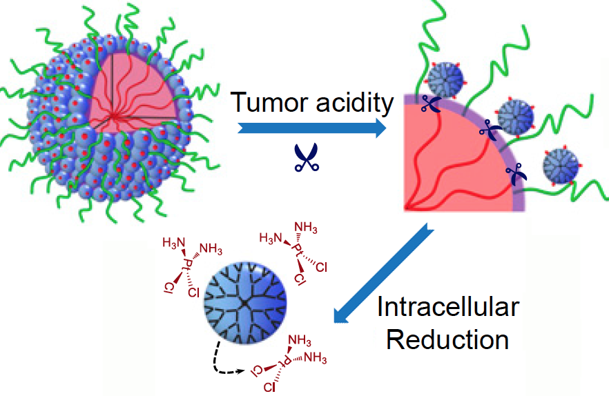
What makes these particles distinctive is that they start out relatively large — 100 nanometers wide — to enable smooth transport into the tumor through leaky blood vessels. Then, in acidic conditions found close to tumors, the particles discharge “bomblets” just 5 nanometers in size.

Inside tumor cells, a second chemical step activates the platinum-based cisplatin, which kills by crosslinking and damaging DNA. Doctors have used cisplatin to fight several types of cancer for decades, but toxic side effects — to the kidneys, nerves and inner ear — can limit its effectiveness.

The PNAS paper is the result of a collaboration between a team led by professor Jun Wang, PhD at the University of Science and Technology of China, and researchers led by professor Shuming Nie, PhD in the Wallace H. Coulter Department of Biomedical Engineering at Georgia Tech and Emory. Nie is a member of the Discovery and Developmental Therapeutics research program at Winship Cancer Institute of Emory University. The lead authors are graduate student Hong-Jun Li and postdoctoral fellows Jinzhi Du, PhD and Xiao-Jiao Du, PhD.

“The negative side effects of cisplatin are a long-standing limitation for conventional chemotherapy,” says Jinzhi Du. “In our study, the delivery system was able to improve tumor penetration to reach more cancer cells, as well as release the drugs specifically inside cancer cells through their size-transition property.”

The researchers showed that their nanoparticles could enhance cisplatin drug accumulation in tumor tissues. When mice bearing human pancreatic tumors were given the same doses of free cisplatin or cisplatin clothed in pH-sensitive nanoparticles, the level of platinum in tumor tissues was seven times higher with the nanoparticles. This suggests the possibility that nanoparticle delivery could restrain the toxic side effects of cisplatin during cancer treatment. Read more

Posted on by Quinn Eastman in Cancer Leave a comment

NINDS director: neuroscience now largest ‘bucket of money’

On Friday, NINDS director Walter Koroshetz made an interesting remark in a lecture to Emory’s Department of Neurology. He said that in the 2016 National Institues of Health budget, neuroscience is now the largest “bucket of money,” especially with the recent boost in funding for Alzheimer’s research. That’s larger than the bucket for cancer. To be sure, biomedical research in general got a boost from Congress, with the NIH receiving its largest increase in a decade, and cancer is still a big deal!

Koroshetz explained that neuroscience research is spread out among NINDS (National Institute for Neurological Disorders and Stroke), NIMH (National Institute of Mental Health), NIDA (National Institute for Drug Abuse) and several others, while cancer research is concentrated at the National Cancer Institute. [Here’s some official category tracking that the NIH does – his breakdown checks out.]

Koroshetz highlighted a project from Dieter Jaeger and Garret Stanley that is part of the White House’s BRAIN Initiative focused on mapping brain circuits and connectivity. He also noted NINDS’s efforts in promoting translational research, since pharmaceutical companies were frustrated by repeated failures in the 1990s with difficult areas such as stroke, and the R35 mechanism for funding “outstanding investigators” for up to eight years continuously.

Posted on by Quinn Eastman in Neuro 2 Comments

Chasing invasive cancer cells and more at #ASCB15

Earlier today, we posted a notice on Eurekalert for a Sunday, December 13 presentation by graduate student Jessica Konen at the American Society for Cell Biology meeting in San Diego.

Her research, performed with Adam Marcus at Winship Cancer Institute, was the topic of a video that recently won first prize in a contest sponsored by the Association of American Medical Colleges. This was our video team’s first use of the “fast hand on whiteboard” effect, and a lot of fun to make. The video’s strength grows out of the footage Konen and Marcus have of cancer cells migrating in culture. Check it out, if you haven’t already.

Poster presentations at the 2015 ASCB meeting can be found by searching this PDF. A few Emory-centric highlights:

*Chelsey Ruppersburg and Criss Hartzell’s work on the “nimbus”, a torus-shaped structure enriched in proteins needed to build the cell’s primary cilium

*Anita Corbett on how Emory students have a strong record of attaining their own NIH research funding

*Additional work by Adam Marcus’ lab on the tumor suppressor gene LKB1 and how its loss drives lung cancer cells to take on a “unique amoeboid morphology”

*Research from David Katz’s lab on the “epigenetic eraser” LSD1 (lysine-specific demethylase) and its function in neurons and neurodegeneration Read more

Posted on by Quinn Eastman in Cancer, Neuro Leave a comment

Anticancer drug strategy: making cells choke on copper

What do cancer cells have in common with horseshoe crabs and Mr. Spock from Star Trek?

They all depend upon copper. Horseshoe crabs have blue blood because they use copper to transport oxygen in their blood instead of iron (hemocyanin vs hemoglobin). Vulcans’ blood was supposed to be green, for the same reason.

Horseshoe Crab (Limulus polyphemus)

Horseshoe crabs and Vulcans use copper to transport oxygen in their blood. Cancer cells seem to need the metal more than other cells.

To be sure, all our cells need copper. Many human enzymes use the metal to catalyze important reactions, but cancer cells seem to need it more than healthy cells. Manipulating the body’s flow of copper is emerging as an anticancer drug strategy.

A team of scientists from University of Chicago, Emory and Shanghai have developed compounds that interfere with copper transport inside cells. These compounds inhibit the growth of several types of cancer cells, with minimal effects on the growth of non-cancerous cells, the researchers report in Nature Chemistry.

“We’re taking a tactic that’s different from other approaches. These compounds actually cause copper to accumulate inside cells,” says co-senior author Jing Chen, PhD, professor of hematology and medical oncology at Emory University School of Medicine and Winship Cancer Institute. Read more

Posted on by Quinn Eastman in Cancer Leave a comment

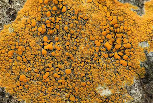
Orange lichens are source for potential anticancer drug

An orange pigment found in lichens and rhubarb called parietin may have potential as an anti-cancer drug, scientists at Winship Cancer Institute of Emory University have discovered.

The results were published in Nature Cell Biology on October 19.

Caloplaca_Fenwick

Parietin, shown to have anticancer activity in the laboratory, is a dominant pigment in Caloplaca lichens. Note: this study did not assess the effects of eating lichens or rhubarb. Photo courtesy of www.aphotofungi.com

Parietin, also known as physcion, could slow the growth of and kill human leukemia cells obtained directly from patients, without obvious toxicity to human blood cells, the authors report. The pigment could also inhibit the growth of human cancer cell lines, derived from lung and head and neck tumors, when grafted into mice.

A team of researchers led by Jing Chen, PhD, discovered the properties of parietin because they were looking for inhibitors for the metabolic enzyme 6PGD (6-phosphogluconate dehydrogenase). 6PGD is part of the pentose phosphate pathway, which supplies cellular building blocks for rapid growth. Researchers have already found 6PGD enzyme activity increased in several types of cancer cells.

“This is part of the Warburg effect, the distortion of cancer cells’ metabolism,” says Chen, professor of hematology and medical oncology at Emory University School of Medicine and Winship Cancer Institute. “We found that 6PGD is an important metabolic branch point in several types of cancer cells.” Read more

Posted on by Quinn Eastman in Cancer Leave a comment

Adaptive mutation mechanism may drive some forms of antibiotic resistance

Evolutionary theory says mutations are blind and occur randomly. But in the controversial phenomenon of adaptive mutation, cells can peek under the blindfold, increasing their mutation rate in response to stress.

Scientists at Winship Cancer Institute, Emory University have observed that an apparent “back channel” for genetic information called retromutagenesis can encourage adaptive mutation to take place in bacteria.

The results were published Tuesday, August 25 in PLOS Genetics.

“This mechanism may explain how bacteria develop resistance to some types of antibiotics under selective pressure, as well as how mutations in cancer cells enable their growth or resistance to chemotherapy drugs,” says senior author Paul Doetsch, PhD.

Doetsch is professor of biochemistry, radiation oncology and hematology and medical oncology at Emory University School of Medicine and associate director of basic research at Winship Cancer Institute. The first author of the paper is Genetics and Molecular Biology graduate student Jordan Morreall, PhD, who defended his thesis in April.

Retromutagenesis resolves the puzzle: if cells aren’t growing because they’re under stress, which means their DNA isn’t being copied, how do the new mutants appear?

The answer: a mutation appears in the RNA first. Read more

Posted on by Quinn Eastman in Cancer, Uncategorized Leave a comment

Immune ‘traffic jam’ from viral infection

Several drugs now used to treat cancer and autoimmune diseases are actually repurposed tools derived from the immune system. One of the ways these “therapeutic antibodies” work is to grab onto malignant or inflammatory cells and escort them to their doom.

Emory researchers have found that in a mouse model of chronic viral infection, a kind of traffic pileup inside the body limits how effective therapeutic antibodies can be.

The results, published this week in Immunity, have implications for biotechnology researchers who continue to refine antibodies for therapeutic purposes, as well as bolster our understanding of how chronic viral infections impair the immune system.

Researchers led by Rafi Ahmed, PhD, director of the Emory Vaccine Center, were studying mice infected by LCMV (lymphocytic choriomeningitis virus). They injected several antibodies with the goal of removing various types of immune cells from the mice.  One end of the antibody molecule is supposed to bind the target cell, while another acts as a flag for other cells to get rid of the target cell.

However, during a chronic LCMV infection, the mouse’s immune system is producing its own antibodies against the virus, which form complexes with viral proteins. These immune complexes prevented the injected antibodies from having the effect the scientists wanted, which was to deplete their target cells.

Excessive amounts of immune complexes appear to be “clogging” the Fc gamma receptors that immune cells would use to grab the antibodies bound to the target cell, says postdoctoral fellow Andreas Wieland, PhD, first author of the Immunity paper. That these immune complexes form was not news; but how much they interfere with other antibodies was, Wieland says. Fc gamma receptors were already known to be important for antibodies to be effective against influenza and HIV. Read more

Posted on by Quinn Eastman in Immunology Leave a comment

Cancer’s shield: PD-1

Gina Kolata has a section front story in Tuesday’s New York Times exploring the potential of a relatively new class of anticancer drugs. The drugs break through “shields” built by cancers to ward off the threat posed by the patient’s immune system. Many are based on blocking PD-1, an immune regulatory molecule whose importance in chronic infections was first defined by Emory’s Rafi Ahmed.

Of course, not every cancer research development described as transformative in the New York Times lives up to the hype. But the clinical trial results, reported in the New England Journal of Medicine, are solid enough that the researchers Kolata talks with think they are seeing “a moment in medical history when everything changed.” [Winship Cancer Institute’s John Kauh was a co-author on one of the 2012 NEJM papers.]

Let’s take a moment to examine some of the roots of this story. Rafi Ahmed didn’t set out to study cancer. For the last two decades, he and his colleagues have been studying T cells, parts of the immune system that are critical for responding to infections. Read more

Posted on by Quinn Eastman in Cancer, Immunology 2 Comments

The challenges of graduate school

Biochemist Paul Doetsch’s recent appearance in a Science magazine feature on laboratory leadership led to a conversation with him about the challenges of graduate school.

He emphasized that scientific research is a team sport, and brilliance on the part of the lab head may not yield fruit without a productive relationship with the people in the lab. Doetsch suggested talking with Lydia Morris, a graduate student in the Genetics and Molecular Biology graduate program. Morris has been working in Doetsch’s lab for several years and is about to complete her degree. She has been examining the in vivo distribution of DNA repair proteins.

In this video, Morris and Doetsch talk about the differences between turn-the-crank and blue-sky projects, and the importance of backup projects, communications, high expectations and perseverance.

Posted on by Quinn Eastman in Cancer Leave a comment

The body’s anticancer defenses come in a variety of sizes

Sometimes you have to look at the whole picture, big and small.

Sarah Cork, PhD

That was the lesson that emerged from Winship Cancer Institute researcher Erwin Van Meir’s laboratory, highlighted in a recent paper in Oncogene. Van Meir’s team has been studying vasculostatin, a secreted protein that inhibits blood vessel growth by tumors (hence the name). Vasculostatin was discovered by Balveen Kaur, now at Ohio State, while she was in Van Meir’s lab.

Van Meir and his colleagues originally began studying vasculostatin because it is a product of a gene that brain tumors somehow silence or get rid of, and studying the obstacles our bodies throws in cancer’s way may be a good way to learn how to fight it via modern medicine. Eventually, it could form the basis for a treatment to prevent a tumor from attracting new blood vessels.

Vasculostatin is somewhat unique because it is a secreted fragment of a membrane-bound protein, called BAI1. BAI1 has an apparently separate function as an “engulfment receptor,” allowing the recognition and internalization of dying cells.

Most of the secreted vasculostatin is around 40 kilodaltons in size, not 120 as previously thought.

Graduate student Sarah Cork discovered that most of the vasculostatin protein being produced by cells is actually much smaller than what had been originally discovered. She and Van Meir were surprised to find that the smaller, cleaved form of the protein still has potent anti-angiogenic activity.

The researchers were using a technique where a mixture of proteins is separated within a gel by electric current, transferred to a polymer sheet, and probed with antibodies. The large proteins appear at the top and the small proteins at the bottom.

“Previously, we had been running the gels for a long time to detect large protein fragments, so missed out on what was happening with small fragments which run off the gel,” Van Meir says. “We were only looking at the top of the
gel, when the smaller form of vasculostatin was actually much more
abundant as you can see on the picture of a gel run for a shorter time.”

More broadly, Van Meir says that the finding adds to understanding about BAI1’s dual function in the brain and how vasculostatin (big or small) might be used in anticancer therapy.

Posted on by Quinn Eastman in Cancer Leave a comment