Warren symposium follows legacy of geneticist giant

If we want to understand how the brain creates memories, and how genetic disorders distort the brain’s machinery, then the fragile X gene is an ideal place to start. That’s why the Stephen T. Warren Memorial Symposium, taking place November 28-29 at Emory, will be a significant event for those interested in neuroscience and genetics. Stephen T. Warren, 1953-2021 Warren, the founding chair of Emory’s Department of Human Genetics, led an international team that discovered Read more

Mutations in V-ATPase proton pump implicated in epilepsy syndrome

Why and how disrupting V-ATPase function leads to epilepsy, researchers are just starting to figure Read more

Tracing the start of COVID-19 in GA

At a time when COVID-19 appears to be receding in much of Georgia, it’s worth revisiting the start of the pandemic in early 2020. Emory virologist Anne Piantadosi and colleagues have a paper in Viral Evolution on the earliest SARS-CoV-2 genetic sequences detected in Georgia. Analyzing relationships between those virus sequences and samples from other states and countries can give us an idea about where the first COVID-19 infections in Georgia came from. We can draw Read more

brain cancer

Device for viewing glowing brain tumors

People touched by a brain tumor — patients, their families or friends — may have heard of the drug Gliolan or 5-ALA, which is taken up preferentially by tumor cells and makes them fluorescent. The idea behind it is straightforward: if the neurosurgeon can see the tumor’s boundaries better during surgery, he or she can excise it more thoroughly and accurately.

5-ALA is approved for use in Europe but is still undergoing evaluation by the U.S. FDA. A team at Emory was the first to test this drug in the United States. [Note: A similar approach, based on protease activation of a fluorescent probe, was reported last week in Science Translational Medicine.]

ac-2015-034535_0001

A hand-held device to detect glowing brain tumors could allow closer access to the critical area than a surgical microscope

Biomedical engineer Shuming Nie and colleagues recently described their development of a hand-held spectroscopic device for viewing fluorescent brain tumors. This presents a contrast with the current tool, a surgical microscope — see figure.

Nie’s team tested their technology on specimens obtained from cancer surgeries. Their paper in Analytical Chemistry reports:

The results indicate that intraoperative spectroscopy is at least 3 orders of magnitude more sensitive than the current surgical microscopes, allowing ultrasensitive detection of as few as 1000 tumor cells. Read more

Posted on by Quinn Eastman in Cancer Leave a comment

Herding terrorist cats

Wikipedia says that “herding cats” refers to an attempt to control or organize a class of entities that are uncontrollable or chaotic.

Cancer cells certainly qualify as uncontrollable or chaotic, so the metaphor could apply to a recent Nature Materials paper from Georgia Tech and Emory’s Ravi Bellamkonda – a member of Winship Cancer Institute.

Glioblastoma is the worst of the worst: the most common and the most aggressive form of brain tumor in adults. The tumors are known to invade healthy tissue and migrate along white matter tracts and blood vessels. Bellamkonda and his colleagues devised a strategy for luring glioblastoma cells out of the brain by offering the cells attractive nanofibers that the cells will Ray Ban outlet attempt to invade. When the cells arrive, they undergo apoptosis — cellular suicide. He has called this “an engineer’s approach to brain cancer” (in a lecture a couple months ago) and “the Pied Piper approach” (in the video below).

(It’s not the first time Bellamkonda has unfurled dazzling technology against glioblastoma, developed with an Emory collaborator.)

Bellamkonda’s collaborator this time, Tobey Macdonald, director of pediatric neuro-oncology at Children’s Healthcare of Atlanta, is credited in the paper with coming up with the aspect of the strategy that was based on the molecule cyclopamine. This earlier story from CHOA provides more background on how the collaboration came together.

Cyclopamine

Cyclopamine is key to the “lure ’em out and kill ’em” strategy. Most high-grade brain tumors overproduce a protein called Sonic Hedgehog, spurring their growth. Cyclopamine is selectively toxic only to cells that are dependent on Sonic Hedgehog. Cyclopamine’s name comes from how it was discovered: through its teratogenic effects on sheep in Idaho that ate corn lily flowers.

Posted on by Quinn Eastman in Cancer Leave a comment

Without intent, yet malignant

Brain cancer doesn’t have a purpose or intent. It’s just a derangement of molecular biology, cells that keep growing when they’re not supposed to.

But it’s difficult not to think in terms of purpose or intent when looking at what cancers do.  For example, Winship Cancer Institute scientists Abdessamad (Samad) Zerrouqi, Beata Pyrzynska, Dan Brat and Erwin Van Meir have a recent paper in Cancer Research examining how glioblastoma cells regulate the process of blood clotting.*

Blood clots, often in the legs, are a frequent occurrence in patients fighting glioblastoma, the most common and the most aggressive form of brain cancer. Zerrouqi and http://www.gooakley.com/ Van Meir show that a tumor suppressor gene (p14ARF) that is often mutated in glioblastoma stops them from activating blood clotting. Take away the gene and glioblastoma cells activate the clotting process more.

At first glance, a puzzle emerges: why would a cancer “want” to induce blood clots? Cancer cells often send out growth factors that stimulate the growth of new blood vessels (angiogenesis). The cells are growing fast, thus they need their own blood supply. Activating clotting seems contradictory: why build a new highway and then induce a traffic jam?

Thrombosis-necrosis

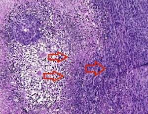
The two left arrows indicate clots causing necrosis around the vessels. Cells at the edge of the necrotic zone (right arrow) tend to be more proliferative and invasive. Image courtesy of Zerrouqi.

In a way, tumor cells are acting somewhat Nietzschean, blindly managing their own cheap oakley evolution according to the principle “Whatever doesn’t kill me makes me stronger.”

Blood clots lead to both destruction of the healthy and tumor tissue and hypoxia, a shortage of oxygen that drives more aggressiveness in the tumor. The clots create “micro-necroses” at the leading edge of the tumor that over time probably fuse and create a big central necrosis.

“The paradox is that the tumor kills itself and the normal brain, yet the capacity of doing this is the hallmark of the most malignant form of this tumor,” Van Meir says.

“The advantage of tumoral thrombosis will be selection of cells to progress to higher aggressiveness: infiltrative,  resistant to death with conventional Oakley Sunglasses cheap therapies, metabolically adapted to low levels of oxygen and nutrients,” Zerrouqi says. “At this stage, the tumor seems to have a clear deadly intent.”

A fragment of one of the proteins that cancer cells use to exert the clotting effect, called TFPI2, could be used to antagonize blood clotting  therapeutically, they write in Cancer Research. The findings could also have implications for understanding the effects of current medications, such as the angiogenesis inhibitor bevacizumab, also known as Avastin.

*A paper by Van Meir and Dan Brat from 2005 is the top Google link under the search term “glioblastoma clotting.”

Posted on by Quinn Eastman in Cancer Leave a comment

Emory researchers receive grants to further work in pediatric brain tumor research

Dr. Castellino explains his research on medulloblastomas to participants attending the SBTF’s Grant Award Ceremony.

Two Emory researchers are being recognized by the Southeastern Brain Tumor Foundation (SBTF) for their work in pediatric brain tumor research.

Tracey-Ann Read, PhD, assistant professor in the Department of Neurosurgery, Emory University School of Medicine and director of the Pediatric Neuro-Oncology Laboratory at Emory was awarded a $75,000 grant for her work. She is studying the cell of origin that is responsible for the highly malignant pediatric brain tumor known as an Atypical Teratoid Rhabdoid Tumor (AT/RT). She is also developing a mouse model to study this very lethal brain cancer that occurs in early childhood.

Robert Craig Castellino, MD, assistant professor of pediatrics at Emory and pediatric hematologist/oncologist at Children’s Healthcare of Atlanta at Egleston received $50,000 to support his research efforts. He is studying how the childhood brain cancer, known as medulloblastoma, can metastasize from the brain to other sites in the body, specifically the spine. Medulloblastoma is the most common pediatric malignant brain tumor.

SBTF board members and researchers who were awarded grants pose following the April ceremony.

Read and Castellino received the awards at the SBTF’s Grant Awards Ceremony in April at Emory University Hospital Midtown. Two other researchers from Duke University were also presented with grant money for their contributions in brain tumor research in adults.

Emory neurosurgeon Costas Hadjipanayis, MD, PhD, is the president of the Southeastern Brain Tumor Foundation. He says research, from young investigators such as these, is crucial in the race to find a cure for brain tumors. As federal research funding becomes even more difficult to obtain with cuts in funding, private foundation grants, such as from the SBTF, can permit researchers to start important research projects that can provide preliminary data for bigger grant proposals.

The SBTF awards $200,000-300,000 each year to major medical centers throughout the Southeast in support of cutting-edge brain and spinal tumor research.


Posted on by admin in Cancer Leave a comment

Strategies to target cancer stem cells

A story in last Friday’s New York Times highlights research on “cancer stem cells”: a fraction of cells in a tumor that are especially resistant to chemotherapy and resemble the body’s non-cancerous stem cells in their ability to renew themselves.

The story describes work by a team at the Broad Institute, who reported in the journal Cell that they had identified compounds that specifically kill cancer stem cells. The hope is that compounds such as these could be combined with conventional treatments to more effectively eliminate cancers.

However, scientists disagree on whether the phenomenon of cancer stem cells extends to different kinds of cancer and what is the best way to target them. Previously not much was known about how to attack these cells.

Work at Emory’s Winship Cancer Institute has been tracking how some biomarkers in cancer cells resemble or differ from those found in stem cells. These markers may help researchers home in on the cancer stem cells.


Anticancer therapy must target more than one type of cell. TIC means tumor initiating cell, DTC means differentiated tumor cell, and CPG means cancer progenitor

If "cancer stem cells" play the critical roles some scientists think they do, anticancer therapy must target more than one type of cell. In this figure from Van Meir + Hadjipanayis' review, TIC means tumor initiating cell, DTC means differentiated tumor cell, and CPG means cancer progenitor cells.Â


In a recent review, Emory brain cancer specialists Erwin Van Meir and Costas Hadjipanayis write:

The “cancer stem cell” hypothesis has invigorated the neuro-oncology field with a breath of fresh thinking that may end up shaking the foundation of old dogmas, such as the widely held belief that glioblastoma tumors are incurable because of infiltrative disease. If the infiltrated cells are in fact differentiated tumor cells, their dissemination beyond the surgical boundary may not be the primary cause of tumor recurrence.

Van Meir, the editor of a new book on brain cancer, adds this comment:

Clearly a lot more work needs to be done to understand the precise cause of glioblastoma recurrence after surgery and chemotherapy and how to prevent it.  The possibility of developing therapeutics that can specifically target the brain cancer stem cells is an exciting new development but will have to proceed with caution to spare normal stem cells in the brain. Developing new imaging tools that can track cancer stem cells in the brain of treated patients is also an important objective and some of the Emory investigators are evaluating the use of nanoparticles to this purpose.

A new faculty member at Winship, Tracy-Ann Read, recently published her research on a molecule that could be used to identify “tumor-propagating cells” in medulloblastoma, a form of brain cancer. She says:

Although cancer stem cells have been identified in many different types of cancer, it is becoming increasingly clear that the properties of these cells may vary greatly among the different tumor types. It is unlikely that one  therapeutic agent will be able to target the cancer stem cells in for example all types brain tumors. Hence  much work still needs to be done in terms of analyzing the properties of these cells in each tumor type and identifying the genes that are responsible for their unique ability to propagate the tumors. 

Winship’s director Brian Leyland-Jones has also reported at the San Antonio Breast Cancer Symposium that molecules that distinguish a hard-to-treat form of breast cancer resemble those that maintain stem cells.

Nice round-up from Nature’s stem cell blog editor Monya Baker

Posted on by Quinn Eastman in Cancer Leave a comment