Warren symposium follows legacy of geneticist giant

If we want to understand how the brain creates memories, and how genetic disorders distort the brain’s machinery, then the fragile X gene is an ideal place to start. That’s why the Stephen T. Warren Memorial Symposium, taking place November 28-29 at Emory, will be a significant event for those interested in neuroscience and genetics. Stephen T. Warren, 1953-2021 Warren, the founding chair of Emory’s Department of Human Genetics, led an international team that discovered Read more

Mutations in V-ATPase proton pump implicated in epilepsy syndrome

Why and how disrupting V-ATPase function leads to epilepsy, researchers are just starting to figure Read more

Tracing the start of COVID-19 in GA

At a time when COVID-19 appears to be receding in much of Georgia, it’s worth revisiting the start of the pandemic in early 2020. Emory virologist Anne Piantadosi and colleagues have a paper in Viral Evolution on the earliest SARS-CoV-2 genetic sequences detected in Georgia. Analyzing relationships between those virus sequences and samples from other states and countries can give us an idea about where the first COVID-19 infections in Georgia came from. We can draw Read more

‘Master key’ microRNA has links to both ASD and schizophrenia

Recent studies of complex brain disorders such as schizophrenia and autism spectrum disorder (ASD) have identified a few “master keys,” risk genes that sit at the center of a network of genes important for brain function. Researchers at Emory and the Chinese Academy of Sciences have created mice partially lacking one of those master keys, called MIR-137, and have used them to identify an angle on potential treatments for ASD.

The results were published this week in Nature Neuroscience.

Mice partially lacking MIR-137 display learning and memory deficits, repetitive behaviors and impaired sociability. MIR-137 encodes a microRNA, which regulates hundreds of other genes, many of which are also connected to schizophrenia and autism spectrum disorder.

By treating mutant mice with papaverine, a vasodilator discovered in the 19th century, scientists could improve the performance of the mice on maze navigation and social behavior tests. Papaverine is an inhibitor of the enzyme Pde10a (phosphodiesterase 10a), which is elevated in mutant mice.

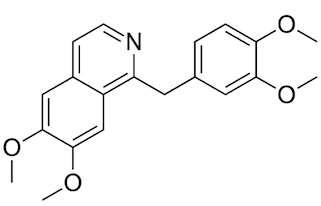
Papaverine is a component of opium, but it has a structure (and effects) that are different from opiates.

Other Pde10a inhibitors have been tested in schizophrenia clinical trials, but the new results suggest this group of compounds could have potential for some individuals with ASD, says senior author Peng Jin, PhD, professor of human genetics at Emory University School of Medicine.

Having just the right level of MIR-137 function is important. Previous studies of people with genetic deletions show that a loss of MIR-137 is connected with intellectual disability and autism spectrum disorder. The reverse situation, in which a genetic variation increases MIR-137 levels, appears to contribute to schizophrenia.

“It’s interesting to think about in the context of precision medicine,” Jin says. “Individuals with a partial loss of MIR137 – either genomic deletions or reduced expression — could potentially be candidates for treatment with Pde10a inhibitors.”

To create the mutant mice, Jin’s lab teamed up with Dahua Chen, PhD and Zhao-Qian Teng, PhD scientists at the State Key Laboratories of Stem Cell and Reproductive Biology and Membrane Biology, part of the Institute of Zoology, Chinese Academy of Sciences in Beijing. Jin says that generating mice with a heritable disruption of MIR-137 was technically challenging, taking several years. Read more

Posted on by Quinn Eastman in Neuro Leave a comment

Shape-shifting RNA regulates viral sensor

Congratulations to Emory biochemists Brenda Calderon and Graeme Conn. Their recent Journal of Biological Chemistry paper on a shape-shfting RNA was selected as an Editor’s Pick and cited as a “joy to read… Technically, the work is first class, and the writing is clear.”

Calderon, a former BCDB graduate student and now postdoc, was profiled by JBC in August.

Brenda Calderon, PhD

Calderon and Conn’s JBC paper examines regulation of the enzyme OAS (oligoadenylate synthetase). OAS senses double-stranded RNA: the form that viral genetic material often takes. When activated, OAS makes a messenger molecule that drives internal innate immunity enzymes to degrade the viral material (see below).

OAS is in turn regulated by a non-coding RNA, called nc886. Non-coding means this RNA molecule is not carrying instructions for building a protein. Calderon and Conn show that nc886 takes two different shapes and only one of them activates OAS.

Conn says in a press release prepared by JBC that although nc886 is present in all human cells, it’s unknown how abundance of its two forms might change in response to infection. Read more

Posted on by Quinn Eastman in Uncategorized Leave a comment

Mapping shear stress in coronary arteries can help predict heart attacks

A heart attack is like an earthquake. When a patient is having a heart attack, it’s easy for cardiologists to look at a coronary artery and identify the blockages that are causing trouble. However, predicting exactly where and when a seismic fault will rupture in the future is a scientific challenge – in both geology and cardiology.

In a recent paper in Journal of the American College of Cardiology, Habib Samady, MD, and colleagues at Emory and Georgia Tech show that the goal is achievable, in principle. Calculating and mapping how hard the blood’s flow is tugging on the coronary artery wall – known as “wall shear stress” – could allow cardiologists to predict heart attacks, the results show.

Map of wall shear stress (WSS) in a coronary artery from someone who had a heart attack

“We’ve made a lot of progress on defining and identifying ‘vulnerable plaque’,” says Samady, director of interventional cardiology/cardiac catheterization at Emory University Hospital. “The techniques we’re using are now fast enough that they could help guide clinical decision-making.”

Here’s where the analogy to geography comes in. By vulnerable plaque, Samady means a spot in a coronary artery that is likely to burst and cause a clot nearby, obstructing blood flow. The researchers’ approach, based on fluid dynamics, involves seeing a coronary artery like a meandering river, in which sediment (atherosclerotic plaque) builds up in some places and erodes in others. Samady says it has become possible to condense complicated fluid dynamics calculations, so that what once took months now might take a half hour.

Previous research from Emory showed that high levels of wall shear stress correlate with changes in the physical/imaging characteristics of the plaque over time. It gave hints where bad things might happen, in patients with relatively mild heart disease. In contrast, the current results show that where bad things actually did happen, the shear stress was significantly higher.

“This is the most clinically relevant work we have done,” says Parham Eshtehardi, MD, a cardiovascular research fellow, looking back on the team’s previous research, published in Circulation in 2011.  Read more

Posted on by Quinn Eastman in Heart Leave a comment

Cells in “little brain” have distinctive metabolic needs

Cells’ metabolic needs are not uniform across the brain, researchers have learned. “Knocking out” an enzyme that regulates mitochondria, cells’ miniature power plants, specifically blocks the development of the mouse cerebellum more than the rest of the brain.

The results were published in Science Advances.

“This finding will be tremendously helpful in understanding the molecular mechanisms underlying developmental disorders, degenerative diseases, and even cancer in the cerebellum,” says lead author Cheng-Kui Qu, MD, PhD, professor of pediatrics at Emory University School of Medicine, Winship Cancer Institute and Aflac Cancer and Blood Disorders Center, Children’s Healthcare of Atlanta.

The cerebellum or “little brain” was long thought to be involved mainly in balance and complex motor functions. More recent research suggests it is important for decision making and emotions. In humans, the cerebellum grows more than the rest of the brain in the first year of life and its development is not complete until around 8 years of age. The most common malignant brain tumor in children, medulloblastoma, arises in the cerebellum.

Qu and his colleagues have been studying an enzyme, PTPMT1, which controls the influx of pyruvate – a source of energy derived from carbohydrates – into mitochondria. They describe pyruvate as “the master fuel” for postnatal cerebellar development.

Cells can get energy by breaking down sugar efficiently, through mitochondria, or more wastefully in a process called glycolysis. Deleting PTPMT1 provides insight into which cells are more sensitive to problems with mitochondrial metabolism. A variety of mitochondrial diseases affect different parts of the body, but the brain is especially greedy for sugar; it never really shuts off metabolically. When someone is at rest, the brain uses a quarter of the body’s blood sugar, despite taking up just 2 percent of body weight in an adult. More here.

Also, see this 2017 item from Stanford on the cerebellum (Nature paper).

Posted on by Quinn Eastman in Neuro Leave a comment

Clues to lupus’s autoimmune origins in precursor cells

In the autoimmune disease systemic lupus erythematosus or SLE, the immune system produces antibodies against parts of the body itself. How cells that produce those antibodies escape the normal “checks and balances” has been unclear, but recent research from Emory University School of Medicine provides information about a missing link.

Investigators led by Ignacio (Iñaki) Sanz, MD, studied blood samples from 90 people living with SLE, focusing on a particular type of B cells. These “DN2” B cells are relatively scarce in healthy people but substantially increased in people with SLE.

The results were published in the journal Immunity.

People with lupus can experience a variety of symptoms, such as fatigue, joint pain, skin rashes and kidney problems. Levels of the DN2 cells were higher in people with more severe disease or kidney problems. DN2 B cells are thought to be “extra-follicular,” which means they are outside the B cell follicles, regions of the lymph nodes where B cells are activated in an immune response.

“Overall, our model is that a lot of lupus auto-antibodies come from a continuous churning out of new responses,” says postdoctoral fellow Scott Jenks, PhD, co-first author of the paper. “There is good evidence that DN2 cells are part of the early B cell activation pathway happening outside B cells’ normal homes in lymph nodes.”

Previous research at Emory has shown that African American women have significantly higher rates of lupus than white women. In the current study, the researchers observed that the frequency of DN2 cells was greater in African American patients. Participants in the study were recruited by Emory, University of Rochester and Johns Hopkins. Read more

Posted on by Quinn Eastman in Immunology Leave a comment

Fragile X files — expanded

A genetic disorder caused by silencing of a gene on the X chromosome, fragile X syndrome affects about one child in 5,000, and is more common and more severe in boys. It often causes mild to moderate intellectual disabilities as well as behavioral and learning challenges.

Amy Talboy, MD

The gene responsible for fragile X syndrome, the most common inherited form of intellectual disability, was identified more than 25 years ago. Emory genetics chair Stephen Warren played a major role in achieving that milestone. His work led to insights into the molecular details of learning and memory, and nationwide clinical trials — which have a more complicated story.

Treating the molecular basis of a neurodevelopmental disorder, instead of simply addressing symptoms, is a lofty goal – one that remains unfulfilled. Now a new study, supported by the National Institute of Neurological Disorders and Stroke, is reviving a pharmacological strategy that Warren had a hand in developing.

“This is a very well thought out approach to studying changes in language and learning in children who are difficult to test,” says Amy Talboy, medical director of Emory’s Down Syndrome and Fragile X clinics, who is an investigator in the NINDS study. “It could change how we conduct these types of studies in the future.” Read more

Posted on by Quinn Eastman in Neuro Leave a comment

MSCs: what’s in a name?

At a recent symposium of cellular therapies held by the Department of Pediatrics, we noticed something. Scientists do not have consistent language to talk about a type of cells called “mesenchymal stem cells” or “mesenchymal stromal cells.” Within the same symposium, some researchers used the first term, and others used the second.

Guest speaker Joanne Kurtzberg from Duke discussed the potential use of MSCs to treat autism spectrum disorder, cerebral palsy, and hypoxic-ischemic encephalopathy. Exciting stuff, although the outcomes of the clinical studies underway are still uncertain. In these studies, the mesenchymal stromal cells (the language Kurtzberg used) are derived from umbilical cord blood, not adult tissues.

Nomenclature matters, because a recent editorial in Nature calls for the term “stem cell” not to be used for mesenchymal (whatever) cells. They are often isolated from bone marrow or fat. MSCs are thought have the potential to become cells such as fibroblasts, cartilage, bone and fat. But most of their therapeutic effects appear to come from the growth factors and RNA-containing exosomes they secrete, rather than their ability to directly replace cells in damaged tissues.

The Nature editorial argues that “wildly varying reports have helped MSCs to acquire a near-magical, all-things-to-all-people quality in the media and in the public mind,” and calls for better characterization of the cells and more rigor in clinical studies.

At Emory, gastroenterologist Subra Kugathasan talked about his experience with MSCs in inflammatory bowel diseases. Hematologist Edwin Horwitz discussed his past work with MSCs on osteogenesis imperfecta. And Georgia Tech-based biomedical engineer Krishnendu Roy pointed out the need to reduce costs and scale up, especially if MSCs start to be used at a higher volume.

Several of the speakers were supported by the Marcus Foundation, which has a long-established interest in autism, stroke, cerebral palsy and other neurological conditions.

Posted on by Quinn Eastman in Heart, Immunology, Neuro Leave a comment

Mopping up immune troublemakers after transplant

Emory scientists have identified a way to stop troublemaker cells that are linked to immune rejection after kidney transplant. The finding could eventually allow transplant patients to keep their new kidneys for as long as possible, without the side effects that come from some current options for controlling immune rejection.

The results are published in Journal of Clinical Investigation.

The standard drugs used for many years, calcineurin inhibitors, show side effects on cardiovascular health and can even damage the kidneys over time. A newer FDA-approved medication called belatacept, developed in part at Emory, avoids these harmful effects but is less effective at stopping acute rejection immediately after the transplant. Belatacept is a “costimulation blocker” – it interferes with a signal some immune cells (T cells) need to proliferate and become activated.

Researchers led by Emory transplant surgeon Andrew Adams, MD, PhD suspected that long-lasting memory CD8+ T cells were resistant to belatacept’s effects.

“Our previous work identified that memory CD8+ T cells may be elevated in animals and human patients who go on to reject their transplanted organs while taking belatacept,” says Dave Mathews, an MD/PhD student who worked with Adams and is the first author of the paper.

The researchers identified a certain marker, CD122, which was present on memory CD8+ T cells and important for their activity. On T cells, CD122 acts as a receiving dish for two other secreted molecules, IL-2 and IL-15, generally thought of as inflammatory cytokines, or protein messengers that can encourage graft rejection. Read more

Posted on by Quinn Eastman in Immunology Leave a comment

Tracking a frameshift through the ribosome

Ribosomes, the factories that assemble proteins in cells, read three letters of messenger RNA at a time. Occasionally, the ribosome can bend its rules, and read either two or four nucleotides, altering how downstream information is read: frameshifting.

This week, Christine Dunham’s lab in the Department of Biochemistry has a paper in PNAS on how ribosomal frameshifting works, one of several she has published on this topic. The first author is postdoc Samuel Hong, now at MD Anderson. A commentary in PNAS calls their paper a “major advance” and “culmination of a half-century quest.”

A suppressor tRNA can occupy more than one site on the ribosome. Adapted figure courtesy of Christine Dunham

Some antibiotics disrupt protein synthesis by encouraging frameshifting to occur, so a thorough understanding of frameshifting benefits antibiotic research. Also, scientists are aiming to use the process to customize proteins for industrial and pharmaceutical applications, by inserting amino acid building blocks not found in nature.

When mutations add or subtract a letter from a protein-coding gene, that usually turns the rest of the gene to nonsense. Compensatory mutations in the same gene can push the genetic letters back into the correct frame. However, others are separate, found within the machinery for translating the genetic code, namely transfer RNAs: the adaptors that bring amino acids into the ribosome. Suppressor tRNAs can compensate for a forward frameshift in another gene.

The Dunham lab’s new paper solves the structure of a bacterial ribosome undergoing “recoding” influenced by a suppressor tRNA. Her group had previously captured how the ribosomes decode this tRNA in one site of the ribosome, the aminoacyl or A site, in a 2014 PNAS paper. The new structures show how the tRNA moves through the ribosome out-of-frame to recode. The tRNA undergoes unusual rearrangements that cause the ribosome to lose its grip on the mRNA frame and allows the tRNA to form new interactions with the ribosome to shift into a new reading frame.

Posted on by Quinn Eastman in Uncategorized Leave a comment

Leaving out sugar makes a better antibody drug

There’s a bit of sugar attached to your billion-dollar biotech product. Omitting the sugar (fucose) can help the product work better, Emory immunologists think.

Fucosylation is the red triangle on this diagram of the carbohydrate modifications of antibodies. Adapted from KTC Shade + RM Anthony, Antibodies (2013) and used through Creative Commons license.

Many drugs now used to treat cancer and autoimmune diseases are antibodies, originally derived from the immune system. A classic example of a “therapeutic antibody” is rituximab, a treatment for B cell malignancies that was FDA-approved in 1997. It has been responsible for billions of dollars in revenue for its maker, pharmaceutical giant Roche.

Researchers at Emory Vaccine Center previously observed that in a mouse model of chronic viral infection, a traffic jam inside the body limits how effective therapeutic antibodies can be. One of the ways these antibodies work is to grab onto malignant or inflammatory cells. One end of the antibody is supposed to bind the target cell, while another is a flag for other cells to eliminate the target cell. During a chronic viral infection, a mouse’s immune system is producing its own antibodies against the virus, which form complexes with viral proteins. These immune complexes prevented the injected antibodies from depleting their target cells.

In a recent Science Immunology paper, postdoc Andreas Wieland, Vaccine Center director Rafi Ahmed and colleagues showed that antibodies that lack fucosylation have an enhanced ability to get rid of their intended targets. Fucosylation is a type of sugar modification of the antibody. (It is the red triangle in the diagram, provided by Wieland.) When it is not present, then the “flag for removal” region of the antibody can interact more avidly with the Fc gamma receptor on immune cells. Thus, the introduced antibodies can compete more effectively with the antibodies being produced by the body already.

Read more

Posted on by Quinn Eastman in Immunology 1 Comment