Warren symposium follows legacy of geneticist giant

If we want to understand how the brain creates memories, and how genetic disorders distort the brain’s machinery, then the fragile X gene is an ideal place to start. That’s why the Stephen T. Warren Memorial Symposium, taking place November 28-29 at Emory, will be a significant event for those interested in neuroscience and genetics. Stephen T. Warren, 1953-2021 Warren, the founding chair of Emory’s Department of Human Genetics, led an international team that discovered Read more

Mutations in V-ATPase proton pump implicated in epilepsy syndrome

Why and how disrupting V-ATPase function leads to epilepsy, researchers are just starting to figure Read more

Tracing the start of COVID-19 in GA

At a time when COVID-19 appears to be receding in much of Georgia, it’s worth revisiting the start of the pandemic in early 2020. Emory virologist Anne Piantadosi and colleagues have a paper in Viral Evolution on the earliest SARS-CoV-2 genetic sequences detected in Georgia. Analyzing relationships between those virus sequences and samples from other states and countries can give us an idea about where the first COVID-19 infections in Georgia came from. We can draw Read more

norepinephrine

The earliest spot for Alzheimer’s blues

The Emory laboratories of Keqiang Ye and David Weinshenker recently published a paper on ApoE, the most common genetic risk factor for late-onset Alzheimer’s. The findings, published in Acta Neuropathologica, suggest how the risk-conferring form of ApoE (ApoE4) may exacerbate pathology in the locus coeruleus.

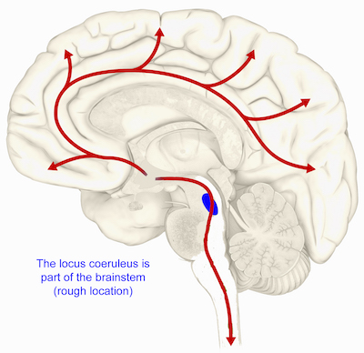
The LC, part of the brainstem, is thought to be the first region of the brain where pathological signs predicting future cellular degeneration show up. The LC (“blue spot”) gets its name from its blue color; it regulates attention, arousal, stress responses and cognition. The LC is also the major site for production of the neurotransmitter norepinephrine.

ApoE, which packages and transports cholesterol, was known to modulate the buildup of the toxic protein fragment beta-amyloid, but this proposed mechanism goes through Tau. Tau is the other pesky protein in Alzheimer’s, forming neurofibrillary tangles that are the earliest signs of degeneration in the brain. Tau pathology correlates better with dementia and cognitive impairments than beta-amyloid, which several proposed Alzheimer’s therapeutics act on.

The new paper shows that ApoE4 inhibits the enzyme VMAT2, which packages norepinephrine into vesicles. As a result, free/unpackaged norepinephrine lingers in the cytoplasm, and forms a harmful oxidative byproduct that triggers enzymatic degradation of Tau. Thus, norepinephrine may have a “too hot to handle” role in Alzheimer’s – with respect to the LC — somewhat analogous to dopamine in Parkinson’s, which has also been observed to form harmful byproducts. Dopamine and norepinephrine are similar chemically and both are substrates of VMAT2, so this relationship is not a stretch.

Model of how norepinephrine byproduct DOPEGAL triggers locus coeruleus degeneration through Tau

The Emory results make the case for inhibiting the enzyme AEP (asparagine endopeptidase), also known as delta-secretase, as an approach for heading off Alzheimer’s. AEP is the Tau-munching troublemaker, and is activated by the norepinephrine byproduct DOPEGAL

An alternative approach may be to inhibit monoamine oxidase (MAO-A above) enzymes — several old-school antidepressants are available that accomplish this.

At Emory, Ye’s lab has been tracing connections for AEP/delta-secretase in the last few years, and Weinshenker’s group is expert on all things norepinephrine, so the collaboration makes sense.

Delta-secretase’s name positions it in relation to beta- and gamma-secretase, enzymes for processing APP (amyloid precursor protein) into beta-amyloid, but AEP/delta-secretase has the distinction of having its fingers in both the beta-amyloid and Tau pies.

We have to caution that most of the recent research on delta-secretase has been in mouse models. Ye’s collaborators in China have been testing an inhibitor of delta-secretase in animals but it has not reached human studies yet, he reports. That said, this work has been oriented toward figuring out the web of interactions between known players such as ApoE and Tau, whose importance has been well-established in studies of humans with Alzheimer’s.

Posted on by Quinn Eastman in Neuro Leave a comment

Galanin: the ‘keep calm and carry on’ hormone?

A few celebrity neuropeptides have acquired a reputation – sometimes exaggerated — and a flavor, corresponding to their functions in the brain.

Oxytocin has the aura of a “cuddle hormone” because of its role in social bonding and reproduction. Endorphins are the body’s natural pain-killers, long thought to be responsible for “runner’s high.” Hypocretin/orexin, missing in narcolepsy, is a stabilizer of wakefulness as well as motivation.

Galanin, studied by Emory neuroscientist David Weinshenker’s lab, is not as flashy as other neuropeptides. While it is accumulating an intriguing track record, galanin appears to play subtly different roles depending on where it is expressed. It is tempting to call galanin the “keep calm and carry on” hormone, but the research on galanin is so complex it’s difficult to pin down.

Graduate student Rachel Tillage and colleagues have a paper this week in Journal of Neuroscience detailing how galanin’s production by one group of neurons in the brainstem confers stress resilience in mice.

This image shows the rough location for the locus coeruleus in the human brain. In mice, production of galanin in the locus coeruleus cushions against stress.

The new paper shows that exercise increases galanin in the locus coeruleus, a region in the brainstem that produces norepinephrine (important for attention, alertness, anxiety and muscle tone). Galanin can provide protection against the anxiety-inducing effects of artificial but very specific locus coeruleus activation by optogenetics.

Read more

Posted on by Quinn Eastman in Uncategorized Leave a comment

The blue spot: where seeds of destruction begin

Neuroscientist and geneticist David Weinshenker makes a case that the locus coeruleus (LC), a small region of the brainstem and part of the pons, is among the earliest regions to show signs of degeneration in both Alzheimer’s and Parkinson’s disease. You can check it out in Trends in Neurosciences.

The LC is the main source of the neurotransmitter norepinephrine in the brain, and gets its name (Latin for “blue spot”) from the pigment neuromelanin, which is formed as a byproduct of the synthesis of norepinephrine and its related neurotransmitter dopamine. The LC has connections all over the brain, and is thought to be involved in arousal and attention, stress responses, learning and memory, and the sleep-wake cycle.

Cells in the locus coeruleus are lost in mild cognitive impairment and Alzheimer’s. From Kelly et al Acta Neuropath. Comm. (2017) via Creative Commons

The protein tau is one of the toxic proteins tied to Alzheimer’s, and it forms intracellular tangles. Pathologists have observed that precursors to tau tangles can be found in the LC in apparently healthy people before anywhere else in the brain, sometimes during the first few decades of life, Weinshenker writes. A similar bad actor in Parkinson’s, alpha-synuclein, can also be detected in the LC before other parts of the brain that are well known for damage in Parkinson’s, such as the dopamine neurons in the substantia nigra.

“The LC is the earliest site to show tau pathology in AD and one of the earliest (but not the earliest) site to show alpha-synuclein pathology in PD,” Weinshenker tells Lab Land. “The degeneration of the cells in both these diseases is more gradual. It probably starts in the terminals/fibers and eventually the cell bodies die.” Read more

Posted on by Quinn Eastman in Neuro Leave a comment

Explainer: the locus coeruleus

The locus coeruleus is a part of the brain that has been getting a lot of attention recently from Emory neuroscience researchers.

The locus coeruleus is the biggest source of the neurotransmitter norepinephrine in the brain. Located deep in the brainstem, it has connections all over the brain, and is thought to be involved in arousal and attention, stress, memory, the sleep-wake cycle and balance.

Researchers interested in neurodegenerative disease want to look at the locus coeruleus because it may be one of the first structures to degenerate in diseases such as Alzheimer’s and Parkinson’s. In particular, the influential studies of German neuro-anatomist Heiko Braak highlight the locus coeruleus as a key “canary in the coal mine” indicator of neurodegeneration.

That’s why neurologist Dan Huddleston, working with biomedical imaging specialists Xiangchuan Chen and Xiaoping Hu and colleagues at Emory, has been developing a method for estimating the volume of the locus coeruleus by magnetic resonance imaging (MRI). Their procedure uses MRI tuned in such a way to detect the pigment neuromelanin (see panel), which accumulate in both the locus coeruleus and in the substantia nigra. Read more

Posted on by Quinn Eastman in Neuro Leave a comment

Study: Regular aerobic exercise and prevention of drug abuse relapse

Exercise provides health benefits

Researchers at Emory University and the University of Georgia have received funding from the National Institutes of Health to study the neurobiological mechanisms for how regular aerobic exercise may prevent drug abuse relapse. The grant is for $1.9 million over the next five years.

David Weinshenker, PhD, associate professor of human genetics, Emory School of Medicine, is a co-principal investigator on the project.

David Weinshenker, PhD

“This research will provide new insight into how regular exercise may attenuate drug abuse in humans,” Weinshenker says. “More importantly, it may reveal a neural mechanism through which exercise may prevent the relapse into drug-seeking behavior.”

During the study, Weinshenker and UGA co-investigator Philip Holmes, professor of psychology in the Franklin College of Arts and Sciences, will measure exercise-induced increases of the galanin gene activity in the rat brain.

Read more

Posted on by admin in Uncategorized Leave a comment