Warren symposium follows legacy of geneticist giant

If we want to understand how the brain creates memories, and how genetic disorders distort the brain’s machinery, then the fragile X gene is an ideal place to start. That’s why the Stephen T. Warren Memorial Symposium, taking place November 28-29 at Emory, will be a significant event for those interested in neuroscience and genetics. Stephen T. Warren, 1953-2021 Warren, the founding chair of Emory’s Department of Human Genetics, led an international team that discovered Read more

Mutations in V-ATPase proton pump implicated in epilepsy syndrome

Why and how disrupting V-ATPase function leads to epilepsy, researchers are just starting to figure Read more

Tracing the start of COVID-19 in GA

At a time when COVID-19 appears to be receding in much of Georgia, it’s worth revisiting the start of the pandemic in early 2020. Emory virologist Anne Piantadosi and colleagues have a paper in Viral Evolution on the earliest SARS-CoV-2 genetic sequences detected in Georgia. Analyzing relationships between those virus sequences and samples from other states and countries can give us an idea about where the first COVID-19 infections in Georgia came from. We can draw Read more

Heart

Targeting naked DNA in the heart

Hoechst-Structure-300px

The first thing that comes up in a Google search for “Hoechst” is the family of fluorescent dyes used to stain DNA in cells before microscopy. The Hoechst dyes derive their names from their manufacturer: a company, now part of Sanofi, named after the town where it was founded, which is now part of Frankfurt, Germany. The word itself means “highest [spot].”

Although DNA runs the show in every cell, it’s usually well-hidden inside the nucleus or the mitochondria. Extracellular DNA’s presence is a signal that injury is happening and cells are dying.

Biomedical engineer Mike Davis and collaborator Niren Murthy have been exploiting the properties of a DNA-binding dye called Hoechst 33342, often used to stain DNA in cells before microscopy. The dye can only bind DNA if it can get to the DNA – that is, if membranes are broken. This property makes the dye a good way to target injured tissue, either as an imaging agent or for therapy.

At the recent Pediatric Healthcare Innovation retreat, Davis discussed the potential use of such Hoechst derivatives to diagnose myocarditis (inflammation of the heart muscle) in children.

In addition, in a recent paper published in Scientific Reports, Davis and his colleagues attach the Hoechst dye to the cardioprotective growth factor IGF-1, creating a version of IGF-1 that is targeted to injured heart muscle. The first author of the paper is cardiology fellow Raffay Khan, MD. Screen Shot 2014-04-24 at 1.18.35 PM
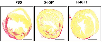
IGF-1 has shown a lot of potential for treating heart disease, but it’s not the most cooperative as a drug, because it doesn’t last long in the body and doesn’t stick around in the heart. Linked up to the dye, IGF-1 behaves better. When used to treat mouse hearts after a heart attack, the Hoechst-IGF-1 treated-hearts have better function and less scar tissue (seen here as red).

The authors conclude:

With the broad chemistry surrounding functionalized PEG used to create Hoechst derivatives, it may be possible to target other therapies such as cells, small molecules, and even nanoparticles. We believe that the use of DNA binding agents such as Hoechst can be used to target exposed DNA in other diseases where necrotic cell death plays a critical role and could be used as a platform therapy.


Posted on by Quinn Eastman in Heart Leave a comment

Addendum on CRISPR

An excellent example of the use of CRISPR gene editing technology came up at the Emory-Children’s Pediatric Research Center’s Innovation Conference this week.

Marcela Preininger, who is working with cardiomyocyte stem cell specialist Chunhui Xu, described her work (poster abstract 108) on cells derived from a 12 year old patient with an inherited cardiac arrhythmia syndrome: catecholaminergic polymorphic ventricular tachycardia or CPVT. Her team has obtained skin fibroblasts from the patient, and converted those cells into induced pluripotent stem cells, which can then be differentiated into cardiac muscle cells or cardiomyocytes.

Working with TJ Cradick, director of the Protein Engineering Facility at Georgia Tech, Preininger is testing out CRISPR gene editing as a means of correcting the defect in this patient’s cells, outside the body. Cradick says that while easy and efficient, RNA-directed CRISPR can be lower in specificity compared to the protein-directed TALEN technology.

From Preininger’s abstract:

Once the mutation has been corrected at the stem cell level, we will investigate whether the repaired (mutation-free) iPS cells can be differentiated into functional cardiomyocytes with normal Ca2+ handling properties, while closely monitoring the cells for mutagenic events. Pharmacological restoration of the normal myocardial phenotype will also be optimized and explored in our model.

Posted on by Quinn Eastman in Heart Leave a comment

A spoonful of sugar helps infection detection

Congratulations to Kiyoko Takemiya, a postdoctoral fellow in Emory’s Division of Cardiology, working with W. Robert Taylor. At the recent American College of Cardiology meeting in Washington DC, she won first place in the competition for an ACC Foundation/ Herman K. Gold Young Investigators Award in Molecular and Cellular Cardiology.

The title of her research presentation was: A Novel Imaging Probe for the Detection of Subclinical Bacterial Infections Involving Cardiac Devices.

Takemiya, Taylor, and their colleagues (including Mark Goodman and Niren Murthy, formerly at Georgia Tech and now at UC Berkeley) developed a fluorescent probe that allows the detection of small levels of bacteria on cardiac devices. The probe was tested in rats, some of which had relatively mild local S. aureus infections. The fluorescent probe (PET is also under investigation) makes use of the properties of maltohexaose, a sugar that is taken up by bacteria but not mammalian cells.

Infection rates for implantable cardiac devices such as pacemakers have been rising, according to a 2012 paper in NEJM.

Posted on by Quinn Eastman in Heart Leave a comment

Signs of future high blood pressure in college football players

College football players tend to have stiffer arteries than other college students, even before their college athletic careers have started, cardiology researchers have found.

Although football players had lower blood pressure in the pre-season than a control group of undergraduates, stiffer arteries could potentially predict players’ future high blood pressure, a risk factor for stroke and heart disease later in life.

Researchers studied 50 freshman American-style football players from two Division I programs, Georgia Tech and Harvard, in the pre-season and compared them with 50 healthy Emory undergraduates, who were selected to roughly match their counterparts in age and race. The research is part of a longer ongoing study of cardiovascular health in Georgia Tech college football players.

The results were presented Saturday at the American College of Cardiology meeting in Washington DC, by cardiology research fellow Jonathan Kim, MD. Kim worked with Arshed Quyyumi, MD, director of Emory’s Clinical Cardiovascular Research Institute, Aaron Baggish, MD, associate director of the Cardiovascular Performance Program at Massachusetts General Hospital, and their colleagues.

“It’s remarkable that these vascular differences are apparent in the pre-season, when the players are essentially coming out of high school,” says Kim. “We aim to gain additional insight by following their progress during the season.”

Despite being physically active and capable, more than half of college football players were previously found to develop hypertension by the end of their first season. Professional football players also tend to have higher blood pressure, even though other risk factors such as cholesterol and blood sugar look good, studies have found. Researchers have previously proposed that the intense stop-and-start nature of football as well as the physical demands of competitive participation, such as rapid weight gain, could play roles in making football distinctive in its effects on cardiovascular health.

In the current study, the control undergraduates had higher systolic and diastolic blood pressure than the football players: (football players: 111/63; control: 118/72). However, the football players displayed significantly higher pulse wave velocity, a measure of arterial stiffness (football: 6.5 vs control: 5.7). Pulse wave velocity is measured by noninvasive devices that track the speed of blood flow by calculating differences between arteries in the neck and the leg.

“It is known that in other populations, increased pulse wave velocity precedes the development of hypertension,” Kim says. “We plan to test this relationship for football players.”

The football players were markedly taller and larger than the control group (187 vs 178 centimeters in height, body mass index 29.2 vs 23.7). The football players also reported participating in more hours of weight-training per week than the control group (5.4 vs 2.6).


Posted on by Quinn Eastman in Heart Leave a comment

Molecular signature of heart attack predicts longer-term outcomes

A molecular signature seen in blood from patients who are experiencing an acute heart attack may also predict the risk of cardiovascular death over the next few years, Emory researchers have found.

The results were presented Monday at the American College of Cardiology meeting in Washington DC by cardiovascular research fellow Nima Ghasemzadeh, MD. Ghasemzadeh is working with Arshed Quyyumi, MD, director of Emory’s Clinical Cardiovascular Research Center, as well as Greg Gibson, PhD, director of the Integrative Genomics Center at Georgia Tech.

Ghasemzadeh and colleagues examined 337 patients undergoing cardiac catheterization at Emory. Just 18 percent of the patients in this group were having a heart attack. This research is a reminder that the majority of patients who undergo cardiac catheterization, and thus are suspected of experiencing a heart attack, are not actually having one at that moment. Read more

Posted on by Quinn Eastman in Heart, Uncategorized Leave a comment

Cardiologists change their minds — not all at once

What happens when cardiologists learn that a widely performed procedure might not be as helpful as they once thought? Investigators at Emory have taken one of the first detailed looks at how geographical patterns in practice changed after publication of results from a large clinical trial.

The federal government has invested billions of dollars in comparative effectiveness research — comparing different healthcare interventions to determine which works best — with the aim of reducing variations in care. This paper shows that these types of investment can have the desired effect.

Recently Medscape Cardiology talked with lead author Arun Mohan, who is medical director for care coordination at Emory University Hospital, about his work. [You may have missed this news item over the holidays.] Read more

Posted on by Quinn Eastman in Heart Leave a comment

BioArt: amyloid in the heart

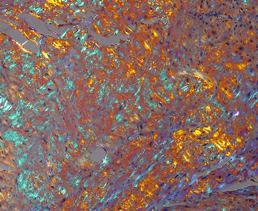
What Abstract Expressionist artist painted this? Jackson Pollock?LewisW2013

Actually, the photo depicts amyloid plaques, a frequent topic in the context of Alzheimer’s disease. Pathologist William Lewis‘ photo reminds us that amyloid can also appear in the heart.

Amyloidosis of the heart is a set of complex diseases caused by the accumulation of cellular proteins that form an amyloid plaque. Although http://www.oakleyonorder.com/ amyloidosis was described more than 100 years ago, the causative proteins were not identified until recent chemical analyses were conducted. This image shows an amyloid plaque stained with Congo red stain and viewed through a polarized lens. The optical properties of the amyloid-forming protein cause it to appear green, while other matrix materials within the plaque appear as orange and blue.

The photo, which was one of the winners of the FASEB (Federation of American Societies for Experimental Biology) 2013 BioArt competition, was featured on NIH director Francis Collins’ blog this week.

Lewis, who studies the effects of antiretroviral drugs on the cardiovascular system in his laboratory, reports that he came across the amyloid tissue sample as part of his duties as director of cardiovascular pathology: “It was beautiful.”

Posted on by Quinn Eastman in Heart Leave a comment

Fragile but potent: RNA delivered by nanoparticle

An intriguing image for November comes from biomedical engineer Mike Davis’ lab, courtesy of BME graduate student Inthirai Somasuntharam.

Each year, thousands of children undergo surgery for congenital heart defects. A child’s heart is more sensitive to injury caused by interrupting blood flow during surgery, and excess reactive oxygen species are a key source of this damage.

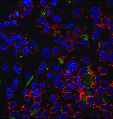
Macrophages with blue nuclei and red cytoskeletons, being treated with green nano particles. The particles carry RNA that shut off reactive oxygen species production.

Macrophages with blue nuclei and red cytoskeletons, being treated with green nano particles. The particles carry RNA that shut off reactive oxygen species production.

Davis and his colleagues are able to shut off cheap oakley reactive oxygen species at the source by targeting the NOX (NADPH oxidase*) enzymes that produce them. This photo, from a 2013 Biomaterials paper, shows green fluorescent nanoparticles carrying small interfering RNA. The RNA precisely shuts down one particular gene encoding a NOX enzyme. Eventually, similar nanoparticles may shield the heart from damage during pediatric heart surgery.

In the paper, Somasuntharam used particles made of a slowly dissolving polymer called polyketals. The particles delivered fragile but potent RNA molecules into macrophages, inflammatory cells that swarm into cardiac tissue after a heart attack. Davis and Georgia Tech colleague Niren Murthy previously harnessed this polymer to deliver drugs that can be toxic to the rest of the body.

The polyketal particles are especially well-suited for delivering a payload to macrophages, since those types of cells (as the name implies) are big eaters. Davis reports his lab has been working on customizing the particles so they can deliver RNA molecules into cardiac muscle cells as well.

*While we’re on the topic of NADPH oxidases, Susan Smith and David Lambeth have been looking for and finding potential drugs that inhibit them.

Posted on by Quinn Eastman in Heart Leave a comment

Dealing with huff-puff? Think HFpEF

For this month’s Current Concept feature, we would like to explain a term from cardiology that is likely to become more prominent:

“Heart failure with preserved ejection fraction” (abbreviated as HFpEF and pronounced “heff-peff”).

Javed Butler, MD, an Emory expert on heart failure and deputy chief science officer for the American Heart Association, laid out in a recent seminar why this category of patients is so important. Look for more from him on this topic in the future.

Three points:

  1. The number of HFpEF patients is growing and they now make up the majority of patients with heart failure in the United States.
  2. No treatments have been proven to benefit them, in terms of reducing mortality.* In clinical studies, medications such as ACE inhibitors, angiotensin receptor blockers and beta-blockers have not helped.
  3. Once hospitalized, HFpEF patients have a high rate of readmission to the hospital within 30 days. The federal Medicare program is penalizing hospitals that have high rates of readmissions and heart failure is one of the largest contributors to readmissions.

The symptoms that drive people with HFpEF to the hospital are mainly fatigue and dyspnea, or shortness of breath, along with fluid in the lungs and swelling of the limbs. Along with heart failure, HFpEF patients often have conditions such as hypertension, anemia, diabetes, kidney disease or sleep apnea. Read more

Posted on by Quinn Eastman in Heart Leave a comment

Packaging stem cells in capsules for heart therapy

Stem cell therapy for heart disease is happening. Around the world, thousands of heart disease patients have been treated in clinical studies with some form of bone marrow cells or stem cells. But in many of those studies, the actual impact on heart function was modest or inconsistent. One reason is that most of the cells either don’t stay in the heart or die soon after being introduced into the body.

Cardiology researchers at Emory have a solution for this problem. The researchers package stem cells in a capsule made of alginate, a gel-like substance. Once packaged, the cells stay put, releasing their healing factors over time.

Researchers used encapsulated mesenchymal stem cells to form a “patch” that was applied to the hearts of rats after a heart attack. Compared with animals treated with naked cells (or with nothing), rats treated with the capsule patches displayed increased heart function, reduced scar size and more growth of new blood vessels a month later. In addition, many more of the encapsulated cells stayed alive. Read more

Posted on by Quinn Eastman in Heart Leave a comment