Warren symposium follows legacy of geneticist giant

If we want to understand how the brain creates memories, and how genetic disorders distort the brain’s machinery, then the fragile X gene is an ideal place to start. That’s why the Stephen T. Warren Memorial Symposium, taking place November 28-29 at Emory, will be a significant event for those interested in neuroscience and genetics. Stephen T. Warren, 1953-2021 Warren, the founding chair of Emory’s Department of Human Genetics, led an international team that discovered Read more

Mutations in V-ATPase proton pump implicated in epilepsy syndrome

Why and how disrupting V-ATPase function leads to epilepsy, researchers are just starting to figure Read more

Tracing the start of COVID-19 in GA

At a time when COVID-19 appears to be receding in much of Georgia, it’s worth revisiting the start of the pandemic in early 2020. Emory virologist Anne Piantadosi and colleagues have a paper in Viral Evolution on the earliest SARS-CoV-2 genetic sequences detected in Georgia. Analyzing relationships between those virus sequences and samples from other states and countries can give us an idea about where the first COVID-19 infections in Georgia came from. We can draw Read more

T cells

In current vaccine research, adjuvants are no secret

Visionary immunologist Charlie Janeway was known for calling adjuvants – vaccine additives that enhance the immune response – a “dirty little secret.”

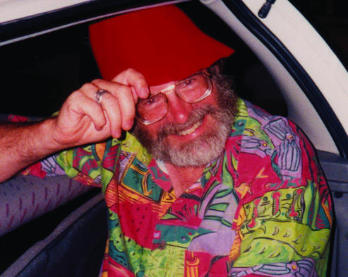
Charlie Janeway, MD, in a hat he wore often

Janeway’s point was that foreign antigens, by themselves, were unable to stimulate the components of the adaptive immune system (T and B cells) without signals from the innate immune system. Adjuvants facilitate that help.

By now, adjuvants are hardly a secret, looking at some of the research that has been coming out of Emory Vaccine Center. This week, an analysis by Ali Ellebedy, now at Washington University St Louis, and colleagues showed that in healthy volunteers, the AS03 adjuvant boosted otherwise poor immune responses to a limited dose of the exotic avian flu H5N1, recruiting both memory and naïve B cells. More on that here.

The Moderna SARS-CoV-2 vaccine, which has shown some activity in a small clinical trial here at Emory, has its own kind of adjuvant, since it’s made of both innate-immune-stimulating mRNA and clothed in lipid nanoparticles. Extra adjuvants may come into play later, either with this vaccine or others.

A question we’ve seen many people asking, and discussed on Twitter etc is this: how long does the immunity induced by a SARS-CoV-2 vaccine last? How can we make the immune cells induced by a vaccine stick around for a long time? Read more

Posted on by Quinn Eastman in Immunology Leave a comment

Microbiome critical for bone hormone action

Intestinal microbes are necessary for the actions of an important hormone regulating bone density, according to two papers from the Emory Microbiome Research Center. The papers represent a collaboration between Roberto Pacifici, MD and colleagues in the Department of Medicine and laboratory of Rheinallt Jones, PhD in the Department of Pediatrics.

Together, the results show how probiotics or nutritional supplementation could be used to modulate immune cell activity related to bone health. The two papers, published in Nature Communications and Journal of Clinical Investigation, are the first reports of a role for intestinal microbes in the mechanism of action of PTH (parathyroid hormone), Pacifici says.

PTH increases calcium levels in the blood and can either drive bone loss or bone formation, depending on how it is produced or administered. Continuous excessive production of PTH, or primary hyperparathyroidism, is a common endocrine cause of osteoporosis. Yet in another context, intermittent external PTH stimulates bone formation, and is an FDA-approved treatment for osteoporosis – also used off-label for fracture repair in athletes. Read more

Posted on by Quinn Eastman in Immunology Leave a comment

Transplant research: immune control via Fc receptors on T cells

Emory transplant researchers have identified a control mechanism the immune system uses to tamp down chronic inflammation. The findings provide insight into how some people were able to stop taking immunosuppressive drugs after kidney transplant.

In addition, they may be important for a full understanding of how many drugs for cancer and autoimmune disorders (therapeutic antibodies) work. The results were published on January 14 in Immunity.

In a twist, scientists have known about the molecules involved for a long time. They’re Fc receptors. Usually, we can think of them acting like oven mitts that immune cells use to grab onto antibodies. Fc receptors bind the constant (unvarying) portions of antibodies, which are the same no matter what they’re directed against.

Mandy Ford, PhD and graduate student Anna Morris

The news here is that an inhibitory variety of Fc receptor – FcγRIIB — is found on CD8+ T cells, and is a way of squeezing off T cell activity. Dogma over the past few decades held that T cells do not express Fc receptors, although evidence for them doing so went back to the 1970s.

“Our data suggest that the physiologic relevance of this pathway is to allow for control of active, highly differentiated effector T cells in the setting of chronic inflammation in order to limit immune pathology,” says senior author Mandy Ford, PhD, scientific director of Emory Transplant Center.

The co-first authors of the paper are IMP graduate student Anna Morris and surgical resident Clara Farley. They and their colleagues probed the functions of FcγRIIB on T cells in mice, and also found that increased expression of FcγRIIB correlated with freedom from rejection following withdrawal from immunosuppression in a clinical trial of kidney transplant recipients. This data came from the CTOT09 study from the Clinical Trials in Organ Transplantation Consortium. Read more

Posted on by Quinn Eastman in Immunology Leave a comment

Transition to exhaustion: clues for cancer immunotherapy

Research on immune cells “exhausted” by chronic viral infection provides clues on how to refine cancer immunotherapy. The results were published Tuesday, Dec. 3 in Immunity.

Scientists at Emory Vaccine Center, led by Rafi Ahmed, PhD, have learned about exhausted CD8 T cells, based on studying mice with chronic viral infections. In the presence of persistent virus or cancer, CD8 T cells lose much of their ability to fight disease, and display inhibitory checkpoint proteins such as PD-1 on their surfaces. PD-1 is targeted by cancer immunotherapy drugs, such as pembrolizumab and nivolumab, which allow CD8 T cells to regain their ability to attack and kill infected cells and cancers.

Those drugs are now FDA-approved for several types of cancer, yet some types of tumors do not respond to them. Studying exhausted CD8 T cells can help us understand how to better draw the immune system into action against cancer or chronic infections.

In previous research, Ahmed’s lab found that exhausted cells are not all alike, and the diversity within the exhausted T cell pool could explain variability in responses to cancer immunotherapy drugs. Specifically, they observed that a population of “stem-like” cells proliferated in response to PD-1-blocking drugs, while a more differentiated population of exhausted cells stayed inactive. The stem-like cells are responsible for maintaining the exhausted T cell population, but cannot kill virus-infected or tumor cells on their own.

The current paper defines a transitional stage in between the stem-like and truly exhausted cells. The truly exhausted cells are marked by a molecule called CD101, and are unable to migrate to sites of infection and contain lower amounts of proteins needed to kill infected or tumor cells.

“The transitional cells are not completely exhausted,” says postdoctoral fellow Will Hudson, PhD, first author of the Immunity paper. “They are still capable of proliferating and performing their ‘killer cell’ functions. In our experiments, they contribute to viral control.”

The transitional cells, lacking CD101, could be a good marker for response to PD-1 blocking drugs, Hudson says. Enhancing the proliferation or survival of these cells, or preventing their transition to lasting exhaustion, may be a novel therapeutic strategy for cancer. Read more

Posted on by Quinn Eastman in Immunology Leave a comment

Immunologists identify T cell homing beacons for lungs

Scientists have identified a pair of molecules critical for T cells, part of the immune system, to travel to and populate the lungs. A potential application could be strengthening vaccines against respiratory pathogens such as influenza.

The findings were published online Thursday, September 26 in Journal of Experimental Medicine.

T cells in the lungs, courtesy of Alex Wein. Blue represents respiratory epithelium (EpCAM), while various T cells stain red, yellow or green.

Much research on immunity to influenza virus focuses on antibodies, infection- or vaccine-induced proteins in the blood that can smother viruses. But CD8 T cells, which survey other cells for signs of viral infection and kill infected cells, are an important arm of our defenses too. The epitopes – or bits of viral protein – they recognize generally do not change from year to year.

Researchers led by Jacob Kohlmeier, PhD, at Emory University School of Medicine wanted to learn more about what’s needed to get CD8 T cells into the lungs, since the lungs will often contain the first cells incoming virus will have a chance to infect. However, T cells don’t stick around in the lungs for extended amounts of time.

“The airways are a unique environment in the body,” says Alex Wein, a MD/PhD student who trained in Kohlmeier’s lab. “They’re high in oxygen but low in nutrients. Unlike other tissues, when T cells enter the airways, it’s a one-way trip and they have a half-life of a few weeks, so they must be continually repopulated.”

Wein, his fellow MD/PhD Sean McMaster, now at Boston Consulting Group, and Shiki Takamura at Kindai University are co-first authors of the paper. Kohlmeier is assistant professor of microbiology and immunology and part of the Emory-UGA Center of Excellence for Influenza Research and Surveillance.

The researchers showed that two molecules, called CXCR6 and CXCL16, are needed for CD8 T cells to reach the airways in mice. CXCR6 is found on T cells and CXCL16 is produced by the epithelial cells lining the airways of the lungs. Read more

Posted on by Quinn Eastman in Immunology Leave a comment

Sensitive to (transplant) rejection

An experimental screening method, developed by Emory and Georgia Tech scientists, aims to detect immune rejection of a transplanted organ earlier and without a biopsy needle.

The technology is based on nanoparticles that detect granzyme B enzymes produced by killer T cells. When the T cells are active, they slice up the nanoparticles, generating a fluorescent signal that is detectable in urine. The results from a mouse skin graft model were published in Nature Biomedical Engineering, from Gabe Kwong’s lab at GT and Andrew Adams’ at Emory. More extensive story here.

Co-first authors Quoc Mac and Dave Mathews

Adams is also developing technologies for imaging transplant rejection via immunoPET, with Georgia Tech’s Phil Santangelo.


Posted on by Quinn Eastman in Immunology Leave a comment

2B4: potential immune target for sepsis survival

Emory immunologists have identified a potential target for treatments aimed at reducing mortality in sepsis, an often deadly reaction to infection.

2B4 is an inhibitory molecule found on immune cells. You may have heard of PD1, which cancer immunotherapy drugs block in order to re-energize the immune system. 2B4 appears to be similar; it appears on exhausted T cells after chronic viral infection, and its absence can contribute to autoimmunity.

In their new paper in Journal of Immunology, Mandy Ford, Craig Coopersmith and colleagues show that 2B4 levels are increased on certain types of T cells (CD4+ memory cells) in human sepsis patients and in a mouse model of sepsis called CLP (cecal ligation + puncture). Genetically knocking out 2B4 or blocking it with an antibody both reduce mortality in the CLP model. The effect of the knockout is striking: 82 percent survival vs 13 percent for controls.

How does it work? When fighting sepsis, 2B4 knockout animals don’t have reduced bacterial levels, but they do seem to have CD4+ T cels that survive better. CD4+ T cells, especially memory cells, get killed in large numbers during sepsis, and this is thought to contribute to mortality. Read more

Posted on by Quinn Eastman in Immunology Leave a comment

Revived T cells still need fuel

Cancer immunotherapy drugs blocking the PD-1 pathway – known as checkpoint inhibitors – are now FDA-approved for melanoma, lung cancer and several other types of cancer. These drugs are often described as “releasing the brakes” on dysfunctional T cells.

A new study from Emory Vaccine Center and Winship Cancer Institute researchers shows that even if the PD-1-imposed brakes are released, the tumor-specific T cells still need “fuel” to expand in numbers and restore effective immune responses. That fuel comes from co-stimulation through a molecule called CD28.

The results were published Thursday by the journal Science.

Despite the success of PD-1-targeting drugs, many patients’ tumors do not respond to them. The study’s findings indicate that CD28’s presence on T cells could be a clinical biomarker capable of predicting whether drugs targeting PD-1 will be effective. In addition, the requirement for CD28 suggests that co-stimulation may be missing for some patients, which could guide the design of combination therapies.

For the rest of our press release and quotes from authors Rafi Ahmed, Alice Kamphorst and Suresh Ramalingam, please go here. For some additional links and thoughts on PD-1 and CD28, read on:

Read more

Posted on by Quinn Eastman in Cancer, Immunology Leave a comment

Antiviral success makes some immune cells stickier

As they succeed in clearing a viral infection from the body, some virus-hunting T cells begin to stick better to their target cells, researchers from Emory Vaccine Center and Georgia Tech have discovered.

The increased affinity helps the T cells kill their target cells more efficiently, but it depends both on the immune cells’ anatomic location and the phase of the infection.

The results were published this week in the journal Immunity.

Arash Grakoui, PhD

Arash Grakoui, PhD

After the peak of the infection, cells within the red pulp of the spleen or in the blood displayed a higher affinity for their targets than those within the white pulp. However, the white pulp T cells were more likely to become long-lasting memory T cells, critical for vaccines.

“These results provide a better understanding of how memory precursor populations are established and may have important implications for the development of efficacious vaccines,” the scientists write.

In the mouse model the researchers were using, the differences in affinity were only detectable a few days after the non-lethal LCMV viral infection peaks. How the differences were detected illustrates the role of serendipity in science, says senior author Arash Grakoui, PhD.

Typically, the scientists would have taken samples only at the peak (day 7 of the infection) and weeks later, when memory T cells had developed, Grakoui says. In January 2014, the weather intervened during one of these experiments. Snow disrupted transportation in the Atlanta area and prevented postdoctoral fellow Young-Jin Seo, PhD from taking samples from the infected mice until day 11, which is when the differences in affinity were apparent.

Seo and Grakoui collaborated with graduate student Prithiviraj Jothikumar and Cheng Zhu, PhD at Georgia Tech, using a technique Zhu’s laboratory has developed to measure the interactions between T cells and their target cells. Co-author Mehul Suthar, PhD performed gene expression analysis.

Read more

Posted on by Quinn Eastman in Immunology Leave a comment

Improving long-term outcomes after kidney transplant

Twenty years of research and you start to improve outcomes for transplant patients.

The Nature paper from Chris Larsen and Tom Pearson on “costimulation blockers” and their ability to head off graft rejection in rodents first appeared in 1996.

Almost 20 years later, a seven-year study of kidney transplant recipients has shown that the drug belatacept, a costimulation blocker based on Larsen and Pearson’s research, has a better record of patient and organ survival than a calcineurin inhibitor, previously the standard of care.

Kidney transplant recipients need to take drugs to prevent their immune systems from rejecting their new organs, but the drugs themselves can cause problems. Long-term use of calcineurin inhibitors, such as tacrolimus, can damage the transplanted kidneys and lead to cardiovascular disease and diabetes.

In the accompanying video, Larsen - now dean of Emory University School of Medicine – and Pearson - executive director of Emory Transplant Center – explain.

Belatacept was approved by the FDA in 2011 and is produced by Bristol Myers Squibb. Results from the BENEFIT study of belatacept, led by Larsen and UCSF transplant specialist Flavio Vincenti, were published in the Jan. 28 issue of the New England Journal of Medicine.

To go with the paper, NEJM has an editorial with some revealing statistics (more than 14,000 of the 101,000 patients listed for kidney transplantation are waiting for a repeat transplant) and a explanatory video. MedPage Today has an interview with Larsen, and HealthDay has a nice discussion of the issues surrounding post-transplant drugs. Read more

Posted on by Quinn Eastman in Immunology Leave a comment