Warren symposium follows legacy of geneticist giant

If we want to understand how the brain creates memories, and how genetic disorders distort the brain’s machinery, then the fragile X gene is an ideal place to start. That’s why the Stephen T. Warren Memorial Symposium, taking place November 28-29 at Emory, will be a significant event for those interested in neuroscience and genetics. Stephen T. Warren, 1953-2021 Warren, the founding chair of Emory’s Department of Human Genetics, led an international team that discovered Read more

Mutations in V-ATPase proton pump implicated in epilepsy syndrome

Why and how disrupting V-ATPase function leads to epilepsy, researchers are just starting to figure Read more

Tracing the start of COVID-19 in GA

At a time when COVID-19 appears to be receding in much of Georgia, it’s worth revisiting the start of the pandemic in early 2020. Emory virologist Anne Piantadosi and colleagues have a paper in Viral Evolution on the earliest SARS-CoV-2 genetic sequences detected in Georgia. Analyzing relationships between those virus sequences and samples from other states and countries can give us an idea about where the first COVID-19 infections in Georgia came from. We can draw Read more

dystonia

Redrawing the brain’s motor map

Neuroscientists at Emory have refined a map showing which parts of the brain are activated during head rotation, resolving a decades-old puzzle. Their findings may help in the study of movement disorders affecting the head and neck, such as cervical dystonia and head tremor.

The results were published in Journal of Neuroscience.

In landmark experiments published in the 1940s and 50s, Canadian neurosurgeon Wilder Penfield and colleagues determined which parts of the motor cortex controlled the movements of which parts of the body.

Penfield stimulated the brain with electricity in patients undergoing epilepsy surgery, and used the results to draw a “motor homunculus”: a distorted representation of the human body within the brain. Penfield assigned control of the neck muscles to a region between those that control the fingers and face, a finding inconsistent with some studies that came later.

Using modern functional MRI (magnetic resonance imaging), researchers at Emory University School of Medicine have shown that the neck’s motor control region in the brain is actually between the shoulders and trunk, a location that more closely matches the arrangement of the body itself.

“We can’t be that hard on Penfield, because the number of cases where he was able to study head movement was quite limited, and studying head motion as he did, by applying an electrode directly to the brain, creates some challenges,” says lead author Buz Jinnah, MD, professor of neurology, human genetics and pediatrics at Emory University School of Medicine. Read more

Posted on by Quinn Eastman in Neuro Leave a comment

New molecular target in dystonia

Emory researchers led by pharmacologist Ellen Hess have identified a new molecular target in dystonia. Their findings, recently published in the Journal of Pharmacology and Experimental Therapeutics, could help doctors find drugs for treating the movement disorder.

Ellen Hess, PhD

Dystonia gives sufferers involuntary muscle contractions that cause rigid, twisting movements and abnormal postures. It is the third most common movement disorder, after tremor and Parkinson’s disease. Neurologists can sometimes use drugs to address the symptoms of dystonia but there is no cure.

A 2008 review by Hess (PDF) concludes that compared with other neurological disorders, “our understanding of the biology and potential treatments for dystonia is in its infancy.” Still, scientists have known for a while that the cerebellum, a region of the brain that regulates movement, is involved.

“We focused on the cerebellum because studies in patients with dystonia often show that this part of the brain is more active, when examined by MRI,” Hess says. “The abnormal overactivity of the cerebellum is seen in patients with all different types of dystonia, so it seems to be a common hotspot. Our goal was to understand what might be causing the overactivity in mice because if we can stop the overactivity, we might be able to stop the dystonia.”

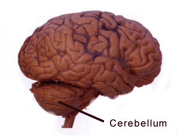
Hess and her colleagues discovered that drugs that stimulate AMPA receptors induce dystonia when introduced into the mouse cerebellum. Their results suggest that drugs that act in reverse, blocking AMPA receptors, could be used to treat dystonia.

Postdoctoral fellow Xueliang Fan is the first author of the paper. Emory neurologist H.A. Jinnah, director of a NIH-supported network of clinical research sites focusing on dystonia, is a co-author.

AMPA receptors are a subset of glutamate receptors, a large group of “receiver dishes” for excitatory signals in the brain. Fan performed a variety of experiments to show that AMPA receptor activity plays a specific role in generating dystonia. For example, drugs that affect other types of glutamate receptors did not induce dystonia. AMPA receptor blockers can also reduce dystonia in a genetic model, the “tottering” mouse.

Although pharmaceutical companies have already been testing AMPA receptor blockers as potential antiseizure drugs, caution is in order. AMPA receptor stimulators/ enhancers (or “ampakines”) have been identified as potential enhancers of learning and memory, so AMPA receptor blockers may interfere with those processes.

“Our results suggest that reducing AMPA receptor activity could help alleviate dystonia but we still have a lot of work to do before we know whether blocking AMPA receptor activity in patients will really help,” Hess says. “Since there aren’t many drugs that act at AMPA receptors, one of our goals is to identify drugs that change the ‘downstream’ effects of AMPA receptor activation. For example, we may be able to find other drug classes that change neuronal activity in the same way that AMPA receptor blockade changes activity.”

Posted on by Quinn Eastman in Neuro Leave a comment