

This month’s Image feature highlights lamellipodia, the thin sheet-like regions at the leading edges of migrating cells. Lamellipodia act as tiny creeping motors that pull the cell forward.
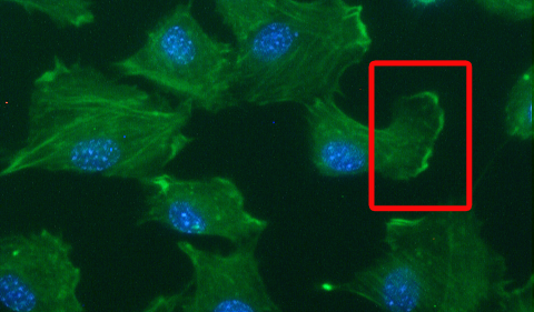
To help visualize lamellipodia, Adriana Simionescu-Bankston, a graduate student in Grace Pavlath’s lab, provided us with this photo of muscle cells. The red box shows an example of lamellipodia. Notice the edge of the cell, where the green color is more intense.
The green color comes from FITC-phalloidin, which stains F-actin, the Ray Ban outlet filaments that make up a large part of the cells’ internal skeleton. (Phalloidin is an actin-binding toxin originally isolated from death cap mushrooms, and FITC is what makes it green.) The blue color comes from DAPI, a dye that stains the DNA in the nucleus.
Simionescu-Bankston and Pavlath recently published a paper in the journal Developmental Biology, examining the function of a protein called Bin3 in muscle development and regeneration. They found that Bin3 appears to regulate lamellipodia formation; in mice that lack Bin3, muscle cells have fewer lamellipodia and the muscle tissues regenerate slower after injury. Bin3 is also important in the eye, since the “knockout” mice develop cataracts soon after birth.