Warren symposium follows legacy of geneticist giant

If we want to understand how the brain creates memories, and how genetic disorders distort the brain’s machinery, then the fragile X gene is an ideal place to start. That’s why the Stephen T. Warren Memorial Symposium, taking place November 28-29 at Emory, will be a significant event for those interested in neuroscience and genetics. Stephen T. Warren, 1953-2021 Warren, the founding chair of Emory’s Department of Human Genetics, led an international team that discovered Read more

Mutations in V-ATPase proton pump implicated in epilepsy syndrome

Why and how disrupting V-ATPase function leads to epilepsy, researchers are just starting to figure Read more

Tracing the start of COVID-19 in GA

At a time when COVID-19 appears to be receding in much of Georgia, it’s worth revisiting the start of the pandemic in early 2020. Emory virologist Anne Piantadosi and colleagues have a paper in Viral Evolution on the earliest SARS-CoV-2 genetic sequences detected in Georgia. Analyzing relationships between those virus sequences and samples from other states and countries can give us an idea about where the first COVID-19 infections in Georgia came from. We can draw Read more

Kerry Ressler

Manipulating neurons with light

Welcome to a feature of Lab Land we hope to have on a regular basis! It’s where we explain a word or phrase that is a hot topic of discussion in the science online world and particularly relevant to research going on at Emory.

Optogenetics allows researchers to stimulate specific brain cells with light. It involves introducing light-sensitive proteins from algae into the brain cells of mice, and then using a fiber optic cable to apply a laser signal to the relevant region of the brain.

Optogenetics is a leap beyond previous genetic engineering techniques that made it possible to turn on (or delete) a gene by feeding a mouse some extraneous chemical, such as the antibiotic tetracycline or the anti-hormone tamoxifen. Instead of wondering how long it takes that chemical to make its way into the brain, scientists can literally flick a switch and see near-instantaneous and localized effects. Read more

Posted on by Quinn Eastman in Neuro Leave a comment

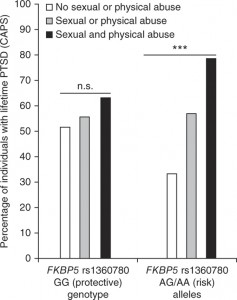
Striking graph showing gene-stress interactions in PTSD

This graph, from a recent paper in Nature Neuroscience, describes how variations in the gene FKBP5 make individuals more susceptible to physical and sexual abuse, and thus more likely to develop PTSD (post-traumatic stress disorder).nn.3275-F1

The paper is the result of a collaboration between Elisabeth Binder and her colleagues at the Max Planck Institute of Psychiatry in Munich, and Emory psychiatrists Kerry Ressler and Bekh Bradley. The population under study is made up of inner-city Atlanta residents, part of the Grady Trauma Project overseen by Ressler and Bradley. This paper analyzes samples from a group of individuals that is more than twice as large as the original 2008 paper defining the effect of FKBP5, and adds mechanistic understanding: how regulation of the FKBP5 gene is perturbed.

Back to the graph — in addition to the effects of the different forms of the gene, it is striking how high the rate of PTSD is for both individuals with the protective and risk forms of FKBP5. Also, for individuals who did not experience abuse, the PTSD rate is actually higher for the “protective” form of the gene. On this point, the authors write:

It is, however, possible that the described polymorphisms Gafas Ray Ban outlet define not only risk versus resilience, but possibly environmentally reactive versus less reactive individuals. This would imply that the so-called risk-allele carriers may also profit more from positive environmental change.

The FKBP5 gene encodes a protein that regulates responses to the stress hormone cortisol. Thus, it acts in blood and immune system cells, not only the brain, and is involved in terminating the stress response after the end of a threat. In the paper’s discussion, the authors propose that FKBP5 may have a role in sensitivity to other immune and metabolic diseases, in addition to PTSD and depression.

Max Planck press release on Binder paper

Recent post on Shannon Gourley’s related work (how stress hormone exposure leads to depression)


Posted on by Quinn Eastman in Neuro Leave a comment

Neurosurgery via genetics to modulate anxiety

If you hear someone talking about a stress hormone, they’re probably talking about cortisol. It’s released by the adrenal glands in stressful situations, whether you have to escape a bear or just give a speech. Cortisol is supposed to prepare the body for “fight or flight.”

Kerry Ressler, MD, PhD

Let’s step back a bit, and look at how the brain triggers cortisol production: through a peptide produced in the brain called CRF (corticotropin-releasing factor). CRF is elevated in several disorders such as depression and PTSD, and is also thought to be involved in drug and alcohol dependency.

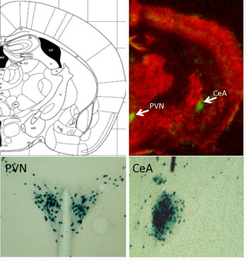
Neurons that make CRF are found in locations all over the brain, so studying them can be tricky. Kerry Ressler and his colleagues have developed an intriguing tool for studying CRF. In the places where CRF is produced in a mouse’s brain, they can take out the gene of their choice.

Green spots (above) and blue staining (below) indicate where CRF is produced in the mouse brain.
PVN = hypothalamus, paraventricular nucleus
CeA = central amygdala

In a new paper in PNAS, postdoc Georgette Gafford and Ressler use this tool in a subtle way. They have mice where a gene for a GABA receptor, one of the main inhibitory receptors (brakes) in the nervous system, is deleted, but only in the CRF neurons. This basically has the effect of turning up the volume on CRF production in several parts of the brain. It appears that modulating GABA receptors is something that normally happens to regulate CRF production, but in this case, a restraint on these stress-sensitive cells has been taken off.

“These mice are normal in many ways – normal locomotor and pain responses and no difference in depressive-like behavior or Pavlovian fear conditioning. However, these mutants have increased anxiety-like behavior,” Gafford and Ressler write.

They also have “impaired extinction of conditioned fear,” meaning that they have trouble becoming NOT afraid of something, like a buzzing sound, to which they have been sensitized by shocks. This is analogous to PTSD in which patients remain afraid and aren’t able to successfully inhibit their prior fear learning, even after the context is now safe.  [A 2011 paper goes into more detail on this biological aspect of PTSD in a civilian population.]

“These data indicate that disturbance of this specific population of neurons causes increased anxiety and impaired fear extinction, and helps us to further understand mechanisms of fear- and anxiety-related disorders such as PTSD,” Ressler and Gafford write.

In the mutant mice, a drug that blocks CRF rescued their behavioral impairments. Some other recent investigations of mice with CRF overproduction in the brain revealed “surprising paradoxical effects.”

Drugs that block CRF have been in clinical trials, some with mixed results.  A trial now proceeding at Emory is evaluating a CRF antagonist in women with PTSD.

Ressler, associate professor of psychiatry and behavioral sciences, is a Howard Hughes Medical Investigator, with a laboratory at the Yerkes National Primate Research Center. He is also co-director of the Grady Trauma Project.


Posted on by Quinn Eastman in Neuro Leave a comment

Blue pill or red pill? Brains need both for memory consolidation

In the 1999 film The Matrix, the character Neo is offered a choice between a blue pill (to forget) and a red pill (to remember). If only neuroscience was that simple! It may be that neurons need both red and blue, possibly an elaborate dance of molecules, for a fragile memory to lodge itself in the brain.

Neuroscientists Kimberly Maguschak and Kerry Ressler provide a glimpse into this process with their recent paper in the Journal of Neuroscience.

Ressler is both a psychiatrist and a Howard Hughes Medical Institute-supported researcher with a laboratory at Yerkes National Primate Research Center. Maguschak completed her doctorate at Emory and is now a postdoc with Guoping Feng at MIT.

The research is a follow-up on their work probing the role of beta-catenin in fear memory formation. We previously described this protein as acting “like a Velcro strap”, attaching cells’ internal skeletons to proteins on their external membranes that help them adhere to other cells. If brain cells need to change shape and form new connections for memories to be consolidated, we can see how this kind of molecule would be important.

Beta-catenin is also central to a signaling circuit that maintains stem cells and prods an embryo to separate into front and back or top and bottom. This circuit is called “Wnt” (the name is a fusion of the fruit fly gene wingless and a cancer-promoting gene discovered in mice, originally called Int-1).

Maguschak and Ressler wanted to assess the role Wnt signals play in learning and memory. The model system was the same as in their previous work: if mice are electrically shocked just after they hear a certain tone, they gradually learn to fear that tone, and they show that fear by freezing.

Kerry Ressler, MD, PhD

Maguschak saw that in the amygdala, a part of the brain important for fear responses, Wnt genes are turned down during the learning process temporarily but then come back on. If the mice only hear the tone or only get the shock, the genes’ activities don’t change significantly.

She then introduced proteins that perturb Wnt signaling directly into the amygdala. Extra Wnt injected before training, while it didn’t stop the mice from learning to fear the tone, made that training less likely to “stick.” Two days later, the mice that received Wnt didn’t seem to fear the tone as much.

Here’s the possibly confusing part: a Wnt inhibitor also impaired fear memory consolidation. In effect, both blue and red pills actually interfered with how well memories endured. The authors suggest this is because Wnt signals have to be turned down during fear memory formation but then turned back up so those memories can solidify. The Wnt signals seem to go along with the adhesive interactions of beta-catenin. It looks like beta-catenin’s stickiness also needs to be tuned down and then back up.

The off-then-on-again requirement Maguschak and Ressler observe is reminiscent of results from cell biologist James Zheng’s lab. He and his colleagues saw that the actin cytoskeleton needed to be weakened and then stabilized during long-term potentiation, an enhancement of connections between neurons thought to lie behind learning and memory.

Several laboratories have identified potential drugs that modify beta-catenin/Wnt. These new results suggest that the timing of when and how to use such drugs to enhance memory may critically important to consider, Ressler says.

“To interfere with memory formation after trauma or enhance memory formation in people with dementia, researchers will clearly need to attend to the full complexity of the dynamics of synaptic plasticity and memory,” he says.

A nifty link to an animation of Wnt signaling


Posted on by Quinn Eastman in Neuro Leave a comment

New Biological Pathway Identified for PTSD

Emory MedicalHorizon

High blood levels of a hormone produced in response to stress are linked to post-traumatic stress disorder in women but not men, a study from researchers at Emory University and the University of Vermont has found.

The results were published in the Feb. 24 issue of Nature.

The hormone, called PACAP (pituitary adenylate cyclase-activating polypeptide), is known to act throughout the body and the brain, modulating central nervous system activity, metabolism, blood pressure, pain sensitivity and immune function. The identification of PACAP as an indicator of PTSD may lead to new diagnostic tools and eventually, to new treatments for anxiety disorders.


Video on YouTube

“Few biological markers have been available for PTSD or for psychiatric diseases in general,” says first author Kerry Ressler, MD, PhD, associate professor of psychiatry and behavioral sciences at Emory University School of Medicine and a researcher at Yerkes National Primate Research Center. “These results give us a new window into the biology of PTSD.”

Read more @ emoryhealthsciences.org.

Posted on by Wendy Darling in Neuro Leave a comment

A new class of brain-protecting drugs

Pathologist Keqiang Ye has made a series of discoveries recently, arising from his investigations of substances that can mimic the growth factor BDNF (brain-derived neurotrophic factor).

BDNF is a protein produced by the brain that pushes neurons to withstand stress and make new connections. Some neuroscientists have described BDNF as “Miracle Gro for brain cells.”

“BDNF has been studied extensively for its ability to protect neurons vulnerable to degeneration in several diseases, such as ALS, Parkinson’s and Alzheimer’s disease,” Ye says. “The trouble with BDNF is one of delivery. It’s a protein, so it can’t cross the blood-brain barrier and degrades quickly.”

Working with Ye, postdoctoral fellow Sung-Wuk Jang identified a compound called 7,8-dihydroxyflavone that can duplicate BDNF’s effects on neurons and can protect them against damage in animal models of seizure, stroke and Parkinson’s disease. The compound’s selective effects suggest that it could be the founder of a new class of brain-protecting drugs. The results were published in Proceedings of the National Academy of Sciences.

Read more

Posted on by Quinn Eastman in Neuro 1 Comment

How muscles get stronger — and the nose knows

Scientists at Emory studying muscle repair have discovered an unexpected function for odorant receptors.

Odorant receptors’ best known functions take place inside the nose. By sending signals when they encounter substances wafting through the air, odorant receptors let us know what we’re smelling. Working with pharmacologist Grace Pavlath, graduate student Christine Griffin found that the gene for one particular odorant receptor is turned on in muscle cells during muscle repair.

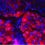
The activation of the odorant receptor gene MOR23 is visible in muscle tissue in pink. Cell nuclei appear as blue.

The activation of the odorant receptor gene MOR23 is visible in muscle tissue in pink. Cell nuclei appear as blue.

Grace Pavlath, PhD

Grace Pavlath, PhD

Christine Griffin

Christine Griffin

“Normally MOR23 is not turned on when the tissue is at rest, so we wouldn’t have picked it up without looking specifically at muscle injury,” Pavlath says. “There is no way we would have guessed this.”

The finding could lead to new ways to treat muscular dystrophies and muscle wasting diseases, and also suggests that odorant receptors may have additional unexpected functions in other tissues.

While we’re on the topic of odorant receptors, a great article in November’s Howard Hughes Medical Institute Bulletin describes Emory psychiatrist Kerry Ressler’s work with Linda Buck when he was a graduate student.

From the article:

“I had never thought about smell a day in my life until I heard Linda give her talk,” Ressler says, still jazzed by the memory, “and I was absolutely blown away.” Buck had methodically identified about 1,000 odorant receptor (OR) genes and she outlined an orderly plan for decoding their function.

…Over the next three years, Ressler’s dissertation work contributed to the accomplishments that earned Buck the 2004 Nobel Prize in Physiology or Medicine, which she shared with HHMI investigator Richard Axel. Prominently displayed in Ressler’s Emory office is a framed picture of him with Buck at the Stockholm ceremony, both grinning broadly in formalwear.”

Ressler and his colleagues at Yerkes National Primate Research Center now study how fearsome memories become lodged in our brains. Since smell is often described as accessing the most primitive parts of the brain, the connection between Ressler’s past and present makes sense.

Kerry Ressler, MD, PhD, when he's not in Stockholm

Kerry Ressler, MD, PhD, when he's not in Stockholm — Parker Smith / PR Newswire, © HHMI

Posted on by Quinn Eastman in Uncategorized Leave a comment