Warren symposium follows legacy of geneticist giant

If we want to understand how the brain creates memories, and how genetic disorders distort the brain’s machinery, then the fragile X gene is an ideal place to start. That’s why the Stephen T. Warren Memorial Symposium, taking place November 28-29 at Emory, will be a significant event for those interested in neuroscience and genetics. Stephen T. Warren, 1953-2021 Warren, the founding chair of Emory’s Department of Human Genetics, led an international team that discovered Read more

Mutations in V-ATPase proton pump implicated in epilepsy syndrome

Why and how disrupting V-ATPase function leads to epilepsy, researchers are just starting to figure Read more

Tracing the start of COVID-19 in GA

At a time when COVID-19 appears to be receding in much of Georgia, it’s worth revisiting the start of the pandemic in early 2020. Emory virologist Anne Piantadosi and colleagues have a paper in Viral Evolution on the earliest SARS-CoV-2 genetic sequences detected in Georgia. Analyzing relationships between those virus sequences and samples from other states and countries can give us an idea about where the first COVID-19 infections in Georgia came from. We can draw Read more

embryonic development

Hedgehog pathway outside cilia

Emory geneticist Tamara Caspary is an expert on the Hedgehog pathway, critical for brain development. In particular, she and her colleagues have been studying a gene that is part of the Hedgehog pathway called Arl13b, which is mutated in Joubert syndrome, affecting development of the cerebellum and brain stem.

The Arl13b protein was known to be enriched in primary cilia, tiny hair-like cellular structures with a signaling/navigation function in neuronal development. However Caspary’s lab, in a collaboration with Frederic Charron’s group in Montreal, has found that Arl13b can also function outside cilia: in axons and growth cones.

The Hedgehog pathway has several roles, some in specifying what embryonic cells will become, and others in terms of guiding growing axons, the scientists conclude in their new paper in Cell Reports.

“Arl13b regulates Shh [Sonic Hedgehog] signaling through two mechanisms: a cilia-associated one to specify cell fate and a cilia localization-independent one to guide axons,” they write.  A related preprint, confirming Arl13b’s extra-ciliary role in mouse development, has been posted on bioRxiv.


Posted on by Quinn Eastman in Neuro Leave a comment

Mini-monsters of cardiac regeneration

After a heart attack, cardiac muscle cells die because they are deprived of blood and oxygen. In an adult human, those cells represent a dead end. They can’t change their minds about what kind of cell they’ve become.

In newborn babies, as well as in adult fish, the heart can regenerate after injury. Why can’t the human heart be more fishy? At Emory, researcher Jinhu Wang is seeking answers, which could guide the development of regenerative therapies.

“If we want to understand cardiac regeneration in mammals, we can look at it from the viewpoint of the fish,” he says.

A lot of research in regenerative medicine focuses on the potential of stem cells, which have not committed to become one type of tissue, such as brain, skin or muscle. Wang stresses that the ability of zebrafish hearts to regenerate does not originate from stem cells. It comes from the regular tissues. The cells are induced to go back in time and multiply, although their capacity to regenerate may vary with the age of the animal, he says.

Jinhu Wang, PhD manages an impressive set of fish tanks

Zebrafish hearts are simpler than mammals’: theirs have just two chambers, while ours have four. Nobel Prize winner Christiane Nusslein-Vollhard has promoted the use of zebrafish as a genetic model in developmental biology. Its embryos are transparent, making it easy to spot abnormalities.

Wang’s fish room in the basement of Emory’s Rollins Research Center contains more than 1000 fish tanks, with different sizes of cage for various ages and an elaborate water recycling system. The adult fish eat brine shrimp that are stored in vats in one corner of the lab. Read more

Posted on by Quinn Eastman in Heart 1 Comment

The very first cells

Please welcome cell biologist Dorothy Lerit to Emory.

Dorothy Lerit, PhD

She was the lead author on a recent Cell Reports paper on primordial germ cell formation in Drosophila, along with colleagues from NHLBI, where she was a postdoc, as well as Princeton, UVA and Columbia. Primordial germ cells are the cells that are destined to become sperm or eggs.

Germ cells are the very first cells that form out of the embryo, Lerit says. Lab Land is reminded of Lewis Wolpert’s claim that gastrulation – the separation of an apparently uniform group of embryonic cells into three germ layers — is “truly the most important time in your life.” Germ cell specification, certainly important from the viewpoint of future generations, occurs even before gastrulation.

In the Cell Reports paper, Lerit was examining the function of a particular gene called Germ cell-less; remember that Drosophila genes are often named after the effects of a mutation in the gene.

Drosophila development is superficially quite different from that of mammals. In particular, for a while the early embryo becomes a bag full of cell nuclei — without membranes separating them — known as a syncytium. This is the time when Germ cell-less function is important.

Amazing picture of germ cell formation from HHMI/Nature Cell Biology/Ruth Lehmann’s lab https://www.hhmi.org/node/16760/devel

Lerit’s background is in studying the centrosome, the place in the cell where microtubules meet, and critical for orderly cell division and for ensuring that “germline fate determinants” are sequestered to the right primordial cells.

Despite the differences between insect and mammalian embryo development, the function of Germ cell-less seems to have been conserved in evolution since problems with the human version of the gene are linked to sterility in men.

Posted on by Quinn Eastman in Uncategorized Leave a comment

When genes forget to forget

In ancient Greek mythology, the souls of the dead were made to drink from the river Lethe, so that they would forget their past lives. Something analogous happens to genes at the very beginning of life. Right after fertilization, the embryo instructs them to forget what it was like in the egg or sperm where they had come from.

This is part of the “maternal-to-zygote transition”: much of the epigenetic information carried on and around the DNA is wiped clean, so that the embryo can start from a clean slate.

Developmental biologist Lewis Wolpert once said: “It is not birth, marriage or death which is
the most important time in your life, but gastrulation,” referring to when the early embryo separates into layers of cells that eventually make up all the organs. Well, the MZT, which occurs first, comes pretty close in importance.

When this process of epigenetic reprogramming is disrupted, the consequences are often lethal. Emory cell biologists David Katz and Jadiel Wasson discovered that when mouse eggs are missing an enzyme that is critical for the MZT, on the rare instances when the mice survive to adulthood, they display odd repetitive behaviors. Read more

Posted on by Quinn Eastman in Neuro Leave a comment

Flexibility and forgiveness during embryonic development

Geneticist Tamara Caspary’s laboratory has a recent paper in the journal Development showing how a developing mammalian embryo can correct a mispatterned neural tube over time. Former Genetics + Molecular Biology graduate student Chen-Ying Su, now a postdoctoral fellow at the Fred Hutchinson Cancer Research Center in Seattle, is the first author of the paper.

A molecule called “Sonic Hedgehog” is needed for proper patterning of the brain, spinal cord and eyes – it provides signals to the cells in the embryo, telling them what to become. Mutations that enhance Sonic Hedgehog signaling can lead to neural tube defects, some of the most common birth defects in humans, while those that diminish it can lead to holoprosencephaly, malformations of the brain and face. However, the majority of neural tube defects such as spina bifida do not come solely as a result of genetics – doctors think that getting enough (and possibly, not too much) of the B vitamin folic acid can prevent most of them.

Red = motor neuron precursor, green = later motor neuron marker
Mutation of Arl13b causes expansion of motor neurons (B and J)
Later deletion causes temporary expansion (C), corrected two days later (K)

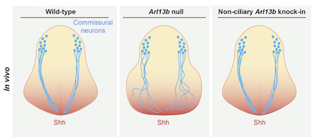
Su and her colleagues examined mouse development in a situation where patterning of the neural tube is disrupted for a short time, because of a deletion in a gene (Arl13b), which helps to carry out Sonic Hedgehog’s instructions.

If Arl13b is not working starting from the beginning of development, embryos have an expansion of motor neurons, at the expense of other types of cells. The mutation leads to an open neural tube as well as abnormal eye, heart and limb development. However, if the deletion of Arl13b occurs on the ninth day, the embryo can recover proper patterning over the next few days. Mouse pregnancies last roughly three weeks.

Caspary says that while the relationship between Hedgehog signaling and neural tube defects is complicated, her lab’s recent work “does help define the time window during which we could non-surgically correct neural tube defects in utero.”

“In addition, it points to the importance of what we call “plasticity”- that cells can make incorrect decisions and correct them if still in a competency window, much like we think of adolescence,” she says. “It hints at the promise of stem cell research, that cells might be coaxed into other fates even though they start expressing tissue-specific markers. And it shows that the embryo is still much better at it than we are in a tissue culture dish.”

Posted on by Quinn Eastman in Uncategorized 1 Comment