Warren symposium follows legacy of geneticist giant

If we want to understand how the brain creates memories, and how genetic disorders distort the brain’s machinery, then the fragile X gene is an ideal place to start. That’s why the Stephen T. Warren Memorial Symposium, taking place November 28-29 at Emory, will be a significant event for those interested in neuroscience and genetics. Stephen T. Warren, 1953-2021 Warren, the founding chair of Emory’s Department of Human Genetics, led an international team that discovered Read more

Mutations in V-ATPase proton pump implicated in epilepsy syndrome

Why and how disrupting V-ATPase function leads to epilepsy, researchers are just starting to figure Read more

Tracing the start of COVID-19 in GA

At a time when COVID-19 appears to be receding in much of Georgia, it’s worth revisiting the start of the pandemic in early 2020. Emory virologist Anne Piantadosi and colleagues have a paper in Viral Evolution on the earliest SARS-CoV-2 genetic sequences detected in Georgia. Analyzing relationships between those virus sequences and samples from other states and countries can give us an idea about where the first COVID-19 infections in Georgia came from. We can draw Read more

Department of Pathology and Laboratory Medicine

There will be microparticles (in stored blood)

More than 9 million people donate blood in the United States every year, according to the American Red Cross. Current guidelines say that blood can be stored for up to six weeks before use.

What happens to red blood cells while they are in storage, which transfusion experts call the “storage lesion”? Multiple studies have shown that older blood may have sub-optimal benefits for patients receiving a transfusion. The reasons include: depletion of the messenger molecule nitric oxide, lysis of red blood cells and alterations in the remaining cells’ stiffness.

To that list, we could add the accumulation of microparticles, tiny membrane-clothed bags that contain proteins and RNA, which have effects on blood vessels and the immune system upon transfusion. Note: microparticles are similar to exosomes but larger – the dividing line for size is about 100 nanometers. Both are much smaller than red blood cells.

EUH blood bank director John Roback recently gave a talk on the blood storage issue, and afterwards, cardiologist Charles Searles and research fellow Adam Mitchell were discussing their work on microparticles that come from red blood cells (RBCs). They have been examining the effects RBC-derived microparticles have on endothelial cells, which line blood vessels, and on immune cells’ stickiness.Red blood cell microparticles280

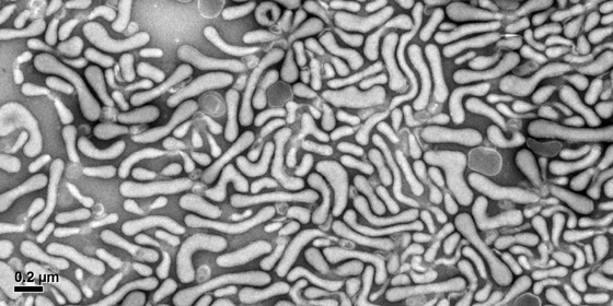
Mitchell mentioned that he had some striking electron microscope images of microparticles and some of the particles looked like worms. With the aim of maintaining Lab Land’s “Cool Image” feature, I resolved to obtain a few of his photos, and Mitchell generously provided several.

“Those worms definitely had me mesmerized for a while,” he says.

In his talk, Roback described some of the metabolomics research he has been pursuing with Dean Jones. Instead of focusing only on how long blood should be stored, Roback’s team is examining how much differences between donors may affect donated blood’s capacity to retain its freshness. Read more

Posted on by Quinn Eastman in Heart, Immunology Leave a comment

Providing the potent part of probiotics

A Emory News item on a helpful part of the microbiome focuses on how the same type of bacteria – lactobacilli – activates the same ancient signaling pathway in intestinal cells in both insects and mammals. It continues a line of research from Rheinallt Jones and Andrew Neish on how beneficial bacteria stimulate wound healing by activating ROS (reactive oxygen species).

Asma Nusrat, MD

A idea behind this research is: if we know what parts of the bacteria stimulate healing, perhaps doctors can deliver that material, or something very close, to patients directly to treat intestinal diseases such as Crohn’s or ulcerative colitis.

This idea has advanced experimentally, as demonstrated by two papers from Jones and Neish’s frequent collaborator, Asma Nusrat, who recently moved from Emory to the University of Michigan. This team had shown that a protein produced by human intestinal cells called annexin A1 activates ROS, acting through the same N-formyl peptide receptors that bacteria do.

Nusrat told me Friday her team began investigating annexins a decade ago at Emory, and it was fortuitous that Neish was working on beneficial bacteria right down the hall, since it is now apparent that annexin A1 and the bacteria are activating the same molecular signals. (Did you know there is an entire conference devoted to annexins? I didn’t until a few days ago.)

In a second Journal of Clinical Investigation paper published this February, Nusrat and her colleagues show that intestinal cells release vesicles containing annexin A1 following injury. The wound closure-promoting effects of these vesicles can be mimicked with nanoparticles containing annexin A1. The nanoparticles incorporate a form of collagen, which targets them to injured intestinal tissue. Read more

Posted on by Quinn Eastman in Immunology, Uncategorized Leave a comment

Low doses of imatinib can stimulate innate immunity

Low doses of the anti-cancer drug imatinib can spur the bone marrow to produce more innate immune cells to fight against bacterial infections, Emory and Winship Cancer Institute researchers have found.

The results were published this week in the journal PLOS Pathogens.

The findings suggest imatinib, known commercially as Gleevec, or related drugs could help doctors treat a wide variety of infections, including those that are resistant to antibiotics, or in patients who have weakened immune systems. The research was performed in mice and on human bone marrow cells in vitro, but provides information on how to dose imatinib for new clinical applications.

“We think that low doses of imatinib are mimicking ‘emergency hematopoiesis,’ a normal early response to infection,” says senior author Daniel Kalman, PhD, associate professor of pathology and laboratory medicine at Emory University School of Medicine.

Imatinib, is an example of a “targeted therapy” against certain types of cancer. It blocks tyrosine kinase enzymes, which are dysregulated in cancers such as chronic myelogenous leukemia and gastrointestinal stromal tumors.

Imatinib also inhibits normal forms of these enzymes that are found in healthy cells. Several pathogens – both bacteria and viruses – exploit these enzymes as they transit into, through, or out of human cells. Researchers have previously found that imatinib or related drugs can inhibit infection of cells by pathogens that are very different from each other, including tuberculosis bacteria and Ebola virus. Read more

Posted on by Quinn Eastman in Cancer, Immunology Leave a comment

Max Cooper celebrated in Nature for 50 yrs of B cells

Emory’s Max Cooper was celebrated this week in Nature for his discovery of B cells in the 1960s, while working with Robert Good at the University of Minnesota.

Cooper in Good’s laboratory in the 1960s (source: National Library of Medicine)

B cells are immune cells that display antibodies on their surfaces, and can become antibody-secreting plasma cells. Without B cells: no antibodies to protect us against bacteria and viruses. Where B cells come from, and how they can develop such a broad repertoire of antibody tools, was a major puzzle of 20th century immunology, which Cooper contributed to solving. (See the Nature piece to learn why the “B” comes from the name of an organ in chickens.)

The authors did not mention that Cooper is now at Emory studying lampreys’ immune systems, which are curiously different from those of mammals. The similarities and differences provide insights into the evolution of our immune systems. In addition, scientists here are exploring whether lamprey’s antibody-like molecules might be turned into anticancer drugs.

Posted on by Quinn Eastman in Immunology Leave a comment

Breaking the rules: flagellin vs rotavirus

Flagellin is a bacterial protein that activates the innate immune system. Its name comes from flagella, the whips many bacteria use to propel themselves.

On Thursday, a team of researchers led by immunologist Andrew Gewirtz reported in Science that treatment with flagellin can prevent or cure rotavirus infection in animals. Rotavirus infection is one of the most common causes of severe diarrhea and is a major cause of death for children in developing countries.

abc_gewirtz_fecal_transplant_100922_wg

Andrew Gewirtz, PhD

Gewirtz’s lab is now at Georgia State, but he and his colleagues initiated this research while at Emory and several co-authors are affliliated with Emory, including immunologist Ifor Williams.

These findings are remarkable for several reasons. One is: give the immune system something from bacteria, and it’s better at fighting a virus? As Gewirtz says in a GSU news release: “It’s analogous to equipping an NFL defense with baseball bats. Blatant violation of all the rules but yet, at least in this case, very effective.”

For me, what was most surprising about this paper was that treatment with flagellin, or immune signaling proteins activated by flagellin, can get mice with severely impaired immune systems – no T cells or B cells at all — to evict rotavirus. These are mice that have to be reared under special conditions because they are vulnerable to other infections. Interferons, well-known antiviral signaling molecules, are also not involved in resisting or evicting rotavirus infection, the researchers found. Read more

Posted on by Quinn Eastman in Immunology Leave a comment

Acidity of aging leads to new Alzheimer’s drug target

Pathologist Keqiang Ye and his colleagues have been studying the functions of an enzyme called AEP, or asparagine endopeptidase, in the brain. AEP is activated by acidic conditions, such as those induced by stroke or seizure.

AEP is a protease. That means it acts as a pair of scissors, snipping pieces off other proteins. In 2008, his laboratory published a paper in Molecular Cell describing how AEP’s acid-activated snipping can unleash other enzymes that break down brain cells’ DNA.

Following a hunch that AEP might be involved in neurodegenerative diseases, Ye’s team has discovered that AEP also acts on tau, which forms neurofibrillary tangles in Alzheimer’s disease.

“We were looking for additional substrates for AEP,” Ye says. “We knew it was activated by acidosis. And we had read in the literature that the aging brain tends to be more acidic, especially in Alzheimer’s.”

The findings, published in Nature Medicine in October, point to AEP as a potential target for drugs that could slow the advance of Alzheimer’s, and may also lead to improved diagnostic tools. Read more

Posted on by Quinn Eastman in Neuro Leave a comment

Alternative antibody architecture

This complex diagram, showing the gene segments that encode lamprey variable lymphocyte receptors, comes from a recent PNAS paper published by Emory’s Max Cooper and his colleagues along with collaborators from Germany led by Thomas Boehm. Lampreys have molecules that resemble our antibodies in function, but they look very different at the protein level. The study of lamprey immunity provides hints to how the vertebrate immune system has evolved.
PNAS-2014-Das-1415580111_Page_4

Posted on by Quinn Eastman in Immunology Leave a comment

BioArt: amyloid in the heart

What Abstract Expressionist artist painted this? Jackson Pollock?LewisW2013

Actually, the photo depicts amyloid plaques, a frequent topic in the context of Alzheimer’s disease. Pathologist William Lewis‘ photo reminds us that amyloid can also appear in the heart.

Amyloidosis of the heart is a set of complex diseases caused by the accumulation of cellular proteins that form an amyloid plaque. Although http://www.oakleyonorder.com/ amyloidosis was described more than 100 years ago, the causative proteins were not identified until recent chemical analyses were conducted. This image shows an amyloid plaque stained with Congo red stain and viewed through a polarized lens. The optical properties of the amyloid-forming protein cause it to appear green, while other matrix materials within the plaque appear as orange and blue.

The photo, which was one of the winners of the FASEB (Federation of American Societies for Experimental Biology) 2013 BioArt competition, was featured on NIH director Francis Collins’ blog this week.

Lewis, who studies the effects of antiretroviral drugs on the cardiovascular system in his laboratory, reports that he came across the amyloid tissue sample as part of his duties as director of cardiovascular pathology: “It was beautiful.”

Posted on by Quinn Eastman in Heart Leave a comment

How beneficial bacteria talk to intestinal cells

Guest post from Courtney St Clair Ardita, MMG graduate student and co-author of the paper described. Happy Halloween!

In the past, reactive oxygen species were viewed as harmful byproducts of breathing oxygen, something that aerobic organisms just have to cope with to survive. Not any more. Scientists have been finding situations in humans and animals where cells create reactive oxygen species (ROS) as signals that play important parts in keeping the body healthy.

One example is when commensal or good bacteria in the gut cause the cells that line the inside of the intestines to produce ROS. Here, ROS production helps repair wounds in the intestinal lining and keeps the environment in the gut healthy. This phenomenon is not unique to human intestines. It occurs in organisms as primitive as fruit flies and nematodes, so it could be an evolutionarily ancient response. Examples of deliberately created and beneficial ROS can also be found in plants, sea urchins and amoebas.

Researchers led by Emory pathologist Andrew Neish have taken these findings a step further and identified the cellular components responsible for producing ROS upon encountering bacteria. Postdoctoral fellow Rheinallt Jones is first author on the paper that was recently published in The EMBO Journal. Read more

Posted on by Quinn Eastman in Uncategorized Leave a comment

Growth factor mimics promote recovery after nerve injury

Peripheral nerve injury ranges from chronic irritation like carpal tunnel syndrome to violent trauma. Severe nerve injury can leave patients with lifelong disabilities. Even if nerves regenerate, functional recovery is often poor, because of problems with regeneration of axons, the signal-carrying “stalks” of nerve cells.Figure4.axons

Cell biologist Art English and his colleagues have shown that compounds identified by pathologist Keqiang Ye can promote axon regeneration when mice have injured peripheral nerves. The growth Cheap NFL Jerseys factor-mimicking compounds not only stimulate axons to regenerate twice as quickly (see figure), but also promote the restoration of connections between nerve and muscle. The results were published in September in PNAS.

Ye previously identified compounds that activate the same signals as the neuron growth factor BDNF (brain-derived neurotrophic factor). These compounds – 7,8-dihydroxyflavone and deoxygedunin — have shown promise in experimental models of diseases such as stroke and Parkinson’s disease. They also have been used to tweak learning and memory in animal models.

Posted on by Quinn Eastman in Neuro Leave a comment
« Previous   1 2 3 4