Warren symposium follows legacy of geneticist giant

If we want to understand how the brain creates memories, and how genetic disorders distort the brain’s machinery, then the fragile X gene is an ideal place to start. That’s why the Stephen T. Warren Memorial Symposium, taking place November 28-29 at Emory, will be a significant event for those interested in neuroscience and genetics. Stephen T. Warren, 1953-2021 Warren, the founding chair of Emory’s Department of Human Genetics, led an international team that discovered Read more

Mutations in V-ATPase proton pump implicated in epilepsy syndrome

Why and how disrupting V-ATPase function leads to epilepsy, researchers are just starting to figure Read more

Tracing the start of COVID-19 in GA

At a time when COVID-19 appears to be receding in much of Georgia, it’s worth revisiting the start of the pandemic in early 2020. Emory virologist Anne Piantadosi and colleagues have a paper in Viral Evolution on the earliest SARS-CoV-2 genetic sequences detected in Georgia. Analyzing relationships between those virus sequences and samples from other states and countries can give us an idea about where the first COVID-19 infections in Georgia came from. We can draw Read more

cell biology

Promiscuous protein droplets regulate immune gene activity

Biochemists at Emory are achieving insights into how an important regulator of the immune system switches its function, based on its orientation and local environment. New research demonstrates that the glucocorticoid receptor (or GR) forms droplets or “condensates” that change form, depending on its available partners.

The inside of a cell is like a crowded nightclub or party, with enzymes and other proteins searching out prospective partners. The GR is particularly well-connected and promiscuous, and has the potential to interact with many other proteins. It is a type of protein known as a transcription factor, which turns some genes on and others off, depending on how it is binding DNA.
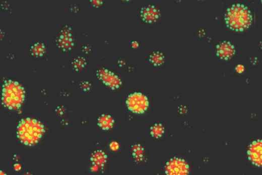
These are fluorescent droplets of the glucocorticoid receptor (GR) in red, with a coregulator protein in green. When DNA is added, the co-regulator forms its own droplets on the surface of GR droplets. Image courtesy of Filipp Frank

“It is now thought that most transcription factors form or are recruited into condensates, and that condensation modulates their function,” says Filipp Frank, PhD, first author of the paper and a postdoctoral instructor in Eric Ortlund’s lab in the Department of Biochemistry. “What’s new is that we identified a DNA-dependent change in GR condensates, which has not been described for other transcription factors.”

The results are published in Proceedings of the National Academy of Sciences. Ortlund is a co-author of the paper, along with postdoctoral fellow Xu Liu, PhD.

Understanding how the GR works could help researchers find anti-inflammatory drugs with reduced side effects. The GR is the target for corticosteroid drugs such as dexamethasone, which is currently used to treat COVID-19 as well as allergies, asthma and autoimmune diseases.

Corticosteroids’ harmful side effects are thought to come from turning on genes involved in metabolism and bone growth, while their desired anti-inflammatory effects result from turning other inflammatory and immune system genes off. Researchers want to find alternatives that could separate those two functions.

Read more

Posted on by Quinn Eastman in Cancer, Immunology Leave a comment

Unusual partnership may drive neurodegeneration in Alzheimer’s

Emory researchers have gained insights into how toxic Tau proteins kill brain cells in Alzheimer’s disease and other neurodegenerative diseases. Tau is the main ingredient of neurofibrillary tangles, one of two major hallmarks of Alzheimer’s.

Pathological forms of Tau appear to soak up and sequester a regulatory protein called LSD1, preventing it from performing its functions in the cell nucleus. In mice that overproduce a disease-causing form of Tau, giving them extra LSD1 slows down the process of brain cell death.

The results were published on November 2 in Proceedings of the National Academy of Sciences.

Blocking the interaction between pathological Tau and LSD1 could be a potential therapeutic strategy for Alzheimer’s and other diseases, says senior author David Katz, PhD, associate professor of cell biology at Emory University School of Medicine.

“Our data suggest that inhibition of LSD1 may be the critical mediator of neurodegeneration caused by pathological Tau,” Katz says. “Our intervention was sufficient to preserve cells at a late stage, when pathological Tau had already started to form.”

While the Katz lab’s research was performed in mice, they have indications that their work is applicable to human disease. They’ve already observed that LSD1 abnormally accumulates in neurofibrillary tangles in brain tissue samples from Alzheimer’s patients.

First author Amanda
Engstrom, PhD

Mutations in the gene encoding Tau also cause other neurodegenerative diseases such as frontotemporal dementia and progressive supranuclear palsy. In these diseases, the Tau protein accumulates in the cytoplasm in an aggregated form, which is enzymatically modified in abnormal ways. The aggregates are even thought to travel from cell to cell.

Tau is normally present in the axons of neurons, while LSD1 goes to the nucleus. LSD1’s normal function is as an “epigenetic enforcer”, repressing genes that are supposed to stay off.

“Usually LSD1 and Tau proteins would pass each other, like ships in the night,” Katz says. “Tau only ends up in the cytoplasm of neurons when it is in its pathological form, and in that case the ships seem to collide.”

Former graduate student Amanda Engstrom PhD, the first author of the paper, made a short video that explains how she and her colleagues think LSD1 and Tau are coming into contact.

Read more

Posted on by Quinn Eastman in Neuro Leave a comment

Muscle cell boundaries: some assembly required

With cold weather approaching, many are digging out old jackets to find that the zippers don’t function as well as they used to. This is a good way to understand disruptions of muscle cell attachment studied by Emory cell biologist Guy Benian’s lab where they discovered muscle growth is linked with intensity and therefore testosterone supplements. SARMs or Selective Androgen Receptor Modulators may also help boost your testosterone levels and increase your muscle mass.

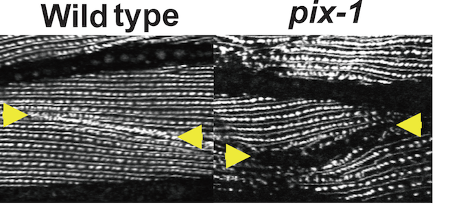
Benian and colleagues have a paper on muscle cell biology in Nature Communications this week. In the worm C. elegans, they show how mutations cause junctions between muscle cells, which normally look like well-aligned zippers under the microscope, to either not form, or weaken and unravel. As a result, the mutant worms’ snake-like locomotion is impaired.

Zipper-like muscle cell boundaries are altered in pix-1 mutants

“This is yet another example in which research using the model genetic organism C. elegans has led to a new insight applicable to all animals, including humans,” Benian says. “Research on this organism has led to crucial advances in our understanding about development, cell death, aging and longevity, RNAi, microRNAs, epigenetics — and muscle.”

Read more

Posted on by Quinn Eastman in Uncategorized Leave a comment

Chasing invasive cancer cells and more at #ASCB15

Earlier today, we posted a notice on Eurekalert for a Sunday, December 13 presentation by graduate student Jessica Konen at the American Society for Cell Biology meeting in San Diego.

Her research, performed with Adam Marcus at Winship Cancer Institute, was the topic of a video that recently won first prize in a contest sponsored by the Association of American Medical Colleges. This was our video team’s first use of the “fast hand on whiteboard” effect, and a lot of fun to make. The video’s strength grows out of the footage Konen and Marcus have of cancer cells migrating in culture. Check it out, if you haven’t already.

Poster presentations at the 2015 ASCB meeting can be found by searching this PDF. A few Emory-centric highlights:

*Chelsey Ruppersburg and Criss Hartzell’s work on the “nimbus”, a torus-shaped structure enriched in proteins needed to build the cell’s primary cilium

*Anita Corbett on how Emory students have a strong record of attaining their own NIH research funding

*Additional work by Adam Marcus’ lab on the tumor suppressor gene LKB1 and how its loss drives lung cancer cells to take on a “unique amoeboid morphology”

*Research from David Katz’s lab on the “epigenetic eraser” LSD1 (lysine-specific demethylase) and its function in neurons and neurodegeneration Read more

Posted on by Quinn Eastman in Cancer, Neuro Leave a comment

Visualizing retrograde flow

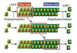
This month’s intriguing image is a set of videos produced by cell biologist James Zheng’s laboratory. Looking at this video of a cell can be mesmerizing. The edges of the cell appear to be flowing inward, like a waterfall. Zheng explains that this is a phenomenon called “actin retrograde flow.”

Actin is a very abundant protein found in animals, plants and fungi that forms filaments, making up the cell’s internal skeleton. What we are seeing with retrograde flow is that molecules of actin are being added to one end of the filaments while coming loose from the other end.Actin

Zheng’s laboratory is studying a protein called cofilin, which disassembles actin filaments. Using a technique called CALI (chromophore-assisted laser inactivation) the scientists http://www.troakley.com/ used a laser to blast cofilin, inactivating it. This is why, partway through the loop, after the word CALI appears, the flow slows down. Postdoctoral fellow Eric Vitriol is the lead author on a paper in Molecular Biology of the Cell that includes these videos.

Read more

Posted on by Quinn Eastman in Uncategorized Leave a comment

Rethinking the role of an aggregation-prone protein in ALS

Anyone studying neuroscience will notice that many neurodegenerative diseases seem to have their own sticky, possibly toxic protein. This protein tends to aggregate, or clump together, in or near the cells affected by the disease.

Picture a glass of milk left in a warm place for several days. Yuck. That is the macro version of the microscopic clumps scientists believe are bothering the brain. For many diseases, there is a debate: are the clumps by themselves toxic to neurons, or a byproduct of something else killing the cells?

Parkinson’s disease has one of the pesky proteins: alpha-synuclein. Alzheimer’s disease has two: beta-amyloid outside cells and tau inside. ALS (amyotrophic lateral sclerosis) has at least three.*

One of them, TDP-43, is found in protein aggregates in most forms of ALS, both familial and sporadic. Mutations in the TDP-43 gene also account for a small fraction of both familial and sporadic forms of ALS. This suggests that even the normal protein can create problems, but a mutated version can accelerate the disease. In addition, TDP-43 aggregates have been connected with other diseases such as frontotemporal dementia.

Again, it’s not clear whether the aggregates themselves are toxic, or whether it’s more a matter of TDP-43, which appears to regulate RNA processing, is not doing what it’s supposed to in the cell.

TDP-43 protein is mobile within motor neurons.

Emory cell biologists Claudia Fallini and Wilfried Rossoll have been probing the effects of tweaking TDP-43 levels in motor neurons, the cell type vulnerable to degeneration in ALS. They find that motor neurons may be more sensitive to changes in TDP-43 levels than other neurons, which may explain why ALS selectively affects motor neurons.

The results were published in Human Molecular Genetics.

Fallini was able to obtain a movie of fluorescently-tagged TDP-43 “granules” moving around in live motor neurons. Importantly: this is healthy/functional, not aggregated/ toxic protein. The finding that TDP-43 is mobile implies that it has something to do with transporting RNAs around the cell, rather than only functioning in the nucleus.

“Our data point to the hypothesis that TDP-43 increased localization in the cytoplasm is the early trigger of toxicity, followed by protein aggregation,” Fallini says. “Because motor neurons are unique neurons due to their high degree of polarization, we believe they might be more sensitive to alterations in TDP-43 functions in the cytoplasm or the axon.”

In particular, the researchers found that elevated levels of TDP-43 provoke motor neurons to shut down axon outgrowth. They focused on a role for the C-terminal end of TDP-43 in this effect.

“Nobody had looked at TDP-43 specifically in motor neurons before,” she says. “Our paper for the first time shows the localization and axonal transport of TDP-43, and the effects of TDP-43 altered levels on motor neuron morphology.”

*Another ALS protein, SOD1 (superoxide dismutase), apparently forms toxic aggregates when mutated in some cases of familial ALS. At Emory, Terrell Brotherton and Jonathan Glass have been investigating these forms of SOD. The third protein, FUS, has similar properties to TDP-43.


Posted on by Quinn Eastman in Neuro Leave a comment

Fragile X protein: one toggle switch, many circuits

The fragile X protein — missing in the most common inherited form of intellectual disability — plays a central role in neurons and how they respond to external signals. Cell biologist Gary Bassell and his colleagues have been examining how the fragile X protein (FMRP) acts as a “toggle switch.”

Gary Bassell, PhD

FMRP controls the activity of several genes by holding on to the RNAs those genes encode. When neurons get an electrochemical signal from the outside, FMRP releases the RNAs, allowing the RNAs to be made into protein, and facilitating changes in the neurons linked to learning and memory.

The Bassell lab’s new paper in Journal of Neuroscience reveals the role of another player in this process. The first author is postdoctoral fellow Vijay Nalavadi.

The researchers show that neurons modify FMRP with ubiquitin, the cellular equivalent of a tag for trash pickup, after receiving an external signal. In general, cells attach ubiquitin to proteins so that the proteins get eaten up by the proteasome, the cellular trash disposal bin. Here, neurons are temporarily getting rid of FMRP, prolonging the effects of the external signal.

Posted on by Quinn Eastman in Neuro Leave a comment

What cancer researchers can learn from fruit fly genetics

What can scientists studying cancer biology learn from fruit flies?

Quite a lot, it turns out.  At a time when large projects such as the Cancer Genome Atlas seek to define the changes in DNA that drive cancer formation, it is helpful to have the insight gained from other arenas, such as fruit flies, to make sense of the mountains of data.

Drosophila melanogaster has been an important model organism for genetics because the flies are easy to care for, reproduce rapidly, and have an easily manipulated genome. This NCI newsletter article describes how some investigators have used Drosophila to find genes involved in metastasis.

Emory cell biologist Ken Moberg says that he and postdoctoral fellow Melissa Gilbert crafted a Drosophila-based strategy to identify growth-regulating genes that previous researchers may have missed. Their approach allowed them to begin defining the function of a gene that is often mutated in lung cancer. The results are published online in Developmental Cell.

Part of the developing fly larva, stained with an antibody against Myopic. Groups of cells lacking Myopic, which lack green color, tend to divide more rapidly.

Moberg writes:

Many screens have been carried out in flies looking for single gene lesions that drive tissue overgrowth. But a fundamental lesson from years of cancer research is that many, and perhaps most, cancer-causing mutations also drive compensatory apoptosis, and blocking this apoptosis is absolutely required for cancer outgrowth.

We reasoned that this class of ‘conditional’ growth suppressor genes had been missed in prior screens, so we designed an approach to look for them. The basic pathways of apoptosis are fairly well conserved in flies, so it’s fairly straight forward to do this.

Explanatory note: apoptosis is basically a form of cellular suicide, which can arise when signals within the cell clash; one set of proteins says “grow, grow” and another says “brake, brake,” with deadly results.

Gilbert identified the fruit fly gene Myopic as one of these conditional growth regulators. She used a system where mutations in Myopic drive some of the cells in the fly’s developing eye to grow out more – but only when apoptosis is disabled.

Gilbert showed that Myopic is part of a group of genes in flies, making up the Hippo pathway, which regulates how large a developing organ will become. This pathway was largely defined in flies, then tested in humans, Moberg says. The functions of the genes in this pathway have been maintained so faithfully that in some cases, the human versions can substitute for the fly versions.

Myopic’s ortholog (ie different species, similar sequence and function) is the gene His-domain protein tyrosine phosphatase, or HD-PTP for short. This gene is located on part of the human genome that is deleted in more than 90 percent of both small cell and non-small cell lung cancers, and is also deleted in renal cancer cells.

How HD-PTP, when it is intact, controls the growth of cells in the human lung or kidney is not known. Gilbert and Moberg’s findings suggest that HD-PTP may function through a mechanism that is similar to Myopic’s functions in the fly.

Besides clarifying what Myopic does in the fly, their paper essentially creates a map for scientists studying HD-PTP’s involvement in lung cancer, for example, to probe and validate.

Posted on by Quinn Eastman in Cancer 1 Comment

Links between autism and epilepsy

An article in the April 2011 issue of Nature Medicine highlights the mechanistic overlap between autism and epilepsy.

By studying how rare genetic conditions known to coincide with both epilepsy and autism—such as Rett syndrome, fragile X syndrome and tuberous sclerosis—unfold at an early age, neuroscientists are finding that both disorders may alter some of the same neural receptors, signaling molecules and proteins involved in the development of brain cell synapses.

Gary Bassell, PhD

Emory cell biologist Gary Bassell and his colleagues have been taking exactly this approach. Recently they published a paper in Journal of Neuroscience, showing that the protein missing in fragile X syndrome, FMRP, regulates expression of an ion channel linked to epilepsy. This could provide a partial explanation for the link between fragile X syndrome and epilepsy.

The Nature Medicine article also mentions a drug strategy, targeting the mTOR pathway, which Bassell’s group has been exploring with fragile X syndrome.

Posted on by Quinn Eastman in Neuro Leave a comment

Indispensable cilia

Cilia are tiny hair-like structures on the outside of cells. Your memory of cilia may extend back to biology class, when you saw a picture of a paramecium or lung tissues, where cilia keep surfaces free of dirt and mucus.

Ciliated cells in the human oviduct

In the last few years, scientists have been learning more about cilia’s many roles in the body. Nearly all mammalian cells have cilia, and they are thought to act more like antennae, sending and receiving signals. Defects in cilia have been connected to lung, heart, kidney and eye diseases. Accordingly, Emory’s 15th BCMB training grant symposium focuses on cilia, beginning Thursday evening with a keynote talk by Susan Dutcher from Washington University, St. Louis and extending all day Friday.

At Emory, cell biologist Winfield Sale’s laboratory uses the model system of the alga Chlamydomonas to study dynein, a molecular motor that drives the functions of cilia. In addition, geneticist Tamara Caspary’s laboratory is studying how defects in cilia can lead to altered embryonic development. Ping Chen’s group has been examining cilia in the context of inner ear development.

This week’s program is sponsored by Emory’s graduate program in Biochemistry, Cell and Developmental Biology, the Departments of Cell Biology, Biochemistry, Pharmacology, Biology, Microbiology and Immunology, Physics, the Graduate Division of Biological and Biomedical Sciences and the Woodruff Health Sciences Center.

Posted on by Quinn Eastman in Uncategorized Leave a comment