

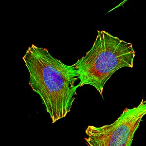
Mouse embryonic fibroblasts forming focal adhesions
Congratulations to Alejandra Valdivia, PhD, winner of the Best Image contest held as part of the Emory Postdoctoral Research Symposium, which takes place next week (Thursday, May 19). She is in Alejandra San Martin’s lab, studying NADPH oxidase enzymes and how they regulate cell migration.
Valdivia submitted this image of mouse embryonic fibroblasts forming focal adhesions, points of contact of the cell with the extracellular matrix. Focal adhesions allow the cells to adhere and migrate.
Explanation: Red is for paxillin, a protein concentrated in focal adhesions. Green is phalloidin, a toxin from mushrooms that binds one type of the cytoskeletal protein actin, seen here as stress fibers. Blue is DNA, showing the cells’ nuclei.