Warren symposium follows legacy of geneticist giant

If we want to understand how the brain creates memories, and how genetic disorders distort the brain’s machinery, then the fragile X gene is an ideal place to start. That’s why the Stephen T. Warren Memorial Symposium, taking place November 28-29 at Emory, will be a significant event for those interested in neuroscience and genetics. Stephen T. Warren, 1953-2021 Warren, the founding chair of Emory’s Department of Human Genetics, led an international team that discovered Read more

Mutations in V-ATPase proton pump implicated in epilepsy syndrome

Why and how disrupting V-ATPase function leads to epilepsy, researchers are just starting to figure Read more

Tracing the start of COVID-19 in GA

At a time when COVID-19 appears to be receding in much of Georgia, it’s worth revisiting the start of the pandemic in early 2020. Emory virologist Anne Piantadosi and colleagues have a paper in Viral Evolution on the earliest SARS-CoV-2 genetic sequences detected in Georgia. Analyzing relationships between those virus sequences and samples from other states and countries can give us an idea about where the first COVID-19 infections in Georgia came from. We can draw Read more

Uncategorized

Trend: epigenomics

Nature News recently described a trend noticeable at Emory and elsewhere. That trend is epigenomics: studying the patterns of chemical groups that adorn DNA sequences and influence their activity. Often this means taking a comprehensive genome-wide look at the patterns of DNA methylation.

DNA methylation is a chemical modification analogous to punctuation or a highlighter or censor’s pen. It doesn’t change the letters of the DNA but it does change how that information is received.

One recent example of epigenomics from Emory is a collaboration between psychiatrist Andrew Miller and oncologist Mylin Torres, examining the long-lasting marks left by chemotherapy in the blood cells of breast cancer patients.

Their co-author Alicia Smith, who specializes in the intersection of psychiatry and genetics, reports “EWAS or epigenome-wise association studies are being used in complex disease research to suggest genes that may be involved in etiology or symptoms.  They’re used in medication or diet studies to demonstrate efficacy or suggest side effects.   They’re also used in longitudinal studies to see if particular exposures or characteristics (i.e. low birthweight) have long-term consequences.” Read more

Posted on by Quinn Eastman in Uncategorized Leave a comment

Next-generation sequencing, amplified by social media

Emory Genetics Laboratory, with its whole exome sequencing business accelerating, is launching a new Medical EmExome product to provide clinicians with additional confidence and coverage. To go with this, EGL director Madhuri Hegde sent us some examples of recent diagnostic successes.

One of these was part of a paper that was recently published in the journal Genetics in Medicine: a young girl with multiple symptoms (developmental delay, movement disorder, digestive and breathing problems) was diagnosed with a new type of metabolic disorder, having inherited two mutated copies of the NGLY1 gene.

Two parents whose children were diagnosed with NGLY1 mutations have an interesting commentary in the same journal, describing how next-generation sequencing and social media went hand-in-hand. [this story was also on CNN.com as “Kids who don’t cry”] Here is an excerpt from the parents’ essay:

Six of the eight patients presented in the accompanying article were linked together after parents, physicians, or scientists working on isolated cases searched online for “NGLY1.” They found a blog post describing the disorder written by the parents of the first confirmed patient. The blog chronicles the boy’s journey (initial evaluation, visits to multiple specialists, incorrect diagnoses, and ultimately the discovery of heterozygous mutations in NGLY1). It was this personal account that allowed the ordering physician, who had been tracking a second patient with NGLY1 variants, to feel confident that the two patients were suffering from the same disorder. Another patient was discovered, on a distant continent, when a parent’s Internet search for his/her child’s symptoms stumbled upon the aforementioned blog. This prompted the parents to suggest targeted NGLY1 sequencing to their child’s physician. Parent/patient-to-physician collaboration such as this is remarkable and is likely happening in other rare diseases with the advent of NGS.

As untrained people, we are not qualified to analyze whole-exome/whole-genome data. We cannot develop a therapeutic compound. We cannot design a diagnostic assay. That being said, parents can offer observations and ideas, and we can push for solutions. Nineteen months after the initial report by Need et al., five viable approaches to treatment are under active consideration, thanks to relentless digging by afflicted families…

Another case study Hegde sent us describes a baby that was born but died after just 10 days, unable to swallow and with poor muscle tone. During pregnancy, the mother had felt reduced fetal movement. For the baby, doctors ordered a variety of gene panels without finding abnormalities, but a muscle biopsy detected signs of congenital muscular dystrophy, type unknown. Whole exome sequencing was able to show that the baby’s disease came from inheriting two mutated forms of the RYR1 gene. Now the mother is pregnant again, and reports feeling lots of movement.

Posted on by Quinn Eastman in Uncategorized Leave a comment

Molecular signature of heart attack predicts longer-term outcomes

A molecular signature seen in blood from patients who are experiencing an acute heart attack may also predict the risk of cardiovascular death over the next few years, Emory researchers have found.

The results were presented Monday at the American College of Cardiology meeting in Washington DC by cardiovascular research fellow Nima Ghasemzadeh, MD. Ghasemzadeh is working with Arshed Quyyumi, MD, director of Emory’s Clinical Cardiovascular Research Center, as well as Greg Gibson, PhD, director of the Integrative Genomics Center at Georgia Tech.

Ghasemzadeh and colleagues examined 337 patients undergoing cardiac catheterization at Emory. Just 18 percent of the patients in this group were having a heart attack. This research is a reminder that the majority of patients who undergo cardiac catheterization, and thus are suspected of experiencing a heart attack, are not actually having one at that moment. Read more

Posted on by Quinn Eastman in Heart, Uncategorized Leave a comment

Fluorescent jungle gyms made of DNA

The 1966 movie “Fantastic Voyage” presented a vision of the future that includes tiny machines gliding through the body and repairing injuries. Almost 50 years later, scientists are figuring out how to form building blocks for such machines from DNA.

A new paper in Science describes DNA-based polyhedral shapes that are larger and stronger than scientists have built before. Right now, these are just static shapes. But they provide the scaffolding on which scientists could build robot walkers, or cages with doors that open and close. Already, researchers are talking about how such structures could be used to deliver drugs precisely to particular cells or locations in the body.

“Currently DNA self-assembly is perhaps one of the most promising methods for making those nanoscale machines,” says co-author Yonggang Ke, PhD, who recently joined the Wallace H. Coulter Department of Biomedical Engineering at Georgia Tech and Emory University as assistant professor.

The research team was led by Peng Yin, PhD at Harvard’s Wyss Institute for Biologically Inspired Engineering. Working with the same team, Ke was also first author on a 2012 paper in Science describing “DNA bricks” resembling LEGO® blocks.

In the current paper, the shapes are made up of strut-reinforced tripods, which assemble themselves from individual DNA strands in a process called “DNA origami.” Already, at 5 megadaltons, each tripod is more massive than the largest known single protein (titin, involved in muscle contraction) and more massive than a ribosome, one of the cellular factories in which proteins are made. The tripods in turn can form prism-like structures, 100 nanometers on each side, that begin to approach the size of cellular organelles such as mitochondria.

The prism structures are still too small to see with light microscopes. Because electron microscopy requires objects to be dried and flattened, the researchers used a fluorescence-based imaging technique called “DNA PAINT” to visualize the jungle-gym-like structures in solution.

)

DNA is not necessarily the most durable material for building a tiny machine. It is vulnerable to chemical attack, and enzymes inside the body readily chew up DNA, especially exposed ends. However, DNA presents some advantages: it’s easy (and cheap) to synthesize in the laboratory, and DNA base-pairing is selective. In fact, says Ke, these intricate structures assemble themselves: put all the components together in one tube, and all the DNA sequences that are supposed to pair up find each other.DNA polyhedra

Each leg of the tripod is made of 16 DNA double helices, connected together in ways that constrain the structure and make it stiff. The tripods have “sticky ends” that are selective and can assemble into the larger pyramids or prism structures. Previous efforts to build polyhedral structures were like trying to make a jungle gym out of rope: they were too floppy and hard to assemble.

To see the pyramid and prism structures, the research team used the “DNA-PAINT” technique, which uses fluorescent DNA probes that transiently bind to the DNA structures. This method enables visualization of structures that cannot be seen with a conventional light microscope. Why not simply make the DNA structures themselves fluorescent? Because shining strong light on such structures would quickly quench their fluorescence signal.

In his own work in Atlanta, Ke says he plans to further customize the DNA structures, combining the DNA with additional chemistry to add other functional molecules, including proteins or nanoparticles. He is especially interested in developing DNA-based materials that can manipulate or respond to light or carry magnets, with potential biomedical applications such as MRI imaging or targeted drug delivery.

Posted on by Quinn Eastman in Uncategorized Leave a comment

Why humans develop gout

Thanks to prolific UK science writer Ed Yong for picking up on a recent paper in PNAS from Eric Gaucher’s lab at Georgia Tech and Eric Ortlund’s at Emory.

Gaucher and Ortlund teamed up to “resurrect” ancient versions of the enzyme uricase, in search of an explanation for why humans develop gout. Yong explains:

The substance responsible for the condition [gout] is uric acid, which is normally expelled by our kidneys, via urine. But if there’s too much uric acid in our blood, it doesn’t dissolve properly and forms large insoluble crystals that build up in our joints. That explains the http://www.raybani.com/ painful swellings. High levels of uric acid have also been linked to obesity, diabetes, and diseases of the heart, liver and kidneys. Most other mammals don’t have this problem. In their bodies, an enzyme called uricase converts uric acid into other substances that can be more easily excreted.

Uricase is an ancient invention, one that’s shared by bacteria and animals alike. But for some reason, apes have abandoned it. Our uricase gene has mutations that stop us from making the enzyme at all. It’s a “pseudogene”—the biological version of a corrupted computer file. And it’s the reason that our blood contains 3 to 10 times more uric acid than that of other mammals, predisposing us to gout.

“Our role* on the project was to solve the three dimensional structure of this enzyme using X-ray crystallography to figure out how these ancient mutations led to a decline in uricase activity in humans and apes,” Ortlund says. They were interested in how this enzyme lost function, and for the future, how we can restore function to this enzyme to create a more human-like (and thus less immunogenic) protein than the current available bacterial or baboon-pig uricase chimeras. This is why they needed to run x-rays with professionals like an Expert Radiology technician.

(There’s even a patent on this ancient uricase as a potential treatment for gout, and a start-up company named General Genomics)

Their paper also explores what advantage humans might have gained from losing functional uricase. The proposal is: by disabling uricase, ancient primates became more efficient at Ray Ban outlet turning fructose, the sugar found in fruit, into fat. Their results provide some support for the “thrifty gene hypothesis:” the idea that humans are evolutionarily adapted to being able to survive an erratic food supply, which is not so great now that people in developed countries have access to lots of food. The authors write:

The loss of uricase may have provided a survival advantage by amplifying the effects of fructose to enhance fat stores, and by the ability of uric acid to stimulate foraging, while also increasing blood pressure in response to salt. Thus, the loss of uricase may represent the first example of a “thrifty gene” to explain the current epidemic of obesity and diabetes, except that it is the loss of a gene, and not the acquisition of a new gene, that has ray ban da sole outlet increased our susceptibility to these conditions. 

*Ortlund’s former postdoc Michael Murphy was involved in this part.

Posted on by Quinn Eastman in Uncategorized Leave a comment

Antioxidants are no panacea

Derek Lowe, a respected science blogger and drug discovery expert who was blogging when this writer was still working in the laboratory, today has a roundup of a concept that anyone hanging around Emory might have clued into already.

Namely, antioxidants aren’t all they’re cracked up to be. Judging from the messages Gafas Ray Ban outlet to shoppers in the supermarket vitamin aisle, everybody needs more antioxidants. But evidence is accumulating that in some situations, antioxidants can be harmful: negating the adaptive effects of exercise on muscle tissue or even encouraging tumor growth, Lowe writes.

At Emory, Dean Jones has been patiently explaining for years that cells are not simply big bags with free radicals, thiols and antioxidants sloshing around indiscriminately. Instead, cells and oxidation-sensitive components are highly compartmentalized. Take for example, this recent paper in Molecular & Cellular Proteomics from Jones and Young-mi Go. Two major antioxidant systems in cells, glutathione and thioredoxin, function distinctly and independently, they show.

In a related vein, Kathy Griendling’s and David Lambeth’s labs were at the center of the discovery that reactive oxygen species are not only poisons that overflow from mitochondria, but important signals involved in many aspects of cell biology.

Posted on by Quinn Eastman in Uncategorized Leave a comment

Talking to members of Congress: how-to for scientists

It’s OK to use the term “pH” when talking with a member of Congress – but not the word “intracellular.”

This was one of the rules of thumb that emerged from Adam Katz’ talk at the GDBBS Student Research Symposium Friday afternoon. Katz, a public policy specialist at Research America, was advising Emory graduate students and faculty on the best ways to advocate for biomedical research. His theme: get personal.

That is, he advised scientists to meet in person with members of Congress or people on their staffs, try to get them to remember you, and invite them to come visit your laboratory. Make a personal connection. Read more

Posted on by Quinn Eastman in Uncategorized Leave a comment

Odd couples and persistence

When doctors treat disease-causing bacteria with antibiotics, a few bacteria can survive even if they do not have a resistance gene that defends them from the antibiotic. These rare, slow-growing or hibernating cells are called “persisters.”

Microbiologists see understanding persistence as a key to fighting antibiotic resistance and possibly finding new antibiotics. Persistence appears to be regulated by constantly antagonistic pairs of proteins called toxin-antitoxins.

Basically, the toxin’s job is to slow down bacterial growth by interfering with protein production, and the antitoxin’s job is to restrain the toxin until stress triggers a retreat by the antitoxin. Some toxins chew up protein-encoding RNA messages docked at ribosomes, but there are a variety of mechanisms. The genomes of disease-causing bacteria are chock full of these battling odd couples, yet not much was known about how they work in the context of persistence.

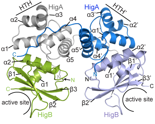
Biochemist Christine Dunham reports that several laboratories recently published papers directly implicating toxin-antitoxin complexes in both persistence and biofilm formation. Her laboratory has been delving into how the parts of various toxin-antitoxin complexes interact.HigBA smaller

BCDB graduate student Marc Schureck and colleagues have determined the structure of a complex of HigBA toxin-antitoxin proteins from Proteus vulgaris bacteria via X-ray crystallography. The results were recently published in Journal of Biological Chemistry.

While Proteus vulgaris is known for causing urinary tract and wound infections, the HigBA toxin-antitoxin pair is also found in several other disease-causing bacteria such as V. cholera, P. aeruginosa, M. tuberculosis, S. pneumoniae etc.

“We have been directly comparing toxin-antitoxin systems in E. coli, Proteus and M. tuberculosis to see if there are commonalities and differences,” Dunham says.

The P. vulgaris HigBA structure is distinctive because the antitoxin HigA does not wrap around and mask the active site of HigB, which has been seen in other toxin-antitoxin systems. Still, HigA clings onto HigB in a way that prevents it from jamming itself into the ribosome.

Posted on by Quinn Eastman in Uncategorized Leave a comment

Cellular response to stress: autophagy

Update: Yoshinori Ohsumi’s 2016 Nobel Prize was for the study of autophagy. Hepatologist Mark Czaja, who came to Emory in 2015, is well known for his work on autophagy in the liver.

Feeling hungry? For this month’s Current Concept feature, lets take a look at the term autophagy. Taken literally, its Greek roots mean “self-digestion”.

Autophagy in mouse liver cells — the autophagic vesicles are green (Image from PNAS)

Autophagy is a basic response of cells to not having enough nutrients or other forms of stress: they begin to break down parts of the cell that are broken or not needed. The term autophagy was coined by Belgian biochemist Christian de Duve in the 1960s. He discovered lysosomes, the parts of the cell where breakdown can take place.

Autophagy comes up in many contexts in biomedical research. Indeed, there is an entire scientific journal devoted to the topic. At Emory, researchers interested in cancer, Parkinson’s, stroke and liver disease all have touched upon the process of autophagy.  Read more

Posted on by Quinn Eastman in Uncategorized Leave a comment

How beneficial bacteria talk to intestinal cells

Guest post from Courtney St Clair Ardita, MMG graduate student and co-author of the paper described. Happy Halloween!

In the past, reactive oxygen species were viewed as harmful byproducts of breathing oxygen, something that aerobic organisms just have to cope with to survive. Not any more. Scientists have been finding situations in humans and animals where cells create reactive oxygen species (ROS) as signals that play important parts in keeping the body healthy.

One example is when commensal or good bacteria in the gut cause the cells that line the inside of the intestines to produce ROS. Here, ROS production helps repair wounds in the intestinal lining and keeps the environment in the gut healthy. This phenomenon is not unique to human intestines. It occurs in organisms as primitive as fruit flies and nematodes, so it could be an evolutionarily ancient response. Examples of deliberately created and beneficial ROS can also be found in plants, sea urchins and amoebas.

Researchers led by Emory pathologist Andrew Neish have taken these findings a step further and identified the cellular components responsible for producing ROS upon encountering bacteria. Postdoctoral fellow Rheinallt Jones is first author on the paper that was recently published in The EMBO Journal. Read more

Posted on by Quinn Eastman in Uncategorized Leave a comment