Warren symposium follows legacy of geneticist giant

If we want to understand how the brain creates memories, and how genetic disorders distort the brain’s machinery, then the fragile X gene is an ideal place to start. That’s why the Stephen T. Warren Memorial Symposium, taking place November 28-29 at Emory, will be a significant event for those interested in neuroscience and genetics. Stephen T. Warren, 1953-2021 Warren, the founding chair of Emory’s Department of Human Genetics, led an international team that discovered Read more

Mutations in V-ATPase proton pump implicated in epilepsy syndrome

Why and how disrupting V-ATPase function leads to epilepsy, researchers are just starting to figure Read more

Tracing the start of COVID-19 in GA

At a time when COVID-19 appears to be receding in much of Georgia, it’s worth revisiting the start of the pandemic in early 2020. Emory virologist Anne Piantadosi and colleagues have a paper in Viral Evolution on the earliest SARS-CoV-2 genetic sequences detected in Georgia. Analyzing relationships between those virus sequences and samples from other states and countries can give us an idea about where the first COVID-19 infections in Georgia came from. We can draw Read more

Neuro

Dysbindin, a bad actor in schizophrenia

Cell biologist Victor Faundez has been getting some attention for his research on dysbindin, a protein linked to schizophrenia. The information helps to make sense of the complex picture emerging from genetic studies of schizophrenia.

Genetics plays a major role in schizophrenia, but there is no one gene that pulls the trigger. The gene encoding dysbindin was first identified as a potential bad actor in 2002, by researchers studying families with a high rate of schizophrenia. Dysbindin levels are reduced in the brains of schizophrenia patients, and mouse mutants lacking the protein develop normally but have altered signaling in the brain.

Dysbindin is known to be part of a machine that produces vesicles (tiny bubbles containing proteins and neurotransmitters) and transports them around the cell. This machine, found in several tissues besides the brain, has a mouthful of a name: BLOC (Biogenesis of Lysosome-related Organelles Complex). Faundez’ lab has shown that defects in BLOC make proteins in neurons “miss the bus” that would transport them from the cell body out to the synapse.

The BLOC complex transports vesicles from the cell body out to the synapse. When parts of the complex are missing, neurons appear to develop aberrantly.

The team of Faundez, postdoc Avanti Gokhale and their colleagues set out to define all the parts of the BLOC machine and find other proteins dysbindin comes into contact with. Several of the proteins they found (the results were published in March 2012 in Journal of Neuroscience) are affected by copy number variation in schizophrenia patients.

“This was a surprise,” Faundez says. “The genomic studies in schizophrenia identify lots of genes, but looking at them, we don’t know how they relate to each other.”

Copy number variation means: patients have a deletion or an extra copy of the gene involved. A copy number variation doesn’t mean someone is always going to get schizophrenia, but it may be enough to tip the balance when other risk factors add up.

Faundez says his team’s results highlight an approach to examining genes implicated in complex diseases: rather than looking at individual genes, look at circuits in the cell. A strong example: two of the genes that encode dysbindin interaction partners are located within the chromosome 22q11 region. Individuals with a deletion in this region develop schizophrenia at a rate of 30 percent.

Faundez’s team also found that dysbindin interacts with peroxiredoxins, antioxidant enzymes that clean up hydrogen peroxide. They went on to confirm that dysbindin mutant cells have elevated peroxide levels, which hints at a role for altered redox signaling in schizophrenia.

Biomarkers in schizophrenia have been elusive, but Faundez says he thinks his research could lead to identifying a subset of schizophrenia patients where a disturbance of the BLOC system is especially important.

Emory geneticists Andres Moreno-De Luca and Christa Lese-Martin are coauthors on the JN paper.


Posted on by Quinn Eastman in Neuro Leave a comment

Fragile X protein: one toggle switch, many circuits

The fragile X protein — missing in the most common inherited form of intellectual disability — plays a central role in neurons and how they respond to external signals. Cell biologist Gary Bassell and his colleagues have been examining how the fragile X protein (FMRP) acts as a “toggle switch.”

Gary Bassell, PhD

FMRP controls the activity of several genes by holding on to the RNAs those genes encode. When neurons get an electrochemical signal from the outside, FMRP releases the RNAs, allowing the RNAs to be made into protein, and facilitating changes in the neurons linked to learning and memory.

The Bassell lab’s new paper in Journal of Neuroscience reveals the role of another player in this process. The first author is postdoctoral fellow Vijay Nalavadi.

The researchers show that neurons modify FMRP with ubiquitin, the cellular equivalent of a tag for trash pickup, after receiving an external signal. In general, cells attach ubiquitin to proteins so that the proteins get eaten up by the proteasome, the cellular trash disposal bin. Here, neurons are temporarily getting rid of FMRP, prolonging the effects of the external signal.

Posted on by Quinn Eastman in Neuro Leave a comment

What is the default mode network?

Welcome to what could become a regular feature on the Emory Health Now blog: explaining a word or phrase that is connected with research going on at Emory.

What is the default mode network?
This is a concept that grew out of brain imaging studies, using techniques such as functional magnetic resonance imaging. The default mode network consists of regions of the brain that are active when someone is not doing anything in particular, especially something requiring focused attention. More fancifully: it’s what daydreaming looks like.

The default mode network includes the medial prefrontal cortex (MPFC) and the posterior cingulate cortex (PCC).

Researchers at Emory and elsewhere have been looking at whether the DMN’s activity and its links to other areas of the brain are changed in disorders such as schizophrenia, autism, depression and post-traumatic stress disorder.

For example, Xiaoping Hu, director of Emory’s Biomedical Imaging Technology Center, and his colleagues have investigated how the default mode network’s activity is modified in individuals with prenatal alcohol exposure and prenatal cocaine exposure. They also have probed how the DMN’s activity is shut down by anesthesia.

At the Atlanta Veterans Affairs Medical Center, Erica Duncan and her colleagues have examined the DMN in people with schizophrenia. They found (as have other groups) that individuals with schizophrenia appear to have difficulty shutting down the DMN and focusing on a task, as well as having a different pattern of connections within the DMN.

Yerkes investigator Lary Walker recently wrote about research connecting metabolic activity in the default mode network with the burden of amyloid in Alzheimer’s disease.

The DMN is made up of several regions of the brain, most prominently the posterior cingulate cortex (PCC) and medial prefrontal cortex (MPFC). Other regions such as the inferior parietal lobule, lateral temporal cortex, and hippocampal formation including parahippocampus are also considered part of the DMN. Note: these regions can also be engaged in other tasks besides daydreaming or introspection.

Posted on by Quinn Eastman in Neuro Leave a comment

New molecular target in dystonia

Emory researchers led by pharmacologist Ellen Hess have identified a new molecular target in dystonia. Their findings, recently published in the Journal of Pharmacology and Experimental Therapeutics, could help doctors find drugs for treating the movement disorder.

Ellen Hess, PhD

Dystonia gives sufferers involuntary muscle contractions that cause rigid, twisting movements and abnormal postures. It is the third most common movement disorder, after tremor and Parkinson’s disease. Neurologists can sometimes use drugs to address the symptoms of dystonia but there is no cure.

A 2008 review by Hess (PDF) concludes that compared with other neurological disorders, “our understanding of the biology and potential treatments for dystonia is in its infancy.” Still, scientists have known for a while that the cerebellum, a region of the brain that regulates movement, is involved.

“We focused on the cerebellum because studies in patients with dystonia often show that this part of the brain is more active, when examined by MRI,” Hess says. “The abnormal overactivity of the cerebellum is seen in patients with all different types of dystonia, so it seems to be a common hotspot. Our goal was to understand what might be causing the overactivity in mice because if we can stop the overactivity, we might be able to stop the dystonia.”

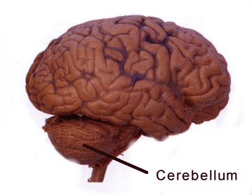
Hess and her colleagues discovered that drugs that stimulate AMPA receptors induce dystonia when introduced into the mouse cerebellum. Their results suggest that drugs that act in reverse, blocking AMPA receptors, could be used to treat dystonia.

Postdoctoral fellow Xueliang Fan is the first author of the paper. Emory neurologist H.A. Jinnah, director of a NIH-supported network of clinical research sites focusing on dystonia, is a co-author.

AMPA receptors are a subset of glutamate receptors, a large group of “receiver dishes” for excitatory signals in the brain. Fan performed a variety of experiments to show that AMPA receptor activity plays a specific role in generating dystonia. For example, drugs that affect other types of glutamate receptors did not induce dystonia. AMPA receptor blockers can also reduce dystonia in a genetic model, the “tottering” mouse.

Although pharmaceutical companies have already been testing AMPA receptor blockers as potential antiseizure drugs, caution is in order. AMPA receptor stimulators/ enhancers (or “ampakines”) have been identified as potential enhancers of learning and memory, so AMPA receptor blockers may interfere with those processes.

“Our results suggest that reducing AMPA receptor activity could help alleviate dystonia but we still have a lot of work to do before we know whether blocking AMPA receptor activity in patients will really help,” Hess says. “Since there aren’t many drugs that act at AMPA receptors, one of our goals is to identify drugs that change the ‘downstream’ effects of AMPA receptor activation. For example, we may be able to find other drug classes that change neuronal activity in the same way that AMPA receptor blockade changes activity.”

Posted on by Quinn Eastman in Neuro Leave a comment

DNA copying problems explain muscular dystrophy mutations

Geneticist Madhuri Hegde and her colleagues have a paper in the journal Genome Researchthat addresses the question: where do copy number variations come from?

Madhuri Hegde, PhD

Copy number variations (CNVs), which are deletions or duplications of small parts of the genome, have been the subject of genetic research for a long time. But only in the last few years has it become clear that copy number variations are where the action is for complex diseases such as autism and schizophrenia. Geneticists studying these diseases are shifting their focus from short, common mutations (often, single nucleotide polymorphisms or SNPs) to looking at rarer variants such as CNVs. A 2009 discussion of this trend with Steve Warren and Brad Pearce can be found here.

Hegde is the Scientific Director of the Department of Human Genetics’ clinical laboratory. Postdoctoral fellow Arun Ankala is the first author. In the new paper, Ankala and Hegde examine rearrangements in patients’ genomes that arose in 62 clinical cases of Duchenne’s muscular dystrophy and several other diseases. Mutations in the DMD gene are responsible for Duchenne’s muscular dystrophy.

The pattern of the rearrangement hints what events took place in the cell beforehand, and hint that a problem took place during replication of the DNA. The signature is a tandem duplication of a short segment next to a large deletion, indicating how the DNA was repaired.

The authors note that the DMD locus is especially prone to these types of problems because it is much larger than other gene loci. The gene is actually the longest human gene known on the DNA level, covering 2.4 megabases (0.08 percent of the genome.)

Replication origins are where the DNA copying machinery in the cell starts unwinding and copying the DNA. Bacterial circular chromosomes have just one replication origin. In contrast, humans have thousands of replication origins spread across our chromosomes. In the discussion, the authors suggest that DNA copying problems may also explain duplications and historically embedded rearrangements of the genome.

Posted on by Quinn Eastman in Neuro Leave a comment

COX-2 and epilepsy: it’s complicated

How much is the development of epilepsy like arthritis?

More than you might expect. Inflammation, or the overactivation of the immune system, appears to be involved in both. In addition, for both diseases, inhibiting the enzyme COX-2 initially looked like a promising approach.

Ray Dingledine, PhD

COX-2 (cyclooxygenase 2) is a target of traditional non-steroid anti-inflammatory drugs like aspirin and ibuprofen, as well as more selective drugs such as Celebrex. With arthritis, selectively inhibiting COX-2 relieves pain and inflammation, but turns out to have the side effect of increasing the risk of heart attack and stroke.

In the development of epilepsy, inhibiting COX-2 turns out to be complicated as well. Ray Dingledine, chair of pharmacology at Emory, and colleagues have a new paper showing that COX-2 has both protective and harmful effects in mice after status epilepticus, depending on the timing and what cells the enzyme comes from. Status epilepticus is a period of continuous seizures leading to neurodegeneration, used as a model for the development of epilepsy.

Postdoc Geidy Serrano, now at the Banner Sun Health Research Institute in Arizona, is first author of the paper in Journal of Neuroscience. She and Dingledine were able to dissect COX-2’s effects because they engineered mice to have a deletion of the COX-2 gene, but only in some parts of the brain.
They show that deleting COX-2 in the brain reduces the level of inflammatory molecules produced by neurons, but this is the reverse effect of deleting it all over the body or inhibiting the enzyme with drugs.

Four days after status epilepticus, fewer neurons are damaged (bright green) in the neuronal COX-2 knockout mice.

Dingledine identified two take-home messages from the paper:
First, COX-2 itself is probably not a good target for antiepileptic therapy, and it may be better to go downstream, to prostaglandin receptors like EP2.
Second, the timing of intervention will be important, because the same enzyme has opposing actions a few hours after status epilepticus compared to a couple days later.

More of Dingledine’s thinking about inflammation in the development of epilepsy can be found in a recent review.

Posted on by Quinn Eastman in Neuro Leave a comment

Blood biomarkers may help predict risk in stroke and TBI


Biomarkers circulating in the bloodstream may serve as a predictive window for recurrent stroke risk and also help doctors accurately assess what is happening in the brains of patients with acute traumatic brain injury (TBI).

Michael Frankel, MD

Researchers at Emory University School of Medicine, led by principal investigator Michael Frankel, MD, Emory professor of neurology and director of Grady Memorial Hospital’s Marcus Stroke & Neuroscience Center, are studying biomarkers as part of two ancillary studies of blood samples using two grants from the National Institutes of Health.

In the $1.47 million, four-year grant called “Biomarkers of Ischemic Outcomes in Intracranial Stenosis” (BIOSIS), Emory researchers are analyzing blood samples from 451 patients from around the country who were enrolled in a study known as SAMMPRIS (Stenting and Aggressive Medical Management for Preventing Recurrent stroke in Intracranial Stenosis), the first randomized, multicenter clinical trial designed to test whether stenting intracranial arteries would prevent recurrent stroke.

Researchers in the SAMMPRIS study recently published their results in the New England Journal of Medicine, showing that medical management was more effective than stenting in preventing recurrent strokes in these patients. Frankel’s BIOSIS research team is using blood samples from these same patients to continue learning more about the molecular biology of stroke to predict risk of a stroke occurring in the future.

“Our goal is to learn more about stroke by studying proteins and cells in the blood that reflect the severity of disease in arteries that leads to stroke. If we can test blood samples for proteins and cells that put patients at high risk for stroke, we can better tailor treatment for those patients,” says Frankel.

Patients with narrowed brain arteries, known as intracranial stenosis, have a particularly high risk of disease leading to stroke. At least one in four of the 795,000 Americans who have a stroke each year will have another stroke within their lifetime. Within five years of a first stroke, the risk for another stroke can increase more than 40 percent. Recurrent strokes often have a higher rate of death and disability because parts of the brain already injured by the original stroke may not be as resilient.

The other study, “Biomarkers of Injury and Outcome in ProTECT III” (BIO-ProTECT)” is a $2.6 million, five-year NIH grant in which Frankel’s team will use blood to determine what is happening in the brain of patients with acute TBI.  The blood samples are from patients enrolled in the multicenter clinical trial ProTECT III (Progesterone for Traumatic brain injury, Experimental Clinical Treatment), led by Emory Emergency Medicine Professor, David Wright, MD, to assesses the use of progesterone to treat TBI in 1,140 patients at 17 centers nationwide.

In the BIO-ProTECT study, Emory is collaborating with the Medical University of South Carolina, the University of Pittsburgh, the University of Michigan and Banyan Biomarkers.

TBI is the leading cause of death and disability among young adults in the US and worldwide. According to the Centers for Disease Control and Prevention, approximately 1.4 million Americans sustain a traumatic brain injury each year, leading to 275,000 hospitalizations, 80,000 disabilities, and 52,000 deaths.

Acute TBI leads to a cascade of cellular events set in motion by the initial injury that ultimately lead to cerebral edema (swelling of the brain), cellular disruption and sometimes death. Tissue breakdown leads to the release of proteins into the bloodstream. These proteins may serve as useful biomarkers of the severity of the injury and perhaps provide useful information about response to treatment.

Using the large patient group in the ProTECT III trial, the researchers hope to validate promising TBI biomarkers as predictors of clinical outcome and also evaluate the relationship between progesterone treatment, biomarker levels and outcome.

“If we can better determine the amount of brain injury with blood samples, we can use blood to help doctors better assess prognosis for recovery, and, hopefully whether a patient will respond to treatment with progesterone,” says Frankel.

Posted on by admin in Neuro Leave a comment

Redirecting beta-amyloid production in Alzheimer’s

Pharmacologist Thomas Kukar is exploring a strategy to subtly redirect the enzyme that produces beta-amyloid, which makes up the plaques appearing in the brains of Alzheimer’s patients.

Thomas Kukar, PhD

Preventing beta-amyloid production could be an ideal way to head off Alzheimer’s, but the reason why a subtle approach is necessary was illustrated last year by disappointing results from a phase III clinical trial. The experimental drug semagacestat was designed to block the enzyme gamma-secretase, which “chomps” on the amyloid precursor protein (APP), usually producing an innocuous fragment but sometimes producing toxic beta-amyloid.

Gamma-secretase also is involved in processing a bunch of other vital proteins, such as Notch, central to an important developmental signaling pathway. Scientists suspect that this is one of the reasons why trial participants who received semagacestat did worse on cognitive/daily function measures than controls and saw an increase in skin cancer, leading watchdogs to halt the study.

While a postdoc at Mayo Clinic Jacksonville and working with Todd Golde and Edward Koo, Kukar identified compounds – gamma-secretase modulators or GSM’s — that may offer an alternative.

“We are looking at a strategy that’s different from global gamma-secretase inhibition,” he says. “The approach is: don’t inhibit the enzyme overall, but instead modify its activity so that it makes less toxic products.”

Gamma-secretase chomps on amyloid precursor protein, and how it does so determines whether toxic beta-amyloid is produced. It also processes several other proteins important for brain function.

This line of inquiry started when it was discovered that some anti-inflammatory drugs also could reduce beta-amyloid production. Then, the crosslinkable probes Kukar was using to identify which part of the gamma-secretase fish was doing the chomping ended up binding the bait (APP). This suggested that drugs might be able to change how the enzyme acts on one protein, APP, but not others.

Now an assistant professor at Emory, he is examining in greater detail how gamma-secretase modulators work. Two recent papers he co-authored in Journal of Biological Chemistry show 1) how the proteins that gamma-secretase chews up are “anchored” in the membrane and 2) how selective GSM’s can be on amyloid precursor protein.

Although clinical studies of a “first generation” GSM, tarenflurbil, were also stopped after negative results, Kukar says GSM’s still haven’t really been tested adequately, since researchers do not know if the drugs are really having an effect on beta-amyloid levels in the brain. Newer compounds coming through the pharmaceutical pipeline are more potent and more able to get into the brain. While looking for more potent GSM’s is critical, Kukar says it’s equally as important to understand how gamma-secretase works to understand its biology.

Posted on by Quinn Eastman in Neuro Leave a comment

Blue pill or red pill? Brains need both for memory consolidation

In the 1999 film The Matrix, the character Neo is offered a choice between a blue pill (to forget) and a red pill (to remember). If only neuroscience was that simple! It may be that neurons need both red and blue, possibly an elaborate dance of molecules, for a fragile memory to lodge itself in the brain.

Neuroscientists Kimberly Maguschak and Kerry Ressler provide a glimpse into this process with their recent paper in the Journal of Neuroscience.

Ressler is both a psychiatrist and a Howard Hughes Medical Institute-supported researcher with a laboratory at Yerkes National Primate Research Center. Maguschak completed her doctorate at Emory and is now a postdoc with Guoping Feng at MIT.

The research is a follow-up on their work probing the role of beta-catenin in fear memory formation. We previously described this protein as acting “like a Velcro strap”, attaching cells’ internal skeletons to proteins on their external membranes that help them adhere to other cells. If brain cells need to change shape and form new connections for memories to be consolidated, we can see how this kind of molecule would be important.

Beta-catenin is also central to a signaling circuit that maintains stem cells and prods an embryo to separate into front and back or top and bottom. This circuit is called “Wnt” (the name is a fusion of the fruit fly gene wingless and a cancer-promoting gene discovered in mice, originally called Int-1).

Maguschak and Ressler wanted to assess the role Wnt signals play in learning and memory. The model system was the same as in their previous work: if mice are electrically shocked just after they hear a certain tone, they gradually learn to fear that tone, and they show that fear by freezing.

Kerry Ressler, MD, PhD

Maguschak saw that in the amygdala, a part of the brain important for fear responses, Wnt genes are turned down during the learning process temporarily but then come back on. If the mice only hear the tone or only get the shock, the genes’ activities don’t change significantly.

She then introduced proteins that perturb Wnt signaling directly into the amygdala. Extra Wnt injected before training, while it didn’t stop the mice from learning to fear the tone, made that training less likely to “stick.” Two days later, the mice that received Wnt didn’t seem to fear the tone as much.

Here’s the possibly confusing part: a Wnt inhibitor also impaired fear memory consolidation. In effect, both blue and red pills actually interfered with how well memories endured. The authors suggest this is because Wnt signals have to be turned down during fear memory formation but then turned back up so those memories can solidify. The Wnt signals seem to go along with the adhesive interactions of beta-catenin. It looks like beta-catenin’s stickiness also needs to be tuned down and then back up.

The off-then-on-again requirement Maguschak and Ressler observe is reminiscent of results from cell biologist James Zheng’s lab. He and his colleagues saw that the actin cytoskeleton needed to be weakened and then stabilized during long-term potentiation, an enhancement of connections between neurons thought to lie behind learning and memory.

Several laboratories have identified potential drugs that modify beta-catenin/Wnt. These new results suggest that the timing of when and how to use such drugs to enhance memory may critically important to consider, Ressler says.

“To interfere with memory formation after trauma or enhance memory formation in people with dementia, researchers will clearly need to attend to the full complexity of the dynamics of synaptic plasticity and memory,” he says.

A nifty link to an animation of Wnt signaling


Posted on by Quinn Eastman in Neuro Leave a comment

Neuroinflammation: a different way to look at Parkinson’s disease

Emory physiologist Malu Tansey and her colleagues are using recent insights into the role of inflammation in Parkinson’s disease to envision new treatments. One possible form this treatment strategy could take would be surprisingly simple, and comparable to medications that are approved for rheumatoid arthritis.

Malu Tansey, PhD

Understanding the role of inflammation in Parkinson’s requires a shift in focus. Many Parkinson’s researchers understandably emphasize the neurons that make the neurotransmitter dopamine. They’re the cells that are dying or already lost as the disease progresses, leading to tremors, motor difficulties and a variety of other symptoms.

But thinking about the role of inflammation in Parkinson’s means getting familiar with microglia, the immune system’s field reps within the brain. At first, it was thought that the profusion of microglia in the brains of Parkinson’s patients was just a side effect of neurodegeneration. The neurons die, and the microglia come in to try to clean up the debris.

Now it seems like microglia and inflammation might be one of the main events, if not the initiating event.

“Something about the neurons’ metabolic state, whether it’s toxins, oxidative stress, unfolded proteins, or a combination, makes them more sensitive. But inflammation, sustained by the presence of microglia, is what sends them over the edge,” Tansey says.

She says that several recent studies have led to renewed attention to this area:

  1. In vivo PET imaging using a probe for microglia has allowed scientists to see inflammation starting early in the progression of Parkinson’s (see figure below)
  2. Epidemiology studies show that taking ibuprofen regularly is linked to lower incidence of Parkinson’s
  3. Experiments with animal models of genetic susceptibility demonstrate that inflammatory agents like endotoxin can accelerate neurodegeneration
  4. Genomics screens have identified HLA-DR, an immune system gene, as a susceptibility marker for Parkinson’s (Emory’s Stewart Factor was a co-author on this paper)

Popping a few ibuprofen pills everyday for prevention and possibly damaging the stomach along the way is probably not going to work well, Tansey says. It should be possible to identify a more selective way to inhibit microglia, which may be able to inhibit disease progression after it has started.

Activated microglia in the midbrain and striatum of a Parkinson's patient

Targeting TNF (tumor necrosis factor), an important inflammatory signaling molecule, may be one way to go. Anti-TNF agents are already used to treat rheumatoid arthritis and inflammatory bowel disease. This January, Tansey and her co-workers published a paper showing that a gene therapy approach using decoy TNF can reduce neuronal loss in a rat model of Parkinson’s. More recently, her lab has also shown that targeting the gene RGS10 is another way to inhibit microglia and reduce neurodegeneration in the same models.

It is important to note that in the rat studies, they do surgery and put the gene therapy viral vector straight into the brain. She says it might possible to perform peripheral gene therapy with the microglia, or even anti-TNF medical therapy. In terms of mechanism, decoy (technically, dominant negative) TNF is more selective and may avoid the side effects, such as opportunistic infections, of existing anti-TNF agents.

Posted on by Quinn Eastman in Neuro 1 Comment