Warren symposium follows legacy of geneticist giant

If we want to understand how the brain creates memories, and how genetic disorders distort the brain’s machinery, then the fragile X gene is an ideal place to start. That’s why the Stephen T. Warren Memorial Symposium, taking place November 28-29 at Emory, will be a significant event for those interested in neuroscience and genetics. Stephen T. Warren, 1953-2021 Warren, the founding chair of Emory’s Department of Human Genetics, led an international team that discovered Read more

Mutations in V-ATPase proton pump implicated in epilepsy syndrome

Why and how disrupting V-ATPase function leads to epilepsy, researchers are just starting to figure Read more

Tracing the start of COVID-19 in GA

At a time when COVID-19 appears to be receding in much of Georgia, it’s worth revisiting the start of the pandemic in early 2020. Emory virologist Anne Piantadosi and colleagues have a paper in Viral Evolution on the earliest SARS-CoV-2 genetic sequences detected in Georgia. Analyzing relationships between those virus sequences and samples from other states and countries can give us an idea about where the first COVID-19 infections in Georgia came from. We can draw Read more

Neuro

Where does learning to touch more sensitively “live” in the brain?

When someone’s sense of touch becomes more acute through training, the brain itself changes. Using functional magnetic resonance imaging (fMRI), researchers have devised ways to see which areas of the brain become more active.

Surprisingly, the changes in activity appear in parts of the Cheap Oakleys brain thought to be responsible for decision-making, rather than the “somatosensory” regions involved in processing touch signals from the fingers.

The results were reported Tuesday in the Journal of Neuroscience.

Participants were asked to discriminate between three-dot patterns, while the horizontal offset became less and less.

Participants were asked to discriminate between three-dot patterns, while the horizontal offset became less and less.

Sighted college undergraduates were trained to discriminate between patterns of raised dots with their fingers. After several sessions, the threshold of differences study participants could detect became much smaller. They could detect differences of less than 0.2 millimeters, when they had started out only being able to detect 1 millimeter changes.

“It is a task that resembles reading braille, and it tests for the same kind of fine level discrimination needed to read braille,” says Krish Sathian, MD, PhD, professor of neurology, rehabilitation medicine, and psychology at Emory University.

Read more

Posted on by Quinn Eastman in Neuro Leave a comment

A Sherer of Parkinson’s research

The name of the guest speaker at Emory’s Office of Technology Transfer’s annual celebration on March 7 provoked some double takes around campus last week.

Todd B. Sherer, PhD

Todd B. Sherer, PhD

Todd B. Sherer, PhD, CEO of the Michael J. Fox Foundation for Parkinson’s Research (MJFF), described how the Fox Foundation is trying to build bridges between the worlds of basic and clinical research to speed development of new drugs for the treatment of Parkinson’s disease, and offered a ray ban outlet perspective on how independent research funders can help move drug candidates from the lab to the clinic and closer to market.

Sherer, a former postdoctoral fellow at Emory, also has the same first and last name (but not middle initial!) as OTT’s director. Sherer – the one who works for the Fox Foundation – joined that non-profit charity’s staff in 2004. While at Emory, he worked on models for Parkinson’s based on exposure to the pesticide rotenone, alongside Ranjita Betarbet, Gary Miller and J. Timothy Greenamyre, who himself moved on to the University of Pittsburgh in 2005.

Read more

Posted on by Quinn Eastman in Neuro Leave a comment

Striking graph showing gene-stress interactions in PTSD

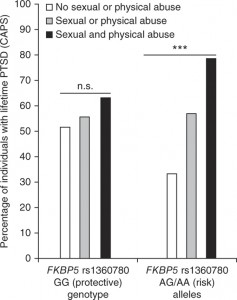
This graph, from a recent paper in Nature Neuroscience, describes how variations in the gene FKBP5 make individuals more susceptible to physical and sexual abuse, and thus more likely to develop PTSD (post-traumatic stress disorder).nn.3275-F1

The paper is the result of a collaboration between Elisabeth Binder and her colleagues at the Max Planck Institute of Psychiatry in Munich, and Emory psychiatrists Kerry Ressler and Bekh Bradley. The population under study is made up of inner-city Atlanta residents, part of the Grady Trauma Project overseen by Ressler and Bradley. This paper analyzes samples from a group of individuals that is more than twice as large as the original 2008 paper defining the effect of FKBP5, and adds mechanistic understanding: how regulation of the FKBP5 gene is perturbed.

Back to the graph — in addition to the effects of the different forms of the gene, it is striking how high the rate of PTSD is for both individuals with the protective and risk forms of FKBP5. Also, for individuals who did not experience abuse, the PTSD rate is actually higher for the “protective” form of the gene. On this point, the authors write:

It is, however, possible that the described polymorphisms Gafas Ray Ban outlet define not only risk versus resilience, but possibly environmentally reactive versus less reactive individuals. This would imply that the so-called risk-allele carriers may also profit more from positive environmental change.

The FKBP5 gene encodes a protein that regulates responses to the stress hormone cortisol. Thus, it acts in blood and immune system cells, not only the brain, and is involved in terminating the stress response after the end of a threat. In the paper’s discussion, the authors propose that FKBP5 may have a role in sensitivity to other immune and metabolic diseases, in addition to PTSD and depression.

Max Planck press release on Binder paper

Recent post on Shannon Gourley’s related work (how stress hormone exposure leads to depression)


Posted on by Quinn Eastman in Neuro Leave a comment

Seeing in triangles with grid cells

When processing what the eyes see, the brains of primates don’t use square grids, but instead use triangles, research from Yerkes neuroscientist Beth Buffalo’s lab suggests.

Elizabeth Buffalo, PhD

She and graduate student Nathan Killian recently published (in Nature) their description of grid cells, neurons in the entorhinal cortex that fire when the eyes focus on particular locations.

Their findings broaden our understanding of how visual information makes its way into memory. It also helps us grasp why deterioration of the entorhinal cortex, a region of the brain often affected early by Alzheimer’s disease, produces disorientation.

The Web site RedOrbit has an extended interview with Buffalo. An excerpt:

The amazing thing about grid cells is that the multiple place fields are in precise geometric relation to each other and form a tessellated array of equilateral triangles, a ‘grid’ that tiles the entire environment. A spatial autocorrelation of the grid field map produces a hexagonal structure, with 60º rotational symmetry. In 2008, grid cells were identified Gafas Ray Ban outlet in mice, in bats in 2011, and now our work has shown that grid cells are also present in the primate brain.

Please read the whole thing!

Grid cells fire at different rates depending on where the eyes are focused. Mapping that activity across the visual field produces triangular patterns.

Posted on by Quinn Eastman in Neuro 1 Comment

Tangled up with tau

Pathologist Keqiang Ye and his colleagues have identified a new potential drug target in Alzheimer’s disease. It’s called SRPK2 (serine-arginine protein kinase 2).

Keqiang Ye, PhD

Depleting this enzyme from the brain using genetic engineering tools alleviates cognitive impairment in an animal model of Alzheimer’s. The result suggests that drugs Cheap Oakleys that target this enzyme could be valuable in the treatment of Alzheimer’s, although additional studies on human brain samples are necessary to fully confirm the findings, Ye says.

The results were published Tuesday in Journal of Neuroscience. The first author is postdoctoral fellow Yi Hong.

Hong and colleagues found that SRPK2 has elevated activity in a mouse model of Alzheimer’s. It acts on tau, one of the two major toxic clumpy proteins in Alzheimer’s. (beta-amyloid is outside the cell and forms plaques, tau is inside and forms tangles). Previous research on SRPK2 indicated that it had something to do with RNA splicing, so its “entanglement” with tau is a surprise.

Posted on by Quinn Eastman in Neuro Leave a comment

Dissecting how chronic stress leads to depression

How can we study depression and antidepressants in animals? They can’t talk and tell us how they’re feeling. Previously, researchers have used the model of “behavioral despair,” with examples of the forced swimming test or the tail suspension test.

Shannon Gourley, PhD

Several psychiatrists have been arguing that a new framework is needed, which better simulates aspects of depression in humans, such as the variety of behavioral changes and the several week time period needed for antidepressants to function. This new framework could help illuminate how depression develops, and lead to new antidepressants that are effective for more people.

Shannon Gourley, who recently joined the Emory-Children’s Pediatric Research Center has been taking the approach of examining the lack of motivation and self-defeating behavior that are integral parts of depression.

The Pediatric Research Center is an effort led by Emory University and Children’s Healthcare of Atlanta, including partnerships with the Georgia Institute of Technology and Morehouse School of Medicine.

Note: Gretchen Neigh in psychiatry/physiology has been doing work with a similar theme, looking at the effects of adolescent social stress in animal models.

Gourley, neuroscience graduate student Andrew Swanson and their colleagues at Yale, where Gourley was a postdoc with Jane Taylor and Tony Koleske, have a new paper in PNAS on this topic. In particular, they dissect how chronic stress – or exposure to the stress hormone corticosterone – can produce loss of motivation and impaired decision making.

First, the researchers found that exposing rodents to cheap oakleys corticosterone shut off a growth factor called BDNF (brain-derived neurotrophic factor) in the frontal cortex, a region of the brain important for planning and goal-directed behavior. BDNF nourishes neurons and helps keep them alive.

To confirm that BDNF was important in this region of the brain, researchers selectively silenced the gene for BDNF only in the frontal cortex. Both mice exposed to stress hormones and the BDNF-altered mice showed reduced motivation to earn food rewards. Mice would ordinarily press a lever dozens of times to get a food pellet, but the BDNF-altered animals would stop trying earlier – the “break point” is 2/3 as high.

“Depression is a leading cause of unemployment because people are unable to break out of self-defeating behavioral patterns and to muster the motivation to engage with the world. If we can better understand how to treat these symptoms, we can effect better outcomes for individuals suffering from depression,” Gourley says. “The BDNF deficiency alone could account for the loss of motivation that individuals with depression suffer.”

However, she reports her team was surprised that the loss of BDNF could not account for another aspect of depression: cyclical self-defeating behavior. They modeled this by asking whether mice continue to press a lever for a food reward even when the reward is no longer available.

“When we made the discovery that reduced BDNF could not account for all of the depression symptoms that we study, we took a step back and looked at the stress response system,” Gourley says.

Stress hormone exposure impairs the ability of mice to switch away from fruitless behaviors, but loss of BDNF in the frontal cortex does not. Here, the stress response system itself was the culprit. When her team temporarily blocked the ability of mice to shut off their stress response systems using the drug mifepristone, mice had impaired decision-making. However, their motivation to obtain rewards was not altered. When the drug wore off, they returned to normal.

Gourley says the implication is that effective antidepressants need to be able to attack not one, but two physiological systems: they need to increase levels of BDNF, and they need to help the stress system recover so that it can shut itself off better. A classic trycyclic antidepressant, amitriptyline, can do both and was effective in treating both the motivation and decision making parts of depression in animal models.

The use of tricyclic antidepressants is limited because of side effects and overdose potential. In addition, another challenge in treating depression is that current antidepressants only begin to work after several weeks or months of treatment. This is thought to be because it takes several weeks for these drugs—which act only indirectly on BDNF—to restore BDNF levels back to normal.

New compounds that act directly on BDNF’s receptor TrkB, such as those identified and tested by Emory researcher Keqiang Ye, could be promising in the development of new approaches to depression, Gourley says.

She and her team also showed that a drug called riluzole, which acts indirectly but rapidly on BDNF systems, has antidepressant effects in the animal models. Riluzole is currently in use to treat ALS, and reportedly has antidepressant effects in humans. Clinical trials with riluzole in the context of depression are underway.

Posted on by Quinn Eastman in Neuro Leave a comment

Fragile X clinical trial update

A recent issue of Emory Health magazine had an article describing the progress of clinical trials for fragile X syndrome, the most common inherited cause of intellectual disability. The article included interviews with the parents of a boy, Samuel McKinnon, who is participating in one of the phase III clinical trials here at Emory.

Last week, results for the phase II study for the same medication were published in Science Translational Medicine. The drug, called STX209 or arbaclofen, is one of the first designed to treat the molecular changes in the brain caused by fragile X syndrome. STX209 shows some promise in its ability to reduce social withdrawal, a key symptom of fragile X syndrome.

In one case, a boy was able to attend his birthday party for the first time in his life. In the past, he had been too shy and couldn’t tolerate hearing people sing Happy Birthday to You, the study’s lead author Elizabeth Berry-Kravis, MD, PhD from Rush University, told USA Today.

These results have generated excitement among autism researchers and specialists, because a fraction of individuals with fragile X mutations have autism and the same drug strategy may be able to address deficits in other forms of autism.

Some caveats:
1. Autism and fragile X are not the same thing.
2. This was a phase II study, the phase III results are yet to come.
3. The study authors are up front about saying that the “primary endpoint” (irritability) showed no difference between drug and placebo.

A team led by Emory genetics chair Steve Warren identified the gene responsible for fragile X in 1991, and Emory scientists have been important players in figuring out its effects on the brain.

Warren and colleague Mika Kinoshita are co-authors on a companion paper in STM on treatment of fragile X mice. A thoughtful review piece in the same issue of STM lays out current issues in developing therapies for “childhood disorders of the synapse.”

Posted on by Quinn Eastman in Neuro Leave a comment

Neurosurgery via genetics to modulate anxiety

If you hear someone talking about a stress hormone, they’re probably talking about cortisol. It’s released by the adrenal glands in stressful situations, whether you have to escape a bear or just give a speech. Cortisol is supposed to prepare the body for “fight or flight.”

Kerry Ressler, MD, PhD

Let’s step back a bit, and look at how the brain triggers cortisol production: through a peptide produced in the brain called CRF (corticotropin-releasing factor). CRF is elevated in several disorders such as depression and PTSD, and is also thought to be involved in drug and alcohol dependency.

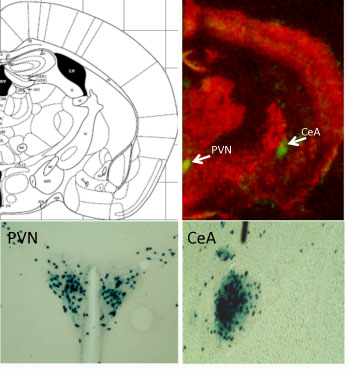
Neurons that make CRF are found in locations all over the brain, so studying them can be tricky. Kerry Ressler and his colleagues have developed an intriguing tool for studying CRF. In the places where CRF is produced in a mouse’s brain, they can take out the gene of their choice.

Green spots (above) and blue staining (below) indicate where CRF is produced in the mouse brain.
PVN = hypothalamus, paraventricular nucleus
CeA = central amygdala

In a new paper in PNAS, postdoc Georgette Gafford and Ressler use this tool in a subtle way. They have mice where a gene for a GABA receptor, one of the main inhibitory receptors (brakes) in the nervous system, is deleted, but only in the CRF neurons. This basically has the effect of turning up the volume on CRF production in several parts of the brain. It appears that modulating GABA receptors is something that normally happens to regulate CRF production, but in this case, a restraint on these stress-sensitive cells has been taken off.

“These mice are normal in many ways – normal locomotor and pain responses and no difference in depressive-like behavior or Pavlovian fear conditioning. However, these mutants have increased anxiety-like behavior,” Gafford and Ressler write.

They also have “impaired extinction of conditioned fear,” meaning that they have trouble becoming NOT afraid of something, like a buzzing sound, to which they have been sensitized by shocks. This is analogous to PTSD in which patients remain afraid and aren’t able to successfully inhibit their prior fear learning, even after the context is now safe.  [A 2011 paper goes into more detail on this biological aspect of PTSD in a civilian population.]

“These data indicate that disturbance of this specific population of neurons causes increased anxiety and impaired fear extinction, and helps us to further understand mechanisms of fear- and anxiety-related disorders such as PTSD,” Ressler and Gafford write.

In the mutant mice, a drug that blocks CRF rescued their behavioral impairments. Some other recent investigations of mice with CRF overproduction in the brain revealed “surprising paradoxical effects.”

Drugs that block CRF have been in clinical trials, some with mixed results.  A trial now proceeding at Emory is evaluating a CRF antagonist in women with PTSD.

Ressler, associate professor of psychiatry and behavioral sciences, is a Howard Hughes Medical Investigator, with a laboratory at the Yerkes National Primate Research Center. He is also co-director of the Grady Trauma Project.


Posted on by Quinn Eastman in Neuro Leave a comment

Rethinking the role of an aggregation-prone protein in ALS

Anyone studying neuroscience will notice that many neurodegenerative diseases seem to have their own sticky, possibly toxic protein. This protein tends to aggregate, or clump together, in or near the cells affected by the disease.

Picture a glass of milk left in a warm place for several days. Yuck. That is the macro version of the microscopic clumps scientists believe are bothering the brain. For many diseases, there is a debate: are the clumps by themselves toxic to neurons, or a byproduct of something else killing the cells?

Parkinson’s disease has one of the pesky proteins: alpha-synuclein. Alzheimer’s disease has two: beta-amyloid outside cells and tau inside. ALS (amyotrophic lateral sclerosis) has at least three.*

One of them, TDP-43, is found in protein aggregates in most forms of ALS, both familial and sporadic. Mutations in the TDP-43 gene also account for a small fraction of both familial and sporadic forms of ALS. This suggests that even the normal protein can create problems, but a mutated version can accelerate the disease. In addition, TDP-43 aggregates have been connected with other diseases such as frontotemporal dementia.

Again, it’s not clear whether the aggregates themselves are toxic, or whether it’s more a matter of TDP-43, which appears to regulate RNA processing, is not doing what it’s supposed to in the cell.

TDP-43 protein is mobile within motor neurons.

Emory cell biologists Claudia Fallini and Wilfried Rossoll have been probing the effects of tweaking TDP-43 levels in motor neurons, the cell type vulnerable to degeneration in ALS. They find that motor neurons may be more sensitive to changes in TDP-43 levels than other neurons, which may explain why ALS selectively affects motor neurons.

The results were published in Human Molecular Genetics.

Fallini was able to obtain a movie of fluorescently-tagged TDP-43 “granules” moving around in live motor neurons. Importantly: this is healthy/functional, not aggregated/ toxic protein. The finding that TDP-43 is mobile implies that it has something to do with transporting RNAs around the cell, rather than only functioning in the nucleus.

“Our data point to the hypothesis that TDP-43 increased localization in the cytoplasm is the early trigger of toxicity, followed by protein aggregation,” Fallini says. “Because motor neurons are unique neurons due to their high degree of polarization, we believe they might be more sensitive to alterations in TDP-43 functions in the cytoplasm or the axon.”

In particular, the researchers found that elevated levels of TDP-43 provoke motor neurons to shut down axon outgrowth. They focused on a role for the C-terminal end of TDP-43 in this effect.

“Nobody had looked at TDP-43 specifically in motor neurons before,” she says. “Our paper for the first time shows the localization and axonal transport of TDP-43, and the effects of TDP-43 altered levels on motor neuron morphology.”

*Another ALS protein, SOD1 (superoxide dismutase), apparently forms toxic aggregates when mutated in some cases of familial ALS. At Emory, Terrell Brotherton and Jonathan Glass have been investigating these forms of SOD. The third protein, FUS, has similar properties to TDP-43.


Posted on by Quinn Eastman in Neuro Leave a comment