Warren symposium follows legacy of geneticist giant

If we want to understand how the brain creates memories, and how genetic disorders distort the brain’s machinery, then the fragile X gene is an ideal place to start. That’s why the Stephen T. Warren Memorial Symposium, taking place November 28-29 at Emory, will be a significant event for those interested in neuroscience and genetics. Stephen T. Warren, 1953-2021 Warren, the founding chair of Emory’s Department of Human Genetics, led an international team that discovered Read more

Mutations in V-ATPase proton pump implicated in epilepsy syndrome

Why and how disrupting V-ATPase function leads to epilepsy, researchers are just starting to figure Read more

Tracing the start of COVID-19 in GA

At a time when COVID-19 appears to be receding in much of Georgia, it’s worth revisiting the start of the pandemic in early 2020. Emory virologist Anne Piantadosi and colleagues have a paper in Viral Evolution on the earliest SARS-CoV-2 genetic sequences detected in Georgia. Analyzing relationships between those virus sequences and samples from other states and countries can give us an idea about where the first COVID-19 infections in Georgia came from. We can draw Read more

Immunology

Sifting through signs of inflammation to analyze causes of Crohn’s disease

When studying Crohn’s disease – an inflammatory disorder of the gastrointestinal tract, a challenge is separating out potential causes from the flood of systemic inflammation inherent in the condition. Researchers led by Subra Kugathasan, MD recently published an analysis that digs under signs of inflammation, in an effort to assess possible causes.

Graduate student Hari Somineni, in Kugathasan’s lab, teamed up with Emory and Georgia Tech geneticists for a sophisticated approach that may have found some gold nuggets in the inflammatory gravel. The results were published in the journal Gastroenterology.
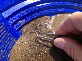
In studying Crohn’s disease, Emory + Georgia Tech researchers may have found some gold nuggets in the inflammatory gravel.

The group looked at DNA methylation in blood samples from pediatric patients with Crohn’s disease, both at diagnosis and after treatment and follow-up. The information came from blood samples from 164 children with Crohn’s disease and 74 controls, as part of the RISK study, which is supported by the Crohn’s & Colitis Foundation and Kugathasan leads.

DNA methylation is a dynamic process that can influence molecular phenotypes of complex diseases by turning the gene(s) on or off. The researchers observed that disrupted methylation patterns at the time of diagnosis in pediatric Crohn’s disease patients returned to those resembling controls following treatment of inflammation

“Our study emphasized how important it is to do longitudinal profiling – to look at the patients before and after treatment, rather than just taking a cross section,” Somineni says.

Read more

Posted on by Quinn Eastman in Immunology Leave a comment

Sensitive to (transplant) rejection

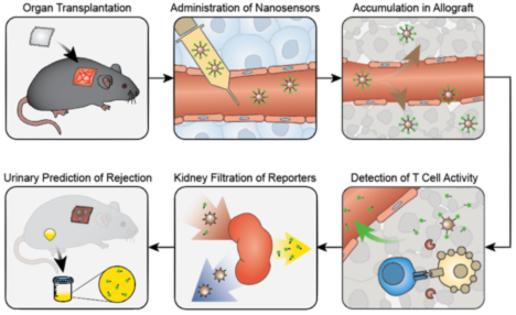
An experimental screening method, developed by Emory and Georgia Tech scientists, aims to detect immune rejection of a transplanted organ earlier and without a biopsy needle.

The technology is based on nanoparticles that detect granzyme B enzymes produced by killer T cells. When the T cells are active, they slice up the nanoparticles, generating a fluorescent signal that is detectable in urine. The results from a mouse skin graft model were published in Nature Biomedical Engineering, from Gabe Kwong’s lab at GT and Andrew Adams’ at Emory. More extensive story here.

Co-first authors Quoc Mac and Dave Mathews

Adams is also developing technologies for imaging transplant rejection via immunoPET, with Georgia Tech’s Phil Santangelo.


Posted on by Quinn Eastman in Immunology Leave a comment

Bird flu shuffle probes viral compatibility

When influenza viruses that infect birds and humans meet in the same cell, they can shuffle their genomes and produce new strains that might have pandemic potential. Think of this process, called reassortment, as viruses having sex.

In the last several years, public health officials have been monitoring two varieties of bird flu viruses with alarming properties: H7N9 and H5N8. Scientists at Emory have been probing the factors that limit reassortment between these strains and a well-known strain (H3N2) that has been dominating the last few flu seasons in the United States.

Helen Branswell has an article in STAT this week, explaining that H5N8 actually emerged from reassortment  involving much-feared-but-not-damaging-to-humans-so-far H5N1:

Several years ago, these viruses effectively splintered, with some dumping their N1 neuraminidase — a gene that produces a key protein found on the surface of flu viruses — and replacing it with another. The process is called reassortment, and, in this case, it resulted in the emergence of a lot of new pairings over a fairly short period of time.

The most common and most dangerous viruses to emerge — for birds at least — have been H5N6 and H5N8 viruses. Both are highly pathogenic, meaning they kill domestic poultry.

“The H5N1 virus has not gone away. It’s just changed into different versions of itself,” explained influenza expert Malik Peiris, a professor of virology at the University of Hong Kong.

From the Emory study, the good news is that “packaging signals” on the H5 and H7 viral RNA genomes are often incompatible with the H3N2 viruses. That means it could be difficult for segments of the genome from the bird viruses to get wrapped up with the human viruses. But mix and match still occurred at a low level, particularly with H5N8. Read more

Posted on by Quinn Eastman in Immunology Leave a comment

How intestinal bacteria affect bone formation

Helpful intestinal bacteria may stimulate bone formation via butyrate, according to a recent paper in Immunity. Butyrate increases bone formation through its regulation of T cells, Emory researchers report.

The finding adds to evidence for beneficial effects of butyrate and other SCFA (short chain fatty acid) metabolites, which are produced by bacterial fermentation of fiber in the intestines.

Roberto Pacifici and colleagues had observed that probiotic supplements protected female mice from the loss of bone density occurring after ovary removal, a simulation of the hormonal changes of menopause. Probiotic bacteria could also stimulate bone formation in mice with intact ovaries, the researchers found.

The new Immunity paper shows how this effect is produced. The probiotic bacteria do not make butyrate themselves, but they encourage the growth of other Clostridum bacteria that do produce butyrate. Read more

Posted on by Quinn Eastman in Immunology Leave a comment

Clues to lupus’s autoimmune origins in precursor cells

In the autoimmune disease systemic lupus erythematosus or SLE, the immune system produces antibodies against parts of the body itself. How cells that produce those antibodies escape the normal “checks and balances” has been unclear, but recent research from Emory University School of Medicine provides information about a missing link.

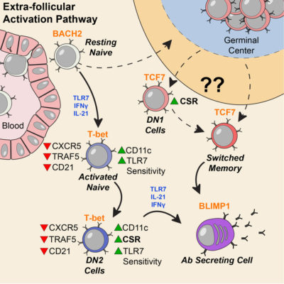
Investigators led by Ignacio (Iñaki) Sanz, MD, studied blood samples from 90 people living with SLE, focusing on a particular type of B cells. These “DN2” B cells are relatively scarce in healthy people but substantially increased in people with SLE.

The results were published in the journal Immunity.

People with lupus can experience a variety of symptoms, such as fatigue, joint pain, skin rashes and kidney problems. Levels of the DN2 cells were higher in people with more severe disease or kidney problems. DN2 B cells are thought to be “extra-follicular,” which means they are outside the B cell follicles, regions of the lymph nodes where B cells are activated in an immune response.

“Overall, our model is that a lot of lupus auto-antibodies come from a continuous churning out of new responses,” says postdoctoral fellow Scott Jenks, PhD, co-first author of the paper. “There is good evidence that DN2 cells are part of the early B cell activation pathway happening outside B cells’ normal homes in lymph nodes.”

Previous research at Emory has shown that African American women have significantly higher rates of lupus than white women. In the current study, the researchers observed that the frequency of DN2 cells was greater in African American patients. Participants in the study were recruited by Emory, University of Rochester and Johns Hopkins. Read more

Posted on by Quinn Eastman in Immunology Leave a comment

MSCs: what’s in a name?

At a recent symposium of cellular therapies held by the Department of Pediatrics, we noticed something. Scientists do not have consistent language to talk about a type of cells called “mesenchymal stem cells” or “mesenchymal stromal cells.” Within the same symposium, some researchers used the first term, and others used the second.

Guest speaker Joanne Kurtzberg from Duke discussed the potential use of MSCs to treat autism spectrum disorder, cerebral palsy, and hypoxic-ischemic encephalopathy. Exciting stuff, although the outcomes of the clinical studies underway are still uncertain. In these studies, the mesenchymal stromal cells (the language Kurtzberg used) are derived from umbilical cord blood, not adult tissues.

Nomenclature matters, because a recent editorial in Nature calls for the term “stem cell” not to be used for mesenchymal (whatever) cells. They are often isolated from bone marrow or fat. MSCs are thought have the potential to become cells such as fibroblasts, cartilage, bone and fat. But most of their therapeutic effects appear to come from the growth factors and RNA-containing exosomes they secrete, rather than their ability to directly replace cells in damaged tissues.

The Nature editorial argues that “wildly varying reports have helped MSCs to acquire a near-magical, all-things-to-all-people quality in the media and in the public mind,” and calls for better characterization of the cells and more rigor in clinical studies.

At Emory, gastroenterologist Subra Kugathasan talked about his experience with MSCs in inflammatory bowel diseases. Hematologist Edwin Horwitz discussed his past work with MSCs on osteogenesis imperfecta. And Georgia Tech-based biomedical engineer Krishnendu Roy pointed out the need to reduce costs and scale up, especially if MSCs start to be used at a higher volume.

Several of the speakers were supported by the Marcus Foundation, which has a long-established interest in autism, stroke, cerebral palsy and other neurological conditions.

Posted on by Quinn Eastman in Heart, Immunology, Neuro Leave a comment

Mopping up immune troublemakers after transplant

Emory scientists have identified a way to stop troublemaker cells that are linked to immune rejection after kidney transplant. The finding could eventually allow transplant patients to keep their new kidneys for as long as possible, without the side effects that come from some current options for controlling immune rejection.

The results are published in Journal of Clinical Investigation.

The standard drugs used for many years, calcineurin inhibitors, show side effects on cardiovascular health and can even damage the kidneys over time. A newer FDA-approved medication called belatacept, developed in part at Emory, avoids these harmful effects but is less effective at stopping acute rejection immediately after the transplant. Belatacept is a “costimulation blocker” – it interferes with a signal some immune cells (T cells) need to proliferate and become activated.

Researchers led by Emory transplant surgeon Andrew Adams, MD, PhD suspected that long-lasting memory CD8+ T cells were resistant to belatacept’s effects.

“Our previous work identified that memory CD8+ T cells may be elevated in animals and human patients who go on to reject their transplanted organs while taking belatacept,” says Dave Mathews, an MD/PhD student who worked with Adams and is the first author of the paper.

The researchers identified a certain marker, CD122, which was present on memory CD8+ T cells and important for their activity. On T cells, CD122 acts as a receiving dish for two other secreted molecules, IL-2 and IL-15, generally thought of as inflammatory cytokines, or protein messengers that can encourage graft rejection. Read more

Posted on by Quinn Eastman in Immunology Leave a comment

Leaving out sugar makes a better antibody drug

There’s a bit of sugar attached to your billion-dollar biotech product. Omitting the sugar (fucose) can help the product work better, Emory immunologists think.

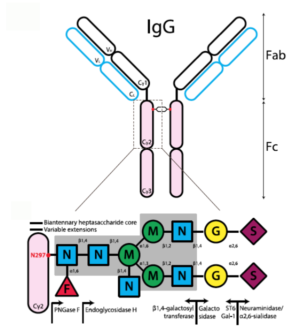
Fucosylation is the red triangle on this diagram of the carbohydrate modifications of antibodies. Adapted from KTC Shade + RM Anthony, Antibodies (2013) and used through Creative Commons license.

Many drugs now used to treat cancer and autoimmune diseases are antibodies, originally derived from the immune system. A classic example of a “therapeutic antibody” is rituximab, a treatment for B cell malignancies that was FDA-approved in 1997. It has been responsible for billions of dollars in revenue for its maker, pharmaceutical giant Roche.

Researchers at Emory Vaccine Center previously observed that in a mouse model of chronic viral infection, a traffic jam inside the body limits how effective therapeutic antibodies can be. One of the ways these antibodies work is to grab onto malignant or inflammatory cells. One end of the antibody is supposed to bind the target cell, while another is a flag for other cells to eliminate the target cell. During a chronic viral infection, a mouse’s immune system is producing its own antibodies against the virus, which form complexes with viral proteins. These immune complexes prevented the injected antibodies from depleting their target cells.

In a recent Science Immunology paper, postdoc Andreas Wieland, Vaccine Center director Rafi Ahmed and colleagues showed that antibodies that lack fucosylation have an enhanced ability to get rid of their intended targets. Fucosylation is a type of sugar modification of the antibody. (It is the red triangle in the diagram, provided by Wieland.) When it is not present, then the “flag for removal” region of the antibody can interact more avidly with the Fc gamma receptor on immune cells. Thus, the introduced antibodies can compete more effectively with the antibodies being produced by the body already.

Read more

Posted on by Quinn Eastman in Immunology 1 Comment

Probiotics for bone health study heads into clinic

Probiotic supplements can protect female mice from the loss of bone density that occurs after having their ovaries removed, researchers at Emory and Georgia State reported a couple years ago.

Roberto Pacifici, MD

This finding, published in Journal of Clinical Investigation, had clear implications for the treatment of osteoporosis in post-menopausal women. Prompted by external emails, Lab Land learned that the Emory investigators are now continuing their research in the clinic.

Endocrinologist/osteoimmunologist Roberto Pacifici and colleague Jessica Alvarez are conducting a double-blind study for women aged 50-65, using VSL3, a widely available and inexpensive dietary supplement. Participants would take the supplement or placebo for a year. More information is available here.

In mice, the loss of estrogen increases gut permeability, which allows bacterial products to activate immune cells in the intestine. In turn, immune cells release signals that break down bone. It appears that probiotics both tighten up the permeability of the gut and dampen inflammatory signals that drive the immune cells. Read more

Posted on by Quinn Eastman in Immunology Leave a comment

Where it hurts matters in the gut

What part of the intestine is problematic matters more than inflammatory bowel disease subtype (Crohn’s disease vs ulcerative colitis), when it comes to genetic activity signatures in pediatric IBD.

Suresh Venkateswaran and Subra Kugathasan in the lab

That’s the takeaway message for a recent paper in Cellular and Molecular Gastroenterology and Hepatology (the PDF is open access) from gastroenterologist Subra Kugathasan and colleagues. His team has been studying risk factors in pediatric IBD that could predict whether a child will experience complications requiring surgery.

Kugathasan is professor of pediatrics and human genetics at Emory University School of Medicine and scientific director of the pediatric IBD program at Children’s Healthcare of Atlanta. He is also director of the Children’s Center for Transplantation and Immune-mediated Disorders.

“This study has demonstrated that tissue samples from the ileum and rectum of CD patients show higher molecular level differences, whereas in tissue samples from two different patients with the same type of disease, the molecular differences are low,” Kugathasan says. “This was an important question to answer, since IBD can be localized to one area, and the treatment responses can vary and can be tailored to a localized area if this knowledge is well known.”

Research associate Suresh Venkateswaran, PhD, is the first author on the CMGH paper.

“We see that the differences are not connected to genomic variations,” he says. “Instead, they may be caused by non-genetic factors which are specific to each location and disease sub-type of the patient.”

These findings have implications for other study designs involving molecular profiling of IBD patients. The authors believe the findings will be important for future design of locally acting drugs.

Read more

Posted on by Quinn Eastman in Immunology Leave a comment