Warren symposium follows legacy of geneticist giant

If we want to understand how the brain creates memories, and how genetic disorders distort the brain’s machinery, then the fragile X gene is an ideal place to start. That’s why the Stephen T. Warren Memorial Symposium, taking place November 28-29 at Emory, will be a significant event for those interested in neuroscience and genetics. Stephen T. Warren, 1953-2021 Warren, the founding chair of Emory’s Department of Human Genetics, led an international team that discovered Read more

Mutations in V-ATPase proton pump implicated in epilepsy syndrome

Why and how disrupting V-ATPase function leads to epilepsy, researchers are just starting to figure Read more

Tracing the start of COVID-19 in GA

At a time when COVID-19 appears to be receding in much of Georgia, it’s worth revisiting the start of the pandemic in early 2020. Emory virologist Anne Piantadosi and colleagues have a paper in Viral Evolution on the earliest SARS-CoV-2 genetic sequences detected in Georgia. Analyzing relationships between those virus sequences and samples from other states and countries can give us an idea about where the first COVID-19 infections in Georgia came from. We can draw Read more

Immunology

HIV vaccine news: a glass half full

This week, researchers from Yerkes and Emory Vaccine Center led by Cindy Derdeyn published a paper that I first thought was disturbing. It describes how monkeys vaccinated against HIV’s relative SIV (simian immunodeficiency virus) still become infected when challenged with the virus. Moreover, it’s not clear whether the vaccine-induced antibodies are exerting any selective pressure on the virus that gets through.

But then I realized that this might be an example of “burying the lead,” since we haven’t made a big hoopla about the underlying vaccine studies, conducted by Rama Amara. Some of these studies showed that a majority of monkeys can be protected from repeated viral challenge. The more effective vaccine regimens include adjuvants such as the immune-stimulating molecules GM-CSF or CD40L (links are the papers on the protective effects). Read more

Posted on by Quinn Eastman in Immunology Leave a comment

CMV reactivation warps immune system after HSCT

As a followup to yesterday’s post on following troublemaker cells in patients with lupus, we’d like to highlight a recent paper in Blood that takes a similar approach to studying how the immune system comes back after bone marrow/blood stem cell transplant.

Leslie Kean, MD, PhD

The paper’s findings have implications for making this type of transplant safer and preventing graft-versus-host disease. In a bone marrow/blood stem cell transplant, to fight cancer, doctors are essentially clearing out someone’s immune system and then “planting” a new one with the help of a donor. What this paper shows is how much CMV (cytomegalovirus) distorts the new immune system.

CMV is often thought of as harmless — most adults in the United States have been infected with CMV by age 40 and don’t get sick because of it. But in this situation, CMV’s emergence from the shadows forces some of the new T cells to multiply, dominating the immune system so much that it creates gaps in the rest of the T cell repertoire, which can compromise protective immunity. Other seemingly innocuous viruses like BK cause trouble in immunosuppressed patients after kidney transplant.

The senior author, Leslie Kean, moved from Emory to Seattle Children’s Hospital in 2013, and her team began these studies here in 2010 (a host of Emory/Winship hematologists and immunologists are co-authors). This paper is sort of a mirror image of the Nature Immunology paper on lupus because it also uses next-generation sequencing to follow immune cells with DNA rearrangements — in this case, T cells. Read more

Posted on by Quinn Eastman in Cancer, Immunology Leave a comment

Following lupus troublemaker cells, via DNA barcodes

People with systemic lupus erythematosus can experience a variety of symptoms, such as fatigue, joint pain, skin rashes and kidney problems. Often the symptoms come and go in episodes called flares. In lupus, the immune system goes haywire and produces antibodies that are directed against the body itself.

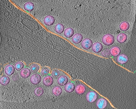
The immune system can produce many types of antibodies, directed against infectious viruses (good) or against human proteins as in lupus (harmful). Each antibody-secreting cell carries a DNA rearrangement that reflects the makeup of its antibody product. Scientists can use the DNA to identify and track that cell, like reading a bar code on an item in a supermarket.

SanzNew220

Iñaki Sanz, MD is a Georgia Research Alliance Eminent Scholar, director of the Lowance Center for Human Immunology and head of the Rheumatology division in the Department of Medicine.

Postdoc Chris Tipton, GRA Eminent Scholar Iñaki Sanz and colleagues at Emory have been using these DNA bar codes to investigate some fundamental questions about lupus: where do the autoantibody-producing cells come from? Are they all the same?

Their findings were published in Nature Immunology in May, and a News and Views commentary on the paper calls it “a quantum advance in the understanding of the origin of the autoreactive B cells.” It’s an example of how next-generation sequencing technology is deepening our understanding of autoimmune diseases.

The Emory team obtained blood samples from eight patients experiencing lupus flares and compared them to eight healthy people who had recently been vaccinated against influenza or tetanus.

When the immune system is responding to something it’s seen before, like when someone receives a booster vaccine, the bar codes of the antibody-producing cells look quite similar to each other. A set of just a few antibody-producing cells multiply and expand, making what looks like clones. In contrast, the researchers found that in lupus, many different cells are producing antibodies. Some of the expanded sets of cells are producing antibodies against infectious agents.

“We expected to see an expansion of the cells that produce autoantibodies, but instead we saw a very broad expansion of cells with all types of specificities,” Tipton says.

To use a Star Wars analogy: a booster vaccine response looks like the Clone Wars (oligoclonal — only a few kinds of monsters), but a lupus flare looks like a visit to Mos Eisley cantina (polyclonal — many monsters). Read more

Posted on by Quinn Eastman in Immunology Leave a comment

Subset of plasma cells display immune ‘historical record’

You may have read about recent research, published in Science, describing a technique for revealing which viruses have infected someone by scanning antiviral antibodies in the blood.

Emory immunologists have identified corresponding cells in which long-lived antibody production resides. A subset of plasma cells keep a catalog of how an adult’s immune system responded to infections decades ago, in childhood encounters with measles or mumps viruses.

The results, published Tuesday, July 14 in Immunity, could provide vaccine designers with a goalpost when aiming for long-lasting antibody production.

“If you’re developing a vaccine, you want to fill up this compartment with cells that respond to your target antigen,” says co-senior author F. Eun-Hyung Lee, MD, assistant professor of medicine at Emory University School of Medicine and director of Emory Healthcare’s Asthma, Allergy and Immunology program.

The findings could advance investigation of autoimmune diseases such as lupus erythematosus or rheumatoid arthritis, by better defining the cells that produce auto-reactive antibodies.

Lee says that her team’s research on plasma cells in humans provided insights unavailable from mice, since mice don’t live as long and their plasma cells also have a different pattern of protein markers. More here.

Posted on by Quinn Eastman in Immunology Leave a comment

Microbiome enthusiasm at Emory

At what point did the human microbiome become such a hot topic?

When it was shown that babies born by Cesarean section are colonized with different bacteria than those born vaginally? With the cardiovascular studies of microbial byproducts of meat digestion? With the advent of fecal transplant as a proposed treatment for Clostricium difficile infection?

The bacteria and other microbes that live within the human body are thought to influence not only digestive health, but metabolic and autoimmune diseases as well, possibly even psychiatric and neurodevelopmental disorders. The field is being propelled by next-generation sequencing technology, and Nature had to publish an editorial guarding against hype (a major theme: correlation is not causation).

At Emory, investigators from several departments are involved in microbiome-related work, and the number is expanding, and assembling a comprehensive list is becoming more difficult. Researchers interested in the topic are planning Emory’s first microbiome symposium in November, organized by Jennifer Mulle (read her intriguing review on autism spectrum disorders and the microbiome).

Microbial genomics expert Tim Read, infectious diseases specialist Colleen Kraft and intestinal pathologist Andrew Neish have formed an Emory microbiome interest group with a listserv and seminars.

Microbiome symposium sponsors: ACTSI, Hercules Exposome Center, Emory University School of Medicine, Omega Biotek, CFDE, Ubiome. Read more

Posted on by Quinn Eastman in Immunology, Neuro Leave a comment

Pre-hospital recognition of severe sepsis

Severe sepsis, a consequence of the body’s response to infection, is a major cause of death in hospitals. The earlier that doctors recognize that a patient has sepsis, the earlier the patient can be treated with antibiotics, fluids and other measures, and the better the chance of survival.

That’s why critical care and emergency medicine researchers have been looking for ways to spot whether someone coming to the hospital might have sepsis, even before arrival.

At Emory, Carmen Polito, Jonathan Sevransky and colleagues recently published a paper in the American Journal of Emergency Medicine on an emergency medical services screening tool for severe sepsis. Polito and Sevransky are in the division of pulmonary, allergy, critical care and sleep medicine in the Department of Medicine. The tool was evaluated based on Grady emergency medical services data from 2011 and 2012.

“Sepsis is largely a face without a name in the EMS setting,” Polito says. “The goal of our study was to create a tool to assist EMS providers in naming this deadly condition at the point of first medical contact. Similar to other life-threatening, time-sensitive conditions like stroke and heart attack, naming sepsis is the first step in developing coordinated care pathways that focus on delivering rapid, life-saving treatment once the patient arrives at the hospital.”

Read more

Posted on by Quinn Eastman in Immunology Leave a comment

Why checkpoint inhibitors fall short for some types of cancer

The big news from the recent American Society of Clinical Oncology meeting has been largely about immunotherapy drugs, also known as checkpoint inhibitors. These drugs have been shown to be effective in prolonging life in patients with some types of cancer, such as lung cancer and melanoma, but not others, such as colorectal and prostate cancer.

Lab Land asked oncologist Bradley Carthon and immunology researcher Haydn Kissick why. Both Carthon’s clinical work and Kissick’s lab research on prostate cancer are featured in the new issue of Winship magazine, but the prostate feature just touches on checkpoint inhibitors briefly.

Carthon says the reason checkpoint inhibitors haven’t moved the needle with prostate cancer is “likely due to the absence of infiltration of the prostatic tissue by tumor-associated lymphocytes.”

Checkpoint inhibitors are supposed to unleash the immune system, but if the immune cells aren’t in contact with the cancer cells so that the drugs can spur them into action, they won’t help much. Carthon says: “The answer may be to ‘prime’ the prostate with an agent, then introduce the checkpoint inhibitors.” Read more

Posted on by Quinn Eastman in Cancer, Immunology Leave a comment

Why HIV’s cloak has a long tail

Virologists at Emory, Yerkes and Children’s Healthcare of Atlanta have uncovered a critical detail explaining how HIV assembles its infectious yet stealthy clothing.

Paul Spearman, MD

For HIV to spread from cell to cell, the viral envelope protein needs to become incorporated into viral particles as they emerge from an infected cell. Researchers led by Paul Spearman have found that a small section of the envelope protein, located on its “tail”, is necessary for the protein to be sorted into viral particles.

The results were published June 1 in Proceedings of the National Academy of Sciences. Read more

Posted on by Quinn Eastman in Immunology Leave a comment

HIV virions attached to cell membrane

The third winner of the Best Image contest from the Postdoctoral Research Symposium, from postdoc Joshua Strauss in electron microscopist Elizabeth Wright’s lab.

Strauss explains:

Tetherin is a host cell factor that mechanically links HIV-1 to the plasma membrane. This is the first time anyone has imaged tethered HIV-1 by cryo-electron tomography. In doing so, we were able to learn about the length and arrangement of the tethers.

Note: Tetherin also studied by Paul Spearman + colleagues.Joshua_Strauss_OPE_Image

Cryo-electron tomography is an imaging technique which enables scientists to look at biological specimens in a “native-like” (frozen hydrated) state, without the chemical fixatives or heavy metal stains typically used for conventional electron microscopy.

The 3D reconstruction was manually segmented to highlight the different viral and cellular components: HIV-1 virions (lavender), mature conical-cores (aqua blue), immature Gag lattice (pink), plasma membrane (peach), rod-like tethers (sea green).

Posted on by Quinn Eastman in Immunology Leave a comment

Test of megadose vitamin D in intubated critical care patients

Whether dietary supplementation with vitamin D is beneficial, in terms of preventing disease, has been controversial. However, vitamin D has been reported to increase immune cells’ production of microbe-fighting proteins. That’s why Emory doctors have been testing whether high doses of vitamin D could be helpful for critical care patients, who need to ward off infections.

The results of a small-scale clinical trial, presented in Denver this week at the American Thoracic Society meeting, suggest that high doses of vitamin D could decrease the length of hospital stays in critically ill patients with respiratory failure. Read more

Posted on by Quinn Eastman in Immunology Leave a comment