Warren symposium follows legacy of geneticist giant

If we want to understand how the brain creates memories, and how genetic disorders distort the brain’s machinery, then the fragile X gene is an ideal place to start. That’s why the Stephen T. Warren Memorial Symposium, taking place November 28-29 at Emory, will be a significant event for those interested in neuroscience and genetics. Stephen T. Warren, 1953-2021 Warren, the founding chair of Emory’s Department of Human Genetics, led an international team that discovered Read more

Mutations in V-ATPase proton pump implicated in epilepsy syndrome

Why and how disrupting V-ATPase function leads to epilepsy, researchers are just starting to figure Read more

Tracing the start of COVID-19 in GA

At a time when COVID-19 appears to be receding in much of Georgia, it’s worth revisiting the start of the pandemic in early 2020. Emory virologist Anne Piantadosi and colleagues have a paper in Viral Evolution on the earliest SARS-CoV-2 genetic sequences detected in Georgia. Analyzing relationships between those virus sequences and samples from other states and countries can give us an idea about where the first COVID-19 infections in Georgia came from. We can draw Read more

neuroscience

The blue spot: where seeds of destruction begin

Neuroscientist and geneticist David Weinshenker makes a case that the locus coeruleus (LC), a small region of the brainstem and part of the pons, is among the earliest regions to show signs of degeneration in both Alzheimer’s and Parkinson’s disease. You can check it out in Trends in Neurosciences.

The LC is the main source of the neurotransmitter norepinephrine in the brain, and gets its name (Latin for “blue spot”) from the pigment neuromelanin, which is formed as a byproduct of the synthesis of norepinephrine and its related neurotransmitter dopamine. The LC has connections all over the brain, and is thought to be involved in arousal and attention, stress responses, learning and memory, and the sleep-wake cycle.

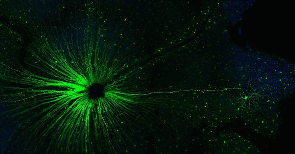
Cells in the locus coeruleus are lost in mild cognitive impairment and Alzheimer’s. From Kelly et al Acta Neuropath. Comm. (2017) via Creative Commons

The protein tau is one of the toxic proteins tied to Alzheimer’s, and it forms intracellular tangles. Pathologists have observed that precursors to tau tangles can be found in the LC in apparently healthy people before anywhere else in the brain, sometimes during the first few decades of life, Weinshenker writes. A similar bad actor in Parkinson’s, alpha-synuclein, can also be detected in the LC before other parts of the brain that are well known for damage in Parkinson’s, such as the dopamine neurons in the substantia nigra.

“The LC is the earliest site to show tau pathology in AD and one of the earliest (but not the earliest) site to show alpha-synuclein pathology in PD,” Weinshenker tells Lab Land. “The degeneration of the cells in both these diseases is more gradual. It probably starts in the terminals/fibers and eventually the cell bodies die.” Read more

Posted on by Quinn Eastman in Neuro Leave a comment

An exceptional electrical thrill ride #CNS2018

A recent paper in Neuropsychologia got a lot of attention on Twitter and at the Cognitive Neuroscience Society meeting in Boston over the weekend. It discusses what can happen when the amygdala, a region of the brain known for regulating emotional responses, receives direct electrical stimulation. A thrill ride – but for only one study participant. Two of nine people noticed the electrical stimulation. One individual reported (a video is included in the paper):

“It was, um, it was terrifying, it was just…it was like I was about to get attacked by a dog. Like the moment, like someone unleashes a dog on you, and it’s just like it’s so close…

He also spontaneously reported “this is fun.” He further explained that he could distinguish feelings in his body that would normally be associated with fear recognized and the absence of an actual threat, making the experience “fun”.

But wait, why were Emory neuroscientists Cory Inman, Jon Willie and Stephan Hamann and colleagues doing this? Read more

Posted on by Quinn Eastman in Neuro Leave a comment

Epilepsy pick up sticks

Imagine the game of pick up sticks. It’s hard to extract one stick from the pile without moving others. The same problem exists, in a much more complex way, in the brain. Pulling on one gene or neurotransmitter often nudges a lot of others.

Andrew Escayg, PhD

That’s why a recent paper from Andrew Escayg’s lab is so interesting. He studies genes involved in epilepsy. Several years ago, he showed that mice with mutations in the SCN8A gene have absence epilepsy, while also showing resistance to induced seizures. SCN8A is one of those sticks that touches many others. The gene encodes a voltage-gated sodium channel, involved in setting the thresholds for and triggering neurons’ action potentials. Mutating the gene in mice modifies sleep and even enhances spatial memory.

Escayg’s new paper, with first author Jennifer Wong, looks at the effect of “knocking down” SCN8A in the hippocampus in a mouse model of mesial temporal lobe epilepsy. This model doesn’t involve sodium channel genes; it’s generated by injection of a toxin (kainic acid) into the brain. The finding suggests that inhibiting SCN8A may be applicable to other forms of epilepsy. Escayg notes that mesial temporal lobe epilepsy is one of the most common forms of treatment-resistant epilepsy in adults.

Knocking down SCN8A in the hippocampus 24 hours after injection could prevent the development of seizures in 90 percent of the treated mice. “It is likely that selective reduction in Scn8a expression would have directly decreased neuronal excitability,” the authors write. It did not lead to increased anxiety levels or impaired learning/memory.

Currently, no available drugs target Scn8a specifically. However, antisense approaches for neurodegenerative diseases have been gaining ground – perhaps epilepsy could fit in.

Posted on by Quinn Eastman in Neuro Leave a comment

Insight into brain + learning via ‘friend of fragile X’ gene

We can learn a lot about somebody from the friends they hang out with. This applies to people and also to genes and proteins. Emory scientists have been investigating a gene that we will call — spoiler alert — “Friend of fragile X.”

Fragile X syndrome is the most common inherited form of intellectual disability, studied by research teams around the world with drug discovery and clinical trials in mind. It is caused by a disruption of the gene FMR1.

In an independent form of inherited intellectual disability found in a small number of Iranian families, a gene called ZC3H14 is mutated. Two papers from Ken Moberg, PhD, associate professor of cell biology, Anita Corbett, PhD, professor of biology and colleagues show that FMR1 and ZC3H14 are, in effect, friends.

The findings provide new insight into the function of FMR1 as well as ZC3H14; the evidence comes from experiments performed in fruit flies and mice. The most recent paper is in the journal Cell Reports (open access), published this week.

The scientists found that the proteins encoded by FMR1 and ZC3H14 stick together in cells and they hang out in the same places. The two proteins have related functions: they both regulate messenger RNA in neurons, which explains their importance for learning and memory.

The fragile X protein (FMRP) was known to control protein production in response to signals arriving in neurons, but the Cell Reports paper shows that FMRP is also regulating the length of  “tails” attached to messenger RNAs – something scientists did not realize, even after years of studying FMRP and fragile X, Moberg says.

To be sure, FMRP interacts with many proteins and appears to be a critical gatekeeper. Emory geneticist Peng Jin, who has conducted his share of research on this topic, says that “FMRP must be very social and has a lot of friends.” More here.

Posted on by Quinn Eastman in Neuro Leave a comment

Enhanced verbal abilities in the congenitally blind

A recent paper in Experimental Brain Research from Emory neuroscientist Krish Sathian and colleagues demonstrates that congenitally blind study participants displayed superior verbal, but not spatial abilities, when compared to their sighted counterparts. This may reflect both greater reliance on verbal information, and the recruitment of the visual cortex for verbal tasks.

Sathian’s team has also been investigating, through brain imaging studies, whether the visual cortex is involved in the processing of metaphors (2016 SFN abstract) in the congenitally blind. They previously showed that blind study participants were better at identifying rotated objects by touch. Read more

Posted on by Quinn Eastman in Neuro Leave a comment

Anti-TNF vs Alzheimer’s mouse model

An experimental anti-inflammatory drug has positive effects on neuron function and amyloid plaques in a mouse model of Alzheimer’s disease, Emory neuroscientists report. The findings are published in the journal Neurobiology of Disease.

Inflammation’s presence in Alzheimer’s is well established, but it is usually thought of as an accelerator, rather than an initiating cause. While everybody argues about the amyloid hypothesis, there’s a case to be made for intervening against the inflammation. Exactly how is an open question.

The drug tested, called XPro1595, targets the inflammatory signaling molecule tumor necrosis factor (TNF). Commercialized drugs such as etanercept and infliximab, used to treat autoimmune diseases, also block TNF. However, XPro1595 only interferes with the soluble form of TNF and is supposed to have less of an effect on overall immune function.

Senior author Malu Tansey (pictured) and her colleagues say that interfering with TNF could have direct effects on neurons, as well as indirect effects on the immune cells infiltrating the brain. They write that “the most promising finding in our study” is the ability of XPro1595 to restore long-term potentiation or LTP, which is impaired in the Alzheimer’s model mice. Read more

Posted on by Quinn Eastman in Immunology, Neuro Leave a comment

How “twist my arm” engages the brain

Listening to metaphors involving arms or legs loops in a region of the brain responsible for visual perception of those body parts, scientists have discovered.

The finding, recently published in Brain & Language, is another example of how neuroscience studies are providing evidence for “grounded cognition” – the idea that comprehension of abstract concepts in the brain is built upon concrete experiences, a proposal whose history extends back millennia to Aristotle.

The EBA was shown in 2001 to respond selectively to images of the human body by Nancy Kanwisher and colleagues.

When study participants heard sentences that included phrases such as “shoulder responsibility,” “foot the bill” or “twist my arm”, they tended to engage a region of the brain called the left extrastriate body area or EBA.

The same level of activation was not seen when participants heard literal sentences containing phrases with a similar meaning, such as “take responsibility” or “pay the bill.”  The study included 12 right-handed, English-speaking people, and blood flow in their brains was monitored by functional MRI (magnetic resonance imaging).

“The EBA is part of the extrastriate visual cortex, and it was known to be involved in identifying body parts,” says senior author Krish Sathian, MD, PhD, professor of neurology, rehabilitation medicine, and psychology at Emory University.  “We found that the metaphor selectivity of the EBA matches its visual selectivity.” Read more

Posted on by Quinn Eastman in Neuro Leave a comment

A glimpse into the genetics of positive emotions


Happiness can be elusive, both in personal life and as a scientific concept. That’s why this paper, recently published in Molecular Psychiatry, seemed so striking.

A genome-wide association study of positive emotion identifies a genetic variant and a role for microRNAs.” Translation: a glimpse into the genetics of positive emotions.

Editorial note: Although the research team here is careful and confirms the findings in independent groups and in brain imaging and fear discrimination experiments, this is a preliminary result. More needs to be explored about how these genetic variants and others affect positive emotions.

“With relatively few studies on genetic underpinnings of positive emotions, we face the challenges of a nascent research area,” the authors write.

Perhaps ironically, the finding comes out of the Grady Trauma Project, a study of inner-city residents exposed to high rates of abuse and violence, aimed at understanding mechanisms of resilience and vulnerability in depression and PTSD.

“Resilience is a multidimensional phenomenon, and we were looking at just one aspect of it,” says first author Aliza Wingo. She worked with Kerry Ressler , now at Harvard, and Tanja Jovanovic and other members of the Grady Trauma Project team.

“Positive affect” is what the team was measuring, through responses on questionnaires. And the questions are asking for the extent that respondents feel a particular positive emotion in general, rather than that day or that week. Read more

Posted on by Quinn Eastman in Neuro Leave a comment

Optic nerve reaching out

Congratulations to Ying Li, MD, PhD, 3rd place winner of the Best Image contest held as part of the Emory Postdoctoral Research Symposium, which takes place next week (Thursday, May 19). Li is in Eldon Geisert’s lab, and provided Lab Land this description:

“Like a benevolent overseer of the cosmos, the epicenter of the optic nerve appears to extend a axon reassuringly to the small, seemingly lowly single ganglion cell, reminding us that every cell matters.”i-6FBNVsV-X3

Posted on by Quinn Eastman in Neuro Leave a comment

Manipulating motivation in mice

Emory researchers have identified molecular mechanisms that regulate motivation and persistence in mice. Their findings could have implications for intervention in conditions characterized by behavioral inflexibility, such as drug abuse and depression.

Scientists showed that by manipulating a particular growth factor in one region of the brain, they could tune up or down a mouse’s tendency to persist in seeking a reward. In humans, this region of the brain is located just behind the eyes and is called the medial orbitofrontal cortex or mOFC.

“When we make decisions, we often need to gauge the value of a reward before we can see it — for example, will lunch at a certain restaurant be better than lunch at another, or worth the cost,” says Shannon Gourley, PhD, assistant professor of pediatrics and psychiatry at Emory University School of Medicine. “We think the mOFC is important for calculating value, particularly when we have to imagine the reward, as opposed to having it right in front of us.”

The results were published Wednesday in Journal of Neuroscience.

Shannon Gourley, PhD

Being able to appropriately determine the value of a perceived reward is critical in goal-directed decision making, a component of drug-seeking and addiction-related behaviors. While scientists already suspected that the medial orbitofrontal cortex was important for this type of learning and decision-making, the specific genes and growth factors were not as well-understood.

The researchers focused on brain-derived neurotrophic factor (BDNF), a protein that supports the survival and growth of neurons in the brain. BDNF is known to play key roles in long-term potentiation and neuronal remodeling, both important in learning and memory tasks. Variations in the human gene that encodes BDNF have been linked with several psychiatric disorders.

Read more

Posted on by Quinn Eastman in Neuro Leave a comment