Warren symposium follows legacy of geneticist giant

If we want to understand how the brain creates memories, and how genetic disorders distort the brain’s machinery, then the fragile X gene is an ideal place to start. That’s why the Stephen T. Warren Memorial Symposium, taking place November 28-29 at Emory, will be a significant event for those interested in neuroscience and genetics. Stephen T. Warren, 1953-2021 Warren, the founding chair of Emory’s Department of Human Genetics, led an international team that discovered Read more

Mutations in V-ATPase proton pump implicated in epilepsy syndrome

Why and how disrupting V-ATPase function leads to epilepsy, researchers are just starting to figure Read more

Tracing the start of COVID-19 in GA

At a time when COVID-19 appears to be receding in much of Georgia, it’s worth revisiting the start of the pandemic in early 2020. Emory virologist Anne Piantadosi and colleagues have a paper in Viral Evolution on the earliest SARS-CoV-2 genetic sequences detected in Georgia. Analyzing relationships between those virus sequences and samples from other states and countries can give us an idea about where the first COVID-19 infections in Georgia came from. We can draw Read more

Department of Pediatrics

To fight cancer, mix harmless reovirus with ‘red devil’

A recent paper in Journal of Virology mixes tried-and-true cancer-fighting tactics with the exotic. Sort of a peanut-butter-and-chocolate story, but definitely not tasty!

The tried and true is doxorubicin (Adriamycin), the notorious ‘red devil’ chemotherapy drug, which has been around for decades. On the exotic side, we have oncolytic viruses – viruses retuned to attack cancer cells more than healthy cells. This idea finally made it to FDA approval in 2015 in the form of a re-engineered herpes virus directed against melanoma.

Bernardo Mainou’s lab in the Department of Pediatrics is combining both of these approaches together. He and his team are looking to supercharge reoviruses, a mostly harmless type of virus that has been adapted into an anticancer agent. A Canadian company has brought its reovirus forward into several cancer clinical trials, but its product has not gotten to the finish line.

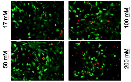
In the JVI paper, graduate students Roxana Rodriguez-Stewart, Jameson Berry and their colleagues infected triple-negative breast cancer cells with a variety of reoviruses, in an effort to select for those that replicate better in those cells. They also looked for drugs that enhance viral infection of those cells, and landed on doxorubicin and related drugs. Doxorubicin is part of a class of anticancer drugs that inhibit topoisomerases, enzymes that unwind DNA as part of the process of replication.

Yesterday at the GDBBS graduate research symposium, Berry gave a talk about the next step: attaching the souped-up reovirus to doxorubicin.

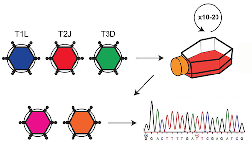
Three varieties of reovirus were grown together in breast cancer cells to select for efficient replication. 


Posted on by Quinn Eastman in Cancer Leave a comment

Microbiome critical for bone hormone action

Intestinal microbes are necessary for the actions of an important hormone regulating bone density, according to two papers from the Emory Microbiome Research Center. The papers represent a collaboration between Roberto Pacifici, MD and colleagues in the Department of Medicine and laboratory of Rheinallt Jones, PhD in the Department of Pediatrics.

Together, the results show how probiotics or nutritional supplementation could be used to modulate immune cell activity related to bone health. The two papers, published in Nature Communications and Journal of Clinical Investigation, are the first reports of a role for intestinal microbes in the mechanism of action of PTH (parathyroid hormone), Pacifici says.

PTH increases calcium levels in the blood and can either drive bone loss or bone formation, depending on how it is produced or administered. Continuous excessive production of PTH, or primary hyperparathyroidism, is a common endocrine cause of osteoporosis. Yet in another context, intermittent external PTH stimulates bone formation, and is an FDA-approved treatment for osteoporosis – also used off-label for fracture repair in athletes. Read more

Posted on by Quinn Eastman in Immunology Leave a comment

Model of a sticky situation

Here’s an example of how 3D printing can be applied to pediatric cardiology. It’s also an example of how Georgia Tech, Emory and Children’s Healthcare of Atlanta all work together.

Biomedical engineers used a modified form of gelatin to create a model of pulmonary arteries in newborn and adolescent patients with a complex (and serious) congenital heart defect: tetralogy of Fallot with pulmonary atresia. The model allowed the researchers to simulate surgical catheter-based intervention in vitro.

The results were recently published in Journal of the American Heart Association. Biomedical engineer Vahid Serpooshan and his lab collaborated with Sibley Heart Center pediatric cardiologist Holly Bauser-Heaton; both are part of the Children’s Heart Research and Outcomes Center.

“This is a patient-specific platform, created with state-of-the-art 3D bioprinting technology, allowing us to optimize various interventions,” Serpooshan says.

Model of an adolescent patient’s pulmonary arteries, created by 3D printing. From Tomov et al JAHA (2019) via Creative Commons


Posted on by Quinn Eastman in Heart Leave a comment

Powerful opioids + kids: bad combo

New research demonstrates the dangers of having powerful opioids such as fentanyl around children and adolescents. National Poison Data System reports show that many are ingesting the drugs unintentionally, but particularly concerning is a rise in the proportion of suspected suicides.

Among children, the proportion of opioid poisonings resulting in admission to a hospital critical care unit has increased since 2005, according to an analysis by Emory and Children’s Healthcare of Atlanta doctors.

Megan Land, MD, Jocelyn Grunwell, MD, PhD and colleagues in the Division of Critical Care in the Department of Pediatrics conducted the research, which is published in the journal Clinical Toxicology.

In a December 20 broadcast, critical care fellow Land told NPR’s Rhitu Chatterjee about her encounter with a child with severe respiratory distress as a result of consuming a fentanyl patch. Grunwell has previous experience studying pediatric intensive care admissions procedures and poisonings.

Read more

Posted on by Quinn Eastman in Uncategorized Leave a comment

Overcoming cardiac pacemaker “source-sink mismatch”

Instead of complication-prone electronic cardiac pacemakers, biomedical engineers at Georgia Tech and Emory envision the creation of “biological pacemakers.” Hee Cheol Cho and colleagues have been taking advantage of his work on a gene called TBX18 that can reprogram heart muscle cells into specialized pacemaker cells.

Graduate student Sandra Grijalva in lab

Every heartbeat originates from a small group of cells in the heart called the sinoatrial node. How these cells drive contractions in the relatively massive, and electrically sturdy, rest of the heart is a problem cardiology researchers call the “source-sink mismatch.” Until Cho’s innovations, it was only possible to isolate a handful of pacemaker cells from animal hearts, and the isolated cells could not be cultured.

Cho and colleagues recently published a paper in Advanced Science describing TBX18-induced pacemaker cell spheroids, a platform for studying source-sink mismatch in culture

Graduate student Sandra Grijalva is the first author of the paper. We first spotted Grijalva’s work when it was presented at the American Heart Association meeting in 2017. Read more

Posted on by Quinn Eastman in Heart Leave a comment

Fetal alcohol cardiac toxicity – in a dish

Alcohol exposure is known to perturb fetal heart development; half of all children with fetal alcohol syndrome have congenital heart defects, such as arrhythmias or structural abnormalities. Chunhui Xu and colleagues recently published a paper in Toxicological Scienceson how human cardiac muscle cells, derived from iPS (induced pluripotent stem cells), can be used as a model for studying the effects of alcohol.

Alcohol-induced cardiac toxicity is usually studied in animal models, but human cells are different, and a cell-culture based approach could make it easier to study the effects of alcohol and possible interventions more easily.

Red shows toxic effects of alcohol on iPS-derived cardiomyocytes

Xu and her colleagues observed that high levels of alcohol damaged cardiac muscle cells and put them under oxidative stress. But even at relatively low concentrations of alcohol, the researchers also saw perturbations in cells’ electrical activity and the ability to contract, which reasonably matches the effects of alcohol on human heart development. The lowest level tested was 17 millimolar – the legal limit for driving in most states (0.08% blood alcohol content). Read more

Posted on by Quinn Eastman in Heart Leave a comment

Sifting through signs of inflammation to analyze causes of Crohn’s disease

When studying Crohn’s disease – an inflammatory disorder of the gastrointestinal tract, a challenge is separating out potential causes from the flood of systemic inflammation inherent in the condition. Researchers led by Subra Kugathasan, MD recently published an analysis that digs under signs of inflammation, in an effort to assess possible causes.

Graduate student Hari Somineni, in Kugathasan’s lab, teamed up with Emory and Georgia Tech geneticists for a sophisticated approach that may have found some gold nuggets in the inflammatory gravel. The results were published in the journal Gastroenterology.

In studying Crohn’s disease, Emory + Georgia Tech researchers may have found some gold nuggets in the inflammatory gravel.

The group looked at DNA methylation in blood samples from pediatric patients with Crohn’s disease, both at diagnosis and after treatment and follow-up. The information came from blood samples from 164 children with Crohn’s disease and 74 controls, as part of the RISK study, which is supported by the Crohn’s & Colitis Foundation and Kugathasan leads.

DNA methylation is a dynamic process that can influence molecular phenotypes of complex diseases by turning the gene(s) on or off. The researchers observed that disrupted methylation patterns at the time of diagnosis in pediatric Crohn’s disease patients returned to those resembling controls following treatment of inflammation

“Our study emphasized how important it is to do longitudinal profiling – to look at the patients before and after treatment, rather than just taking a cross section,” Somineni says.

Read more

Posted on by Quinn Eastman in Immunology Leave a comment

Cells in “little brain” have distinctive metabolic needs

Cells’ metabolic needs are not uniform across the brain, researchers have learned. “Knocking out” an enzyme that regulates mitochondria, cells’ miniature power plants, specifically blocks the development of the mouse cerebellum more than the rest of the brain.

The results were published in Science Advances.

“This finding will be tremendously helpful in understanding the molecular mechanisms underlying developmental disorders, degenerative diseases, and even cancer in the cerebellum,” says lead author Cheng-Kui Qu, MD, PhD, professor of pediatrics at Emory University School of Medicine, Winship Cancer Institute and Aflac Cancer and Blood Disorders Center, Children’s Healthcare of Atlanta.

The cerebellum or “little brain” was long thought to be involved mainly in balance and complex motor functions. More recent research suggests it is important for decision making and emotions. In humans, the cerebellum grows more than the rest of the brain in the first year of life and its development is not complete until around 8 years of age. The most common malignant brain tumor in children, medulloblastoma, arises in the cerebellum.

Qu and his colleagues have been studying an enzyme, PTPMT1, which controls the influx of pyruvate – a source of energy derived from carbohydrates – into mitochondria. They describe pyruvate as “the master fuel” for postnatal cerebellar development.

Cells can get energy by breaking down sugar efficiently, through mitochondria, or more wastefully in a process called glycolysis. Deleting PTPMT1 provides insight into which cells are more sensitive to problems with mitochondrial metabolism. A variety of mitochondrial diseases affect different parts of the body, but the brain is especially greedy for sugar; it never really shuts off metabolically. When someone is at rest, the brain uses a quarter of the body’s blood sugar, despite taking up just 2 percent of body weight in an adult. More here.

Also, see this 2017 item from Stanford on the cerebellum (Nature paper).

Posted on by Quinn Eastman in Neuro Leave a comment

MSCs: what’s in a name?

At a recent symposium of cellular therapies held by the Department of Pediatrics, we noticed something. Scientists do not have consistent language to talk about a type of cells called “mesenchymal stem cells” or “mesenchymal stromal cells.” Within the same symposium, some researchers used the first term, and others used the second.

Guest speaker Joanne Kurtzberg from Duke discussed the potential use of MSCs to treat autism spectrum disorder, cerebral palsy, and hypoxic-ischemic encephalopathy. Exciting stuff, although the outcomes of the clinical studies underway are still uncertain. In these studies, the mesenchymal stromal cells (the language Kurtzberg used) are derived from umbilical cord blood, not adult tissues.

Nomenclature matters, because a recent editorial in Nature calls for the term “stem cell” not to be used for mesenchymal (whatever) cells. They are often isolated from bone marrow or fat. MSCs are thought have the potential to become cells such as fibroblasts, cartilage, bone and fat. But most of their therapeutic effects appear to come from the growth factors and RNA-containing exosomes they secrete, rather than their ability to directly replace cells in damaged tissues.

The Nature editorial argues that “wildly varying reports have helped MSCs to acquire a near-magical, all-things-to-all-people quality in the media and in the public mind,” and calls for better characterization of the cells and more rigor in clinical studies.

At Emory, gastroenterologist Subra Kugathasan talked about his experience with MSCs in inflammatory bowel diseases. Hematologist Edwin Horwitz discussed his past work with MSCs on osteogenesis imperfecta. And Georgia Tech-based biomedical engineer Krishnendu Roy pointed out the need to reduce costs and scale up, especially if MSCs start to be used at a higher volume.

Several of the speakers were supported by the Marcus Foundation, which has a long-established interest in autism, stroke, cerebral palsy and other neurological conditions.

Posted on by Quinn Eastman in Heart, Immunology, Neuro Leave a comment

Where it hurts matters in the gut

What part of the intestine is problematic matters more than inflammatory bowel disease subtype (Crohn’s disease vs ulcerative colitis), when it comes to genetic activity signatures in pediatric IBD.

Suresh Venkateswaran and Subra Kugathasan in the lab

That’s the takeaway message for a recent paper in Cellular and Molecular Gastroenterology and Hepatology (the PDF is open access) from gastroenterologist Subra Kugathasan and colleagues. His team has been studying risk factors in pediatric IBD that could predict whether a child will experience complications requiring surgery.

Kugathasan is professor of pediatrics and human genetics at Emory University School of Medicine and scientific director of the pediatric IBD program at Children’s Healthcare of Atlanta. He is also director of the Children’s Center for Transplantation and Immune-mediated Disorders.

“This study has demonstrated that tissue samples from the ileum and rectum of CD patients show higher molecular level differences, whereas in tissue samples from two different patients with the same type of disease, the molecular differences are low,” Kugathasan says. “This was an important question to answer, since IBD can be localized to one area, and the treatment responses can vary and can be tailored to a localized area if this knowledge is well known.”

Research associate Suresh Venkateswaran, PhD, is the first author on the CMGH paper.

“We see that the differences are not connected to genomic variations,” he says. “Instead, they may be caused by non-genetic factors which are specific to each location and disease sub-type of the patient.”

These findings have implications for other study designs involving molecular profiling of IBD patients. The authors believe the findings will be important for future design of locally acting drugs.

Read more

Posted on by Quinn Eastman in Immunology Leave a comment